

Loading, please wait...
Loading, please wait...
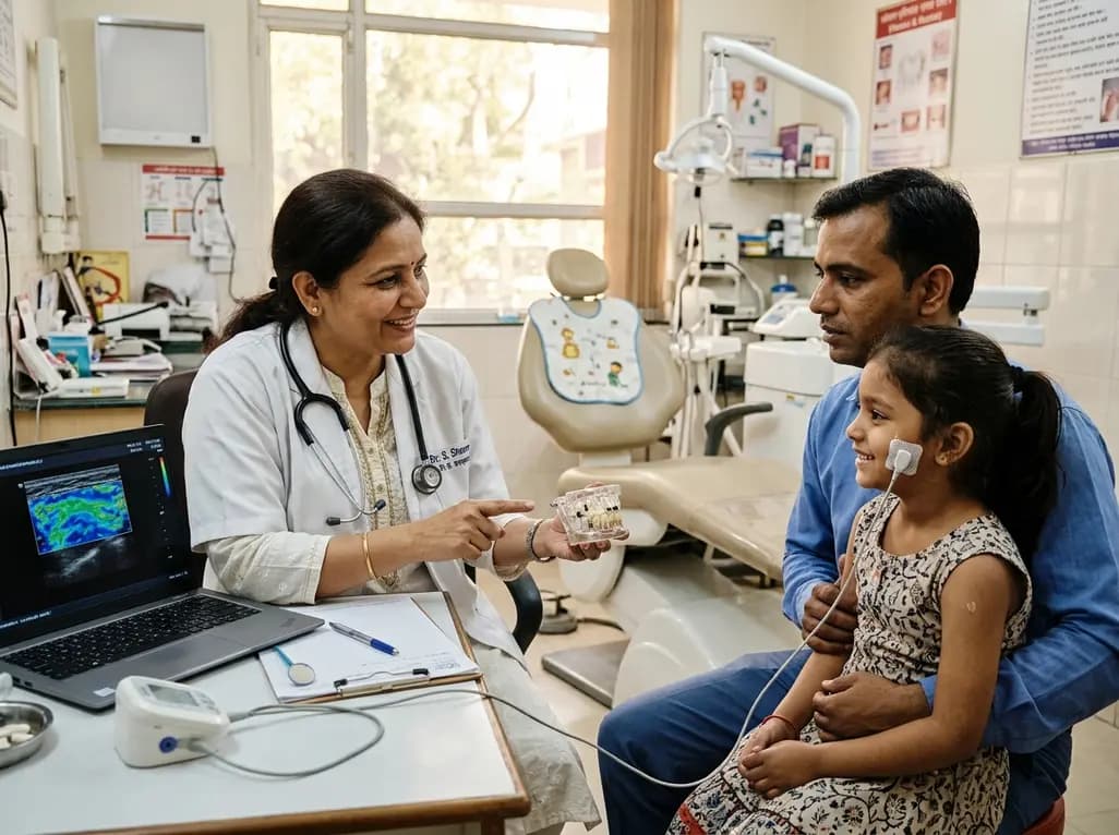
Untreated unilateral dental caries represents a pervasive oral health challenge that frequently forces young patients to adopt asymmetric chewing habits. When children experience localized dental pain or occlusal breakdown, they intuitively favor the healthy side of the arch. Over time, clinicians often wonder how this preferential chewing influences masticatory muscle morphology and facial growth dynamics. Pediatric dentists in India and worldwide regularly encounter children presenting with cavitated primary or permanent molars who demonstrate altered jaw movement. Although restoring anatomical form and function remains the standard intervention, objective data regarding neuromuscular recovery have remained limited. High-resolution ultrasonography now permits non-invasive visualization of superficial facial musculature in clinical pediatric settings. By assessing tissue dimensions at rest and maximal clenching, clinicians can quantify functional symmetry. Consequently, understanding whether restorative care reverses asymmetrical muscular strain is essential for comprehensive pediatric rehabilitation. Clinicians must recognize that anatomical restoration of teeth may initiate subtle physiological adaptations throughout the stomatognathic apparatus. Therefore, evaluating muscular behavior provides crucial insights into complete functional recovery beyond mere cavity filling.
To clarify these neuromuscular patterns, researchers conducted a prospective clinical trial involving twenty-five children between six and twelve years of age. Each participant presented with active unilateral dental caries requiring restorative intervention. The investigators utilized validated diagnostic tools, including the International Caries Detection and Assessment System II and the Decayed, Missing, and Filled Teeth index. Before initiating treatment, the team performed bilateral ultrasonographic examinations to capture baseline anatomical metrics. Specifically, they recorded the thickness of the masseter and temporalis muscles during relaxation and maximal voluntary contraction. In addition, examiners measured the anterior digastric muscle thickness at rest to evaluate lower jaw stabilizers. Following comprehensive restorative therapy that eliminated cavitated lesions, investigators re-evaluated all participants at three and six months postoperatively. This structured longitudinal design enabled clinicians to monitor whether removing unilateral pain and occlusal interference promoted symmetrical bilateral muscle development. Furthermore, by evaluating both the affected and unaffected sides, the researchers established an internal reference standard. Thus, the protocol provided a rigorous framework to assess muscular adaptations during active juvenile growth.
The primary objective centered on determining whether dental restorations altered muscle dimensions across the six-month follow-up window. Surprisingly, the data demonstrated no statistically significant longitudinal changes in muscle thickness on either the carious or non-carious side. Both the masseter and temporalis muscles maintained stable thickness measurements during rest and functional clenching throughout all observation periods. Similarly, the anterior digastric muscle showed no dimensional fluctuation after restorative therapy. These findings indicate that short-term restoration of unilateral dental caries does not trigger rapid structural hypertrophy or atrophy in pediatric jaw muscles. In growing children, baseline muscle dimensions appear remarkably resilient to localized dental decay over intermediate intervals. Moreover, habitual asymmetric mastication might require an extended period to establish measurable morphological asymmetry. Alternatively, bilateral compensatory chewing patterns may develop swiftly without causing macroscopic muscle bulk alterations. Therefore, conventional ultrasound thickness alone cannot fully capture the nuanced physiological adaptations occurring after dental rehabilitation. Clinicians must consequently look beyond macroscopic thickness when assessing masticatory recovery in pediatric patients.
Because conventional ultrasonography showed stable dimensions, researchers applied strain elastography to evaluate internal tissue stiffness and biomechanical elasticity. Strain elastography measures relative tissue deformation under mechanical stress, generating semi-quantitative red, green, and blue pixel distributions. In this color-coding scheme, varying hues represent distinct elasticity gradients within the masseter muscle tissue. On the carious side, the masseter exhibited no significant elastographic alterations following restorative dental therapy. However, the non-carious side revealed remarkable longitudinal shifts in color proportions over the six-month observation timeframe. Specifically, investigators detected significant differences in resting red pixel proportions, as well as contraction green and blue proportions. Pairwise statistical analysis confirmed a significant change in contraction green pixels between baseline and six months, displaying a moderate effect magnitude. Consequently, these findings indicate that the habitual chewing side undergoes noticeable biomechanical modulation once bilateral mastication resumes. Although muscle thickness remained unchanged, internal elasticity patterns adapted to redistributed masticatory loads. Hence, strain elastography successfully detects subtle biomechanical reorganization that standard dimensional imaging completely overlooks.
These clinical findings provide valuable insights for dental practitioners managing unilateral carious lesions in pediatric patients. In daily practice, dentists frequently focus solely on restoring tooth structure and alleviating localized discomfort. However, unilateral dental lesions inherently distort bilateral chewing symmetry, creating functional reliance on the healthy contralateral side. As this prospective investigation demonstrates, restoring occlusal contact prompts measurable changes in masseter elasticity on the previously favored side. Therefore, successful dental treatment does not merely seal margins; it also initiates neuromuscular re-equilibration across the craniofacial complex. Clinicians should educate parents that restorative care restores normal oral physiology and balanced functional stimulation for growing jaw structures. Additionally, the study underscores that strain elastography offers exceptional sensitivity for monitoring soft tissue biomechanics without exposing pediatric patients to ionizing radiation. While macroscopic muscle dimensions remain stable over six months, qualitative tissue changes demonstrate ongoing functional remodeling. Consequently, future pediatric dental protocols might incorporate functional muscle assessments to ensure holistic rehabilitation. Ultimately, early intervention protects balanced mastication and supports optimal long-term craniofacial development.
While these prospective results offer compelling preliminary evidence, several critical questions warrant further scientific exploration. The study evaluated twenty-five pediatric participants over a six-month duration, which represents a relatively concise developmental window. Because juvenile masticatory structures grow dynamically, extended multicenter trials with larger cohorts could clarify long-term neuromuscular outcomes. Furthermore, combining strain elastography with surface electromyography could correlate tissue elasticity directly with bilateral bite force distribution. Researchers should also examine how different restorative materials and full-coverage crowns influence masticatory re-equilibration over time. In India, where childhood caries prevalence remains high across diverse socioeconomic strata, establishing normative ultrasound values could refine interceptive care. Moreover, investigators must validate the clinical relevance of elastographic color shifts in preventing future temporomandibular disorders. Understanding how early unilateral pain modifies permanent motor habits will strengthen preventive oral health guidelines. Thus, continuing interdisciplinary research between pediatric dentistry and functional imaging promises to enhance diagnostic precision. By adopting advanced non-invasive diagnostic tools, clinicians can deliver comprehensive care that protects both dentition and masticatory physiology.
Unilateral dental caries frequently causes pain or mechanical food impaction, prompting children to avoid chewing on the affected side. Consequently, children establish habitual unilateral mastication, directing bite forces almost entirely toward the unaffected side. This asymmetric chewing alters local functional demands on the masseter and temporalis muscles. Over time, these compensatory motor patterns modify internal muscle elasticity and strain distribution, even when macroscopic muscle thickness appears unchanged on ultrasound.
Muscle thickness did not change significantly because a six-month postoperative interval is relatively brief for gross morphological remodeling. In growing pediatric patients, masticatory muscle dimensions exhibit baseline stability despite localized oral pathology. Additionally, children compensate for unilateral chewing through complex bilateral muscle coordination without developing macroscopic hypertrophy. Structural thickness changes generally require prolonged functional loads or extreme atrophy. Therefore, conventional ultrasonographic thickness assessments cannot detect subtle short-term biomechanical adaptations after restorative therapy.
Strain elastography evaluates tissue stiffness and biomechanical elasticity under gentle mechanical compression, whereas conventional B-mode ultrasonography only visualizes anatomical thickness and cross-sectional morphology. By converting deformation patterns into semi-quantitative color-coded pixel maps, elastography reveals functional changes within myofibers. In pediatric patients, this advanced technique detects internal muscular reorganization on the non-carious chewing side before structural dimensional shifts manifest, providing clinicians with sensitive diagnostic information regarding physiological recovery.
Disclaimer: This content is for informational and educational purposes only. It should not be used as a substitute for professional medical advice, diagnosis, or treatment. Always seek the advice of your physician or other qualified healthcare provider with any questions you may have regarding a medical condition. Refer to the latest local and national guidelines for clinical practice.
References
Demiroğlu M et al. Longitudinal Changes in Masticatory Muscle Morphology and Elastographic Characteristics Following Restorative Treatment of Unilateral Dental Caries in Children: A Prospective Clinical Study. J Oral Rehabil. 2026 Oct 30. doi: 10.1111/joor.70362. PMID: 42963132.
Kilic SC, et al. Ultrasonographic measurement of masseter muscle thickness and elasticity in patients with symptomatic apical periodontitis. Clin Oral Investig. 2026;30(6):312. doi: 10.1007/s00784-026-05689-1.
Patil S, et al. Application of Strain Elastography in Dentistry: A Systematic Review. Cureus. 2024;16(10):e70695. doi: 10.7759/cureus.70695.

Read summarized clinical updates, watch expert medical content, and earn CME certifications right from your smartphone.
Open your smartphone camera to scan and install.





A prospective clinical study reveals that restoring unilateral dental caries in children does not alter masticatory muscle thickness over six months. However, strain elastography showed significant functional shifts on the non-carious side, highlighting subtle neuromuscular adaptations after dental rehabilitation.
Today

New evidence demonstrates that ageing and female sex remodel lymphocyte compartments, weakening peripheral T-cell tolerance and lowering the threshold for red blood cell autoantibody production in warm autoimmune haemolytic anaemia.
Today
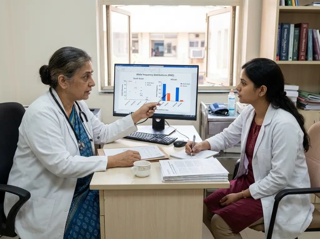
Large genomic databases like gnomAD and All of Us offer crucial insights into the population prevalence of partial RHD genotypes. Identifying these variants helps clinicians mitigate anti-D alloimmunization risks in transfusion medicine and obstetric care across diverse ancestral populations.
Today
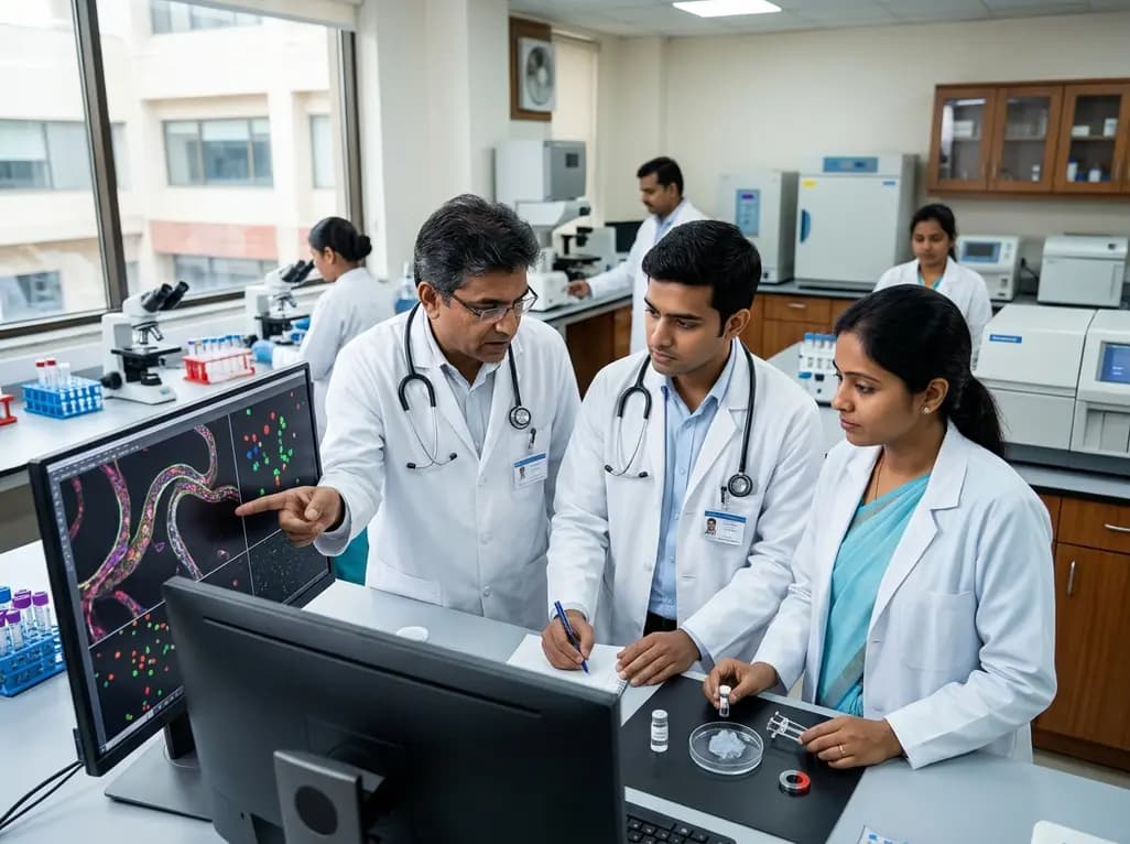
A novel magnetoactive hydrogel platform demonstrates that vascular curvature independently guides endothelial mechanosensing. Elevated curvature triggers nuclear YAP translocation, VE-cadherin junction thinning, and eNOS membrane shifts, offering critical insights into arterial tortuosity and aneurysm pathology.
Today
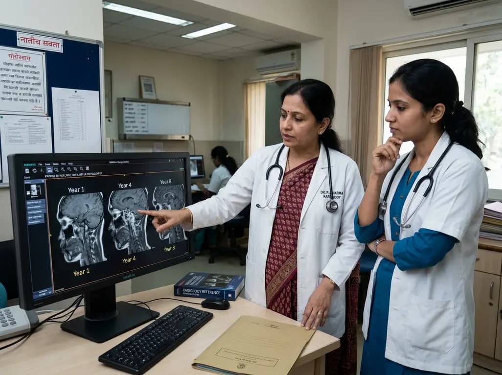
An eight-year longitudinal imaging follow-up reveals severe temporomandibular joint remodeling in nodulosis-arthropathy-osteolysis (NAO) syndrome. Despite extensive osseous destruction, the patient maintained functional jaw opening without TMJ interventions, highlighting the role of conservative management.
Today

Discover essential clinical insights into AGLP1-type amyloidosis, a rare localized complication of glucagon-like peptide-1 analog injections. Learn how histopathology and mass spectrometry differentiate benign iatrogenic amyloidomas from systemic disease, preventing misdiagnosis and unwarranted therapies.
Today
