

Loading, please wait...
Loading, please wait...

Nodulosis-arthropathy-osteolysis syndrome represents an exceptionally rare autosomal recessive skeletal dysplasia within the multicentric osteolysis spectrum. Pathogenic loss-of-function variants in the MMP2 gene cause this progressive disorder. Consequently, affected individuals experience progressive carpal and tarsal osteolysis, subcutaneous nodulosis, generalized osteoporosis, and severe arthropathy. Although peripheral appendicular joint destruction is a hallmark of the condition, clinicians infrequently characterize craniofacial manifestations. Recent clinical findings provide critical insights into longitudinal NAO syndrome TMJ alterations over an eight-year observation period. Researchers evaluated an adult female with genetically confirmed disease using serial panoramic radiography and cone-beam computed tomography. Notably, the patient demonstrated extensive structural degeneration of the craniomandibular articulation without crippling functional impairment. Understanding these anatomical changes allows clinicians to avoid misinterpreting osteolytic remodeling as ordinary temporomandibular joint dysfunction. Furthermore, systemic skeletal diseases frequently mimic juvenile idiopathic arthritis, leading to inappropriate therapeutic delays. Because matrix metalloproteinases regulate extracellular matrix turnover, MMP2 deficiency disrupts physiological osseous homeostasis throughout the entire axial and craniofacial skeleton. Therefore, multidisciplinary teams must recognize how underlying connective tissue disorders uniquely reshape the stomatognathic system over time.
Despite profound anatomical remodeling, the clinical presentation often diverges sharply from classic temporomandibular disorder expectations. Over eight years of comprehensive follow-up, the patient reported only occasional articular sounds without debilitating pain or jaw locking. Moreover, she maintained an active lifestyle with preserved chewing capabilities and unhindered speech. Physical examination revealed mild crepitus over the left condyle during functional excursions. Nevertheless, palpation demonstrated no tenderness across the masticatory muscles, lateral poles, or retrodiscal tissues. Range of motion remained remarkably stable across multiple annual evaluations. Specifically, her maximum comfortable mouth opening reached 36 mm, unassisted opening measured 37 mm, and assisted opening attained 38 mm. Clinicians detected neither opening deviations nor pathological mandibular deflections during dynamic jaw movements. Therefore, jaw functionality remained adequate for daily mastication despite evident articular degeneration. In addition, these physiological observations underscore the capacity of surrounding soft tissues to adapt dynamically to chronic osseous regression. Clinicians frequently encounter severe structural degradation that fails to produce proportional subjective pain. Consequently, dental professionals should refrain from aggressive interventions when physical function remains essentially intact.
Advanced imaging modalities offer crucial insights into the progressive destruction of articular morphology. Baseline cone-beam computed tomography demonstrated marked bilateral condylar flattening, extensive cortical thinning, and pronounced surface irregularity. Additionally, the cross-sectional views revealed abnormal subchondral trabecular architecture extending into the temporal components of the joint. Serial panoramic radiographs taken over eight consecutive years documented persistent, bidirectional osseous remodeling. Specifically, progressive erosive changes gradually eroded the mandibular condyles and severely flattened the articular eminences bilaterally. Consequently, the glenoid fossae transformed into shallow depressions, fundamentally altering normal craniomandibular anatomy. However, the subchondral bone managed to achieve cortical consolidation rather than suffering catastrophic joint collapse. Thus, serial panoramic radiographs served as an effective low-dose monitoring tool for long-term surveillance. Furthermore, high-resolution imaging helped clinicians differentiate these changes from active osteomyelitis, aggressive neoplasia, and inflammatory arthritides. Understanding these characteristic radiological patterns prevents unnecessary surgical biopsies. Therefore, radiologists must synthesize structural imaging findings with serial functional evaluations to guide prudent clinical decisions.
The patient received annual intravenous zoledronic acid throughout the surveillance period to control systemic skeletal osteolysis. Nitrogen-containing bisphosphonates inhibit osteoclast-mediated bone resorption, thereby helping preserve skeletal density in progressive genetic disorders. However, prolonged antiresorptive therapy carries a recognized risk of medication-related osteonecrosis of the jaw. Clinicians must exercise extreme vigilance whenever evaluating osteolytic jaw alterations in patients on potent bisphosphonates. Interestingly, the progressive condylar resorption observed in this patient occurred without mucosal dehiscence, exposed bone, or localized infection. In addition, the antiresorptive treatment may have stabilized subchondral bone remodeling, preventing catastrophic structural fragmentation. Clinicians must meticulously distinguish genetic multicentric osteolysis from bisphosphonate-induced bone necrosis. Consequently, routine oral surveillance and preventative dental care remain essential components of multidisciplinary management. Furthermore, collaborative dialogue between medical geneticists, rheumatologists, and maxillofacial surgeons ensures balanced risk stratification. Ultimately, this careful oversight avoided unnecessary cessation of essential bone-strengthening systemic treatments.
This longitudinal case underscores the foundational clinical principle of treating the patient rather than isolated radiographic images. Because severe radiographic abnormalities can coexist with minimal symptoms, invasive interventions are rarely justified without functional decline. Healthcare providers initially might feel tempted to recommend splint therapy, joint arthrocentesis, or total joint replacement. However, surgical manipulation in patients with genetic matrix disorders carries substantial risks of non-union, accelerated bone lysis, and prosthetic failure. Instead, conservative surveillance preserved the patient's comfortable jaw mobility and prevented unnecessary iatrogenic morbidity. Furthermore, maintaining jaw function through gentle physiological use supported compensatory muscular adaptations. Clinicians should only consider surgical or interventional therapies if true mechanical obstruction, intractable pain, or severe functional limitation arises. In conclusion, managing rare skeletal dysplasias requires diagnostic restraint, serial clinical monitoring, and comprehensive interdisciplinary cooperation. Thus, healthcare providers protect patient well-being by prioritizing functional preservation over radiographic perfection.
NAO syndrome causes progressive osteolysis and structural remodeling within the temporomandibular articulation due to underlying MMP2 deficiency. Radiographic examinations typically reveal bilateral condylar flattening, thinning of cortical margins, erosive changes, and shallow glenoid fossae. Interestingly, patients often experience minimal pain or functional impairment despite severe anatomical deterioration. Clinicians observe preserved jaw opening because compensatory neuromuscular mechanisms adapt to chronic degenerative changes over time.
Conservative management remains preferred because patients frequently maintain normal jaw opening and masticatory function despite alarming radiographic osteolysis. Invasive surgical procedures or aggressive splinting can trigger unpredictable tissue reactions, impaired bone healing, and catastrophic failure in patients with genetic matrix disorders. Therefore, healthcare providers prioritize clinical observation, routine oral hygiene, and physical jaw exercise unless intractable pain, open bites, or severe functional limitations develop.
Physicians frequently prescribe antiresorptive bisphosphonates such as zoledronic acid to treat generalized osteopenia and peripheral osteolysis in NAO syndrome. However, long-term intravenous bisphosphonates increase the potential risk of medication-related osteonecrosis of the jaw. Therefore, clinicians must carefully differentiate benign genetic condylar remodeling from osteonecrotic lesions. Regular non-invasive dental examinations ensure oral mucosal integrity without interrupting essential systemic bone-targeted therapies.
Disclaimer: This content is for informational and educational purposes only and should not be considered professional medical advice. Always consult a qualified healthcare provider for diagnosis and treatment decisions. Refer to the latest local and national guidelines for clinical practice.
References

Read summarized clinical updates, watch expert medical content, and earn CME certifications right from your smartphone.
Open your smartphone camera to scan and install.





An eight-year longitudinal imaging follow-up reveals severe temporomandibular joint remodeling in nodulosis-arthropathy-osteolysis (NAO) syndrome. Despite extensive osseous destruction, the patient maintained functional jaw opening without TMJ interventions, highlighting the role of conservative management.
Today
A user-centered design study outlines a theoretical hybrid COPD care pathway combining remote patient monitoring with standard clinical care. Stakeholders emphasize a dedicated coordinator role to manage device onboarding, data review, and timely alert escalation to curb exacerbations and readmissions.
Today

A prospective study reveals significant first-trimester trace-element alterations in pregnant women with epilepsy. Lower serum selenium independently predicted subsequent seizure recurrence despite prior remission, highlighting the vital clinical role of micronutrient profiling during antenatal care.
Today
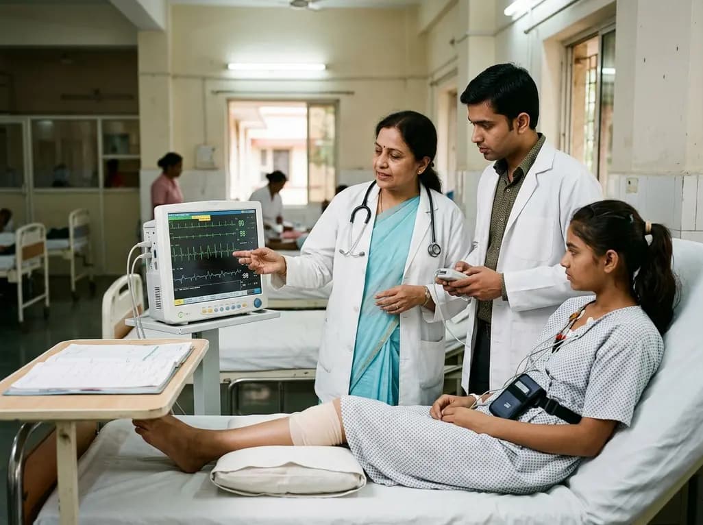
A 15-year-old patient presented with knee arthralgia and acute rheumatic fever, exhibiting complex arrhythmias including AV blocks and non-sustained ventricular tachycardia despite lacking severe valvulitis. This highlights the importance of extended Holter monitoring in pediatric rheumatic carditis.
Today

Discover essential clinical insights into AGLP1-type amyloidosis, a rare localized complication of glucagon-like peptide-1 analog injections. Learn how histopathology and mass spectrometry differentiate benign iatrogenic amyloidomas from systemic disease, preventing misdiagnosis and unwarranted therapies.
Today
