

Loading, please wait...
Loading, please wait...

Human blood vessels exhibit intricate three-dimensional architectures with pronounced nonplanar curvatures. Clinicians frequently encounter severe structural variations during progressive arterial remodeling, such as intracranial aneurysms and arterial tortuosity. Historically, cardiovascular research has focused primarily on hemodynamic shear stress as the main driver of vascular physiology. However, intrinsic tissue geometry also exerts profound biophysical signals. Curvature directly regulates endothelial cell mechanosensing, yet isolating geometric inputs from fluid shear stress has long remained a technical hurdle. Traditional in vitro cell culture models rely on static, rigid substrates that fail to recreate dynamic tissue deformations. Consequently, researchers could not easily determine whether vessel curvature itself directly governs cellular behavior. To address this knowledge gap, investigators developed an innovative magnetoactive biomaterial system. This approach simulates arterial tortuosity in real time without introducing confounding fluid shear stresses. Furthermore, the findings reveal that endothelial cells perceive physical curvature as an autonomous physical instruction. Therefore, physical geometry acts alongside fluid dynamics to orchestrate vascular homeostasis and pathological vascular remodeling. In addition, understanding these isolated geometric pathways provides crucial insight into early endothelial dysfunction across convoluted vascular beds.
Cardiovascular bioengineers designed a magnetoactive hydrogel platform to examine curvature independently of fluid flow. Specifically, the team embedded responsive magnetic microparticles within compliant hydrogel matrices to enable dynamic, on-demand surface deformation. By modulating an external magnetic field, researchers can dynamically curve endothelial monolayers to match clinical tortuosity indices. In clinical practice, clinicians measure tortuosity indices to evaluate aneurysm rupture risk and arterial elongation. Traditional microfluidic devices cannot decouple luminal shear stress from structural topography. In contrast, this magnetoactive system bends cultured endothelial monolayers reversibly without altering surrounding fluid flow. Furthermore, the platform allows real-time observation of single-cell adaptations during mechanical deformation. Investigators can precisely recreate convex and concave microenvironments that mirror human vascular bends. As a result, the technology establishes an unprecedented experimental tool for vascular mechanobiology. Researchers can now disentangle physical curvature from continuous blood flow. Consequently, this innovation offers fundamental insights into how geometric distortions trigger early molecular changes in vascular disease. Moreover, the responsive matrix accurately mimics the viscoelastic modulus observed in human arterial tissue, ensuring physiological translational relevance.
The study demonstrated that mechanical curvature strongly activates Yes-associated protein within endothelial cell sheets. Yes-associated protein, known as YAP, functions as an essential transcriptional coactivator in the Hippo signaling pathway. Under basal resting conditions, healthy endothelial cells keep YAP phosphorylated and sequestered within the cytoplasm. However, mechanical strain causes YAP to dephosphorylate and accumulate inside the cell nucleus. The hydrogel platform showed that increasing substrate curvature sharply elevated nuclear YAP accumulation. Notably, the strongest nuclear translocation occurred in convex regions rather than concave zones. Convex topography places endothelial cells under heightened membrane tension and cytoskeletal stretching. Consequently, stress fibers pull on nuclear pores, which facilitates rapid YAP nuclear entry. In addition, nuclear YAP drives transcriptional programs linked to cellular proliferation, inflammation, and vascular remodeling. Therefore, vessel convexity creates localized biochemical signals that may accelerate vascular wall degeneration. These findings highlight that geometric asymmetry alone triggers pro-remodeling molecular programs across arterial curves. Furthermore, this localized transcriptional activation explains why pathological remodeling consistently localizes to outer vessel curvatures in hypertensive patients.
Vascular endothelial cadherin, or VE-cadherin, maintains strict endothelial barrier integrity throughout human blood vessels. Robust adherens junctions prevent vascular hyperpermeability, macromolecular leakage, and inflammatory cell extravasation into subendothelial layers. Interestingly, the study revealed that elevated curvature caused significant reduction in VE-cadherin junctional thickness. Cells located on convex curved surfaces exhibited disorganized adherens junctions and diminished junctional stability. Consequently, physical curvature compromises the protective endothelial barrier independently of hemodynamic shear forces. In clinical medicine, loss of junctional integrity directly contributes to atherogenesis, lipid infiltration, and mural destabilization. Furthermore, weakened intercellular junctions facilitate plasma protein extravasation that accelerates arterial aneurysm expansion. Researchers observed that mechanical stretching during curvature directly alters junctional remodeling cascades. Thus, vascular bending physically loosens inter-endothelial contacts in predictable spatial patterns. Accordingly, vascular segments with high curvature represent inherently fragile regions where vascular leakage and inflammatory infiltration readily occur. Moreover, therapeutic strategies stabilizing VE-cadherin complexes could potentially protect tortuous arterial bends from accelerated atherosclerotic degeneration.
Endothelial nitric oxide synthase, commonly designated as eNOS, plays an indispensable role in maintaining basal vascular tone. In quiescent endothelium, eNOS resides primarily in intracellular caveolae and Golgi compartments. Upon physical stimulation, the enzyme translocates toward the plasma membrane to produce protective nitric oxide. The study established that acute hydrogel curvature significantly increased eNOS membrane localization. Specifically, convex curvature stimulated rapid translocation of eNOS to the peripheral cell membrane. However, this redistribution coincided with reduced junctional thickness and heightened nuclear YAP activity. Nitric oxide generation in unstable or inflamed microenvironments frequently leads to peroxynitrite formation rather than healthy vasodilation. Moreover, persistent geometric distortion exhausts this compensatory reserve, culminating in local endothelial dysfunction. These findings offer profound implications for managing clinical conditions like arterial tortuosity syndrome and cerebral aneurysms. Clinicians recognize that aneurysms and atherosclerotic plaques develop preferentially along convex vessel arches. By proving that tissue curvature directly drives endothelial barrier failure, this research uncovers novel mechanobiological targets. Consequently, targeting curvature-sensitive pathways may prevent aneurysm progression and optimize future endovascular graft designs.
Vascular curvature alters membrane tension and cytoskeletal filament alignment within endothelial monolayers without requiring hemodynamic flow. Specifically, physical bending stretches the apical and basal plasma membranes over three-dimensional topographies. This localized tensile deformation directly pulls on integrins and adherens junctions. Consequently, the mechanical strain dephosphorylates Yes-associated protein and promotes its translocation into the nucleus. Therefore, cells respond biochemically to geometric deformation as an autonomous biophysical signal.
Convex vessel regions subject endothelial cells to pronounced outward radial stretching and tensile mechanical forces. In contrast, concave regions experience compressive forces that restrain cytoskeletal remodeling. The outward tensile stress on convex surfaces flattens the cell nucleus and opens nuclear pores. Consequently, Yes-associated protein rapidly translocates from the cytoplasm into the nucleus to activate pro-remodeling genes. Therefore, convex vessel arches exhibit heightened mechanotransduction activity and cellular proliferation compared to concave geometries.
Vascular endothelial cadherin preserves junctional tightness and controls vascular permeability across the circulatory system. When geometric curvature reduces VE-cadherin thickness, endothelial barriers become permeable and fragile. Consequently, plasma lipids, inflammatory monocytes, and cytokines infiltrate the subendothelial matrix. Furthermore, this structural breakdown destabilizes the arterial wall, which predisposes tortuous vessels to aneurysm expansion, dissection, and atheroma formation. Thus, curvature-induced junctional loss establishes a vulnerable site for early vascular pathology before clinical symptoms emerge.
Disclaimer: This content is for informational and educational purposes only... Refer to the latest local and national guidelines for clinical practice.
References

Read summarized clinical updates, watch expert medical content, and earn CME certifications right from your smartphone.
Open your smartphone camera to scan and install.





A novel magnetoactive hydrogel platform demonstrates that vascular curvature independently guides endothelial mechanosensing. Elevated curvature triggers nuclear YAP translocation, VE-cadherin junction thinning, and eNOS membrane shifts, offering critical insights into arterial tortuosity and aneurysm pathology.
Today

New evidence demonstrates that ageing and female sex remodel lymphocyte compartments, weakening peripheral T-cell tolerance and lowering the threshold for red blood cell autoantibody production in warm autoimmune haemolytic anaemia.
Today
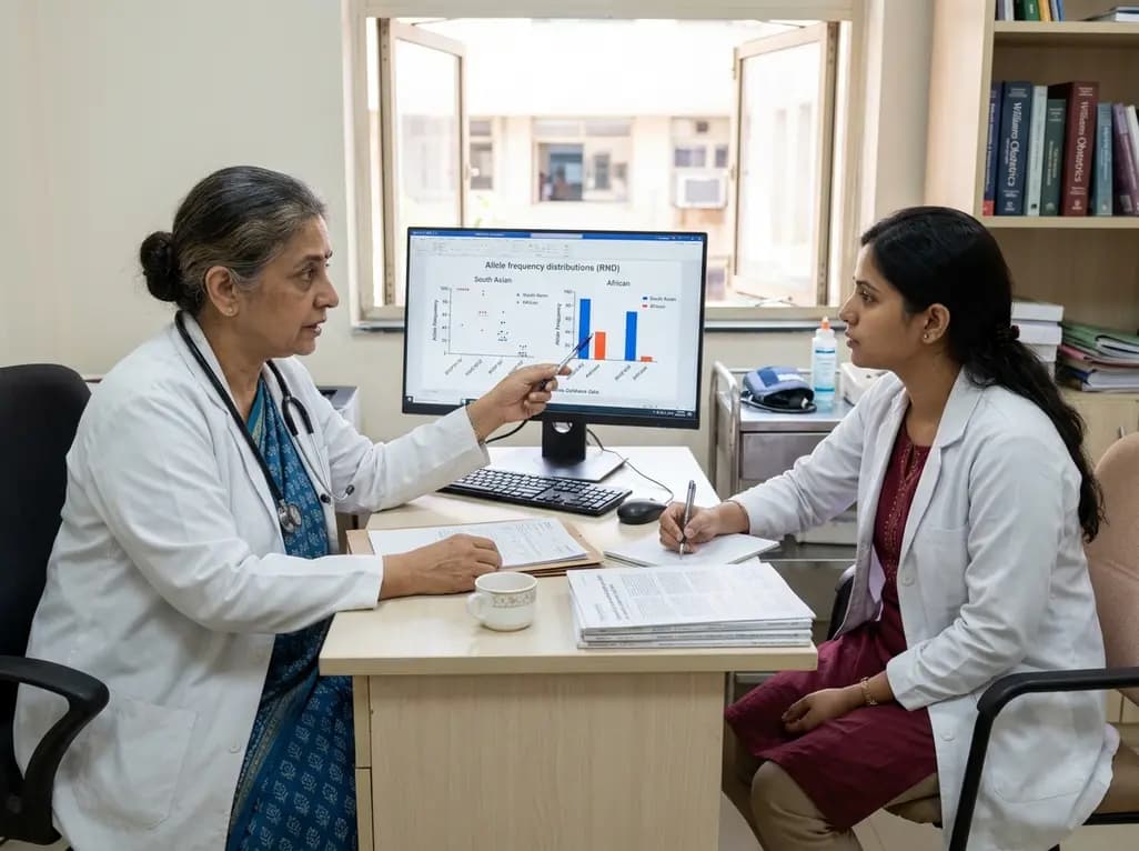
Large genomic databases like gnomAD and All of Us offer crucial insights into the population prevalence of partial RHD genotypes. Identifying these variants helps clinicians mitigate anti-D alloimmunization risks in transfusion medicine and obstetric care across diverse ancestral populations.
Today
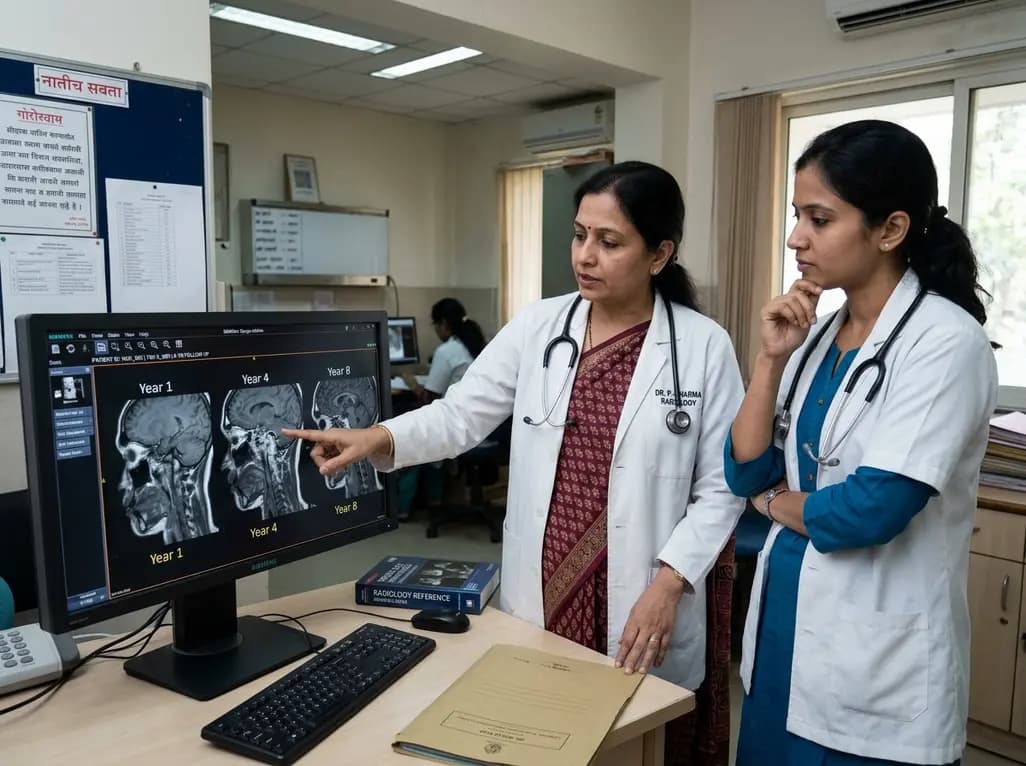
An eight-year longitudinal imaging follow-up reveals severe temporomandibular joint remodeling in nodulosis-arthropathy-osteolysis (NAO) syndrome. Despite extensive osseous destruction, the patient maintained functional jaw opening without TMJ interventions, highlighting the role of conservative management.
Today

Discover essential clinical insights into AGLP1-type amyloidosis, a rare localized complication of glucagon-like peptide-1 analog injections. Learn how histopathology and mass spectrometry differentiate benign iatrogenic amyloidomas from systemic disease, preventing misdiagnosis and unwarranted therapies.
Today
