.webp&w=384&q=75)

Loading, please wait...
Loading, please wait...

The mandibular buccal shelf has emerged as a premier extra-alveolar site for placing temporary skeletal anchorage devices in contemporary orthodontics. Clinicians frequently utilize this anatomical zone to accomplish complex tooth movements without interfering with dental roots. For instance, practitioners achieve reliable mandibular total arch distalization, molar uprighting, and Class III corrections from this site. Successful skeletal anchorage depends directly on local cortical bone thickness, which provides crucial primary stability during force application. Cone beam computed tomography offers unmatched three-dimensional visualization of this bony ridge, allowing dental specialists to evaluate bone quality accurately. Because the buccal shelf consists of dense cortical bone, it readily supports substantial biomechanical loading without loosening. However, local bone dimensions vary significantly among clinical patients. Practitioners must therefore understand how specific anatomical sites, vertical heights, and skeletal variations dictate cortical bone availability. Furthermore, thorough preoperative imaging helps the clinician design a tailored surgical plan that accounts for subtle individual anatomical variations. Consequently, detailed radiographic assessment prior to mini-screw placement remains an indispensable step in modern orthodontic treatment.
Patients exhibit diverse craniofacial morphology, which substantially alters the structural architecture of the mandibular alveolar process. Cephalometric analysis routinely categorizes individuals into horizontal, normal, and vertical growth patterns. Recent cone beam computed tomography investigations demonstrate that horizontal growers consistently display greater cortical bone thickness along the posterior mandible. In contrast, vertical hyperdivergent growers present thinner cortical plates and reduced overall alveolar width across multiple sites. Therefore, clinicians must exercise greater caution when treating high-angle vertical patients, as insufficient bone depth increases implant failure risks. Conversely, hypodivergent horizontal growers generally provide a robust bony foundation that facilitates superior mechanical interlock for cortical screws. Normal growers present intermediate bone dimensions that typically accommodate standard bone screws without extensive modifications. Furthermore, these morphological differences highlight why a standardized insertion protocol cannot fit every skeletal phenotype. Orthodontists should adjust their biomechanical strategies according to the individual vertical facial dimensions. Thus, identifying the skeletal growth pattern allows clinicians to anticipate bone availability accurately before selecting screw dimensions. Additionally, acknowledging these structural variations prevents clinicians from provoking thermal necrosis or localized microfractures during insertion.
Primary stability of extra-alveolar mini-screws correlates strongly with the specific height and angulation chosen during clinical placement. Researchers evaluate cortical bone thickness at distinct heights, specifically five and seven millimeters apical to the alveolar bone crest. Measurements confirm that cortical bone thickness increases significantly as one moves farther apically from the crest. Therefore, an insertion level of seven millimeters provides substantially greater cortical support compared to more coronal positions. In addition, the insertion angulation relative to the occlusal plane markedly affects the total path length of the screw. Insertion at an eighty-degree angle typically engages a deeper cross-section of dense cortex than an entry at seventy degrees. Consequently, steeper insertion angles enhance primary mechanical retention while reducing the danger of root proximity during traction. However, excessively steep angulations may challenge soft tissue clearance and cause mechanical impingement against the movable buccal mucosa. Clinicians must balance these geometric parameters to achieve maximum bony purchase while preserving surrounding mucogingival health. Furthermore, selecting the correct insertion height ensures sufficient clearance from adjacent root concavities during space closure.
Selecting the precise anatomical site along the posterior arch remains essential for predictable skeletal anchorage. Cortical bone thickness varies across different landmarks, extending from the first molar to the distal root of the second molar. Anatomical evaluations consistently indicate that the buccal bone adjacent to the second molar provides the greatest cortical thickness. Specifically, the distobuccal region of the mandibular second molar exhibits the most favorable bone volume for temporary anchorage devices. Clinicians should initially target the interdental bone between the first and second molars or the buccal shelf lateral to the second molar. Moreover, inserting screws into attached gingiva rather than movable mucosa substantially reduces peri-implant inflammation and subsequent screw failure. Orthodontists often use longer mini-screws, such as ten to twelve millimeters in length, to traverse thick soft tissues. In addition, pre-drilling the dense cortical plate prevents microfractures and minimizes excessive insertion torque. Clinicians should also deliver steady, continuous manual force to maintain absolute axial control during driver rotation. By combining optimal site selection with appropriate surgical technique, clinicians ensure exceptional implant survival rates.
Although the posterior mandible offers dense anchorage, practitioners must navigate several critical anatomical boundaries to avoid serious complications. The inferior alveolar neurovascular canal courses through the mandibular body beneath the buccal shelf. Consequently, clinicians must maintain an adequate vertical safety buffer of at least two millimeters above the mandibular canal. Detailed preoperative cone beam computed tomography allows precise measurement of this vertical clearance across different cross-sections. In addition, clinicians must carefully avoid traumatizing adjacent periodontal ligament spaces and molar root structures during screw advancement. If a patient experiences sudden sharp pain during placement under local infiltration, the practitioner must pause immediately and re-evaluate trajectory. Furthermore, post-insertion hygiene plays a pivotal role in preventing soft tissue overgrowth and peri-implant mucositis around the head of the screw. Patients should clean the screw head daily using chlorhexidine swabs or soft interdental brushes. Therefore, combining meticulous anatomical assessment with strict oral hygiene maintenance guarantees predictable orthodontic biomechanics and protects vital neurovascular structures. Similarly, regular clinical monitoring during routine adjustment visits permits prompt identification of any mucosal inflammation before it compromises skeletal stability.
Clinicians generally consider a cortical bone thickness of at least two millimeters necessary for sufficient primary stability of extra-alveolar mini-screws. Thinner cortical bone increases the risk of micro-mobility, which directly contributes to early screw loosening under orthodontic traction. Furthermore, adequate cortical bone thickness provides mechanical resistance against heavy lateral and distalization forces. Therefore, assessing bone volume using cone beam computed tomography ensures that selected insertion sites meet this essential anatomical threshold before device placement.
Skeletal growth patterns directly alter mandibular cortical bone architecture and overall bone density. Horizontal, hypodivergent growers exhibit significantly thicker cortical bone plates, offering superior primary stability for extra-alveolar anchorage devices. In contrast, vertical hyperdivergent growers possess noticeably thinner cortical bone, requiring greater clinical care to prevent anchorage failure. Therefore, orthodontists must customize mini-screw dimensions, insertion depths, and angulations based on individual cephalometric facial patterns to optimize clinical outcomes and prevent biomechanical loosening.
The buccal bone adjacent to the distobuccal root of the mandibular second molar offers the greatest cortical thickness. Placing mini-screws at seven millimeters apical to the alveolar crest engages significantly more bone than shallower placements. In addition, an insertion angulation of eighty degrees to the occlusal plane maximizes cortical contact while safely avoiding dental roots. Consequently, practitioners achieve the highest retention and stability by combining this distobuccal site with steeper insertion angles.
Disclaimer: This content is for informational and educational purposes only. It is not intended to be a substitute for professional medical advice, diagnosis, or treatment. Refer to the latest local and national guidelines for clinical practice.
References

Read summarized clinical updates, watch expert medical content, and earn CME certifications right from your smartphone.
Open your smartphone camera to scan and install.





A CBCT study evaluates mandibular buccal shelf cortical bone thickness across horizontal, normal, and vertical growth patterns. Cortical thickness increases at greater insertion heights and steeper angulations, identifying optimal sites lateral to the second molar for temporary skeletal anchorage devices.
Today

The All India Institute of Medical Sciences has convened national health experts, policymakers, and state nodal officers to transform medical training for cardiometabolic disorders. This collaborative effort under the PEP initiative addresses training gaps to elevate chronic disease care across Indian healthcare.
Today

A recent qualitative study examines how college students seek digital sexual health information under persistent uncertainty. Discover the concept of functional trust, the clinical hazards of search abandonment, and evidence-based approaches clinicians can adopt to guide youth toward verified reproductive healthcare.
Today

A randomized controlled trial demonstrates that curated FOAMed videos and podcasts produce superior gains in high-risk ECG interpretation and acute coronary syndrome clinical decision-making compared with print-based materials among interprofessional emergency care teams.
Today
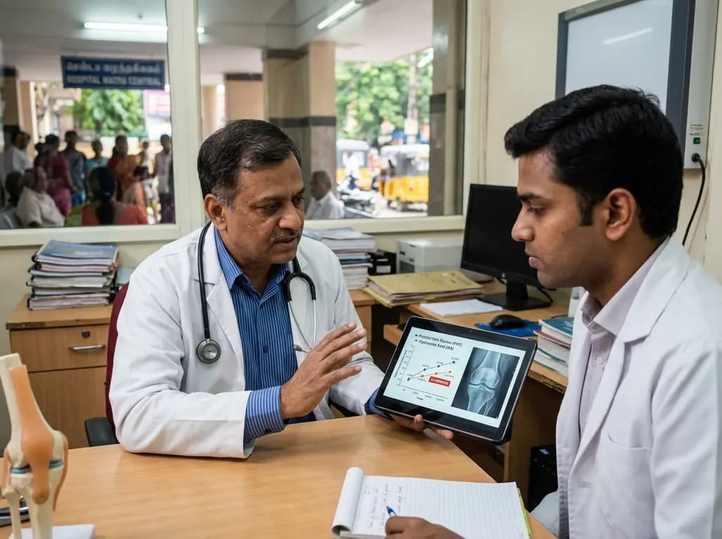
A systematic review and meta-analysis of 21 RCTs reveals that platelet-rich plasma provides small but statistically superior pain relief and functional improvements compared to hyaluronic acid at 12 months in knee osteoarthritis, influenced by platelet concentration and leukocyte enrichment.
Today

A multicenter real-world prospective study in Taiwan demonstrates that liraglutide effectively reduces body weight and BMI standard deviation score in adolescents with obesity. The therapy shows a favorable safety profile with mild gastrointestinal adverse events and zero treatment discontinuations due to intolerance.
Today
