.webp&w=384&q=75)

Loading, please wait...
Loading, please wait...
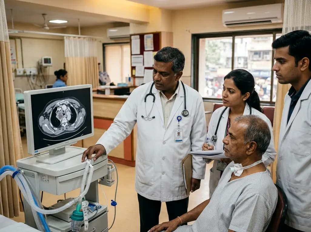
Surgical airway management is essential during complex head and neck oncologic resections, yet it carries rare but life-threatening risks. Among these complications, tracheostomy malignant seeding represents a grave clinical entity that requires urgent identification and aggressive intervention. Although surgeons routinely perform temporary tracheostomies to secure airway patency during major extirpative surgeries, tumor cells can occasionally dislodge and implant directly onto the freshly injured tracheal mucosa. Consequently, oncologists and surgical teams must understand the mechanisms of tumor seeding, recognize early clinical indicators, and implement strict procedural protocols to prevent this challenging oncologic occurrence.
Tracheostomy malignant seeding involves the accidental mechanical implantation or retrograde lymphatic migration of malignant cells to the tracheal lumen or stomal cutaneous margins. Although primary tracheal malignancies are exceedingly uncommon, secondary involvement following head and neck tumor ablation represents a recognized clinical phenomenon. Most recorded occurrences involve squamous cell carcinoma (SCC), with primary lesions located predominantly within the oral cavity, including the mandibular gingiva, floor of mouth, and tongue.
Furthermore, clinical evidence indicates that patients presenting with advanced T-stage disease and regional lymph node involvement demonstrate higher susceptibility to stomal seeding. The physical disruption of aggressive tumors during surgical manipulation provides an opportunity for exfoliated neoplastic cells to travel downward. When these viable cells encounter the raw, vascularized bed of a newly created tracheostomy stoma, they can successfully engraft. In addition, previous studies indicate that stomal recurrences typically manifest months to years after index surgery, making vigilant postoperative monitoring imperative.
The pathophysiology underlying tracheal and stomal tumor seeding involves multiple mechanical and biological factors. First, direct mechanical transplantation occurs when surgical instruments, endotracheal tubes, suction catheters, or gloves carry exfoliated tumor cells into the tracheal wound. Because the tracheal rings undergo surgical trauma during incision, the exposed submucosal tissue provides a fertile microenvironment for neoplastic engraftment.
Second, gravitational shedding of tumor cells during intraoral manipulation allows cancer fragments to pool in the subglottic space, especially if airway manipulation occurs before complete primary tumor resection. Third, retrograde lymphatic dissemination or direct extension from paratracheal lymph nodes (Level VI) can also drive stomal recurrence. In patients with extensive nodal disease, compromised regional lymphatic pathways may direct microscopic disease toward the peristomal lymphatic plexus. Therefore, differentiating between true mechanical tumor seeding, isolated regional recurrence, and contiguous extension requires comprehensive histopathological and radiological correlation.
Patients with stomal seeding frequently present with insidious respiratory symptoms that mimic benign postoperative stenosis or chronic tracheitis. Common initial complaints include progressive shortness of breath, stridor, persistent cough, and hemoptysis. Physical examination may reveal a firm, ulcerative, or exophytic mass around the prior tracheostomy scar, although intraluminal tracheal lesions can exist without visible external skin changes.
Consequently, thorough evaluation demands flexible fiberoptic tracheobronchoscopy to inspect the full extent of the tracheal mucosa and assess the degree of airway narrowing. In addition, contrast-enhanced computed tomography (CT) of the neck and chest, along with positron emission tomography (PET), helps evaluate local invasion, mediastinal extension, and distant metastases. Histological verification through tissue biopsy remains essential to confirm squamous cell carcinoma and exclude inflammatory granulation tissue. Because delayed diagnosis significantly worsens airway compromise, clinicians must maintain a low threshold for diagnostic bronchoscopy in any head and neck cancer survivor presenting with unexplained respiratory difficulty.
Managing malignant tracheal seeding presents formidable therapeutic challenges and requires coordinated multidisciplinary oncology care. Treatment selection depends on the patient's performance status, prior radiation exposure, disease extent, and the feasibility of achieving clear surgical margins. For localized and resectable recurrences, aggressive surgical intervention provides the best opportunity for locoregional disease control.
Surgical options include wide local excision of the stoma, partial tracheal resection, sleeve resection, or mediastinal tracheostomy, often combined with complex reconstructive tissue transfer, such as pectoralis major myocutaneous flaps or free tissue flaps. However, many patients present with unresectable mediastinal involvement or severe comorbidities that preclude extensive salvage surgery. In these challenging scenarios, palliative radiation therapy, concurrent chemoradiation, or systemic immunotherapy provides disease stabilization and symptom control. Furthermore, interventional pulmonology techniques, such as endoscopic laser debulking, airway stenting, and cryotherapy, offer rapid palliative relief for severe airway obstruction.
Given the aggressive nature of stomal recurrences, preventive operative protocols represent the most effective defense against malignant implantation. Surgical teams must adopt meticulous intraoperative precautions to minimize the risk of mechanical tumor contamination. First, surgeons should delay the creation of a temporary tracheostomy until the primary oral or pharyngeal tumor has undergone complete extirpation and resection margins are verified.
Second, operating teams must maintain strict instrument segregation. Instruments, drapes, suction tips, and gloves used during the tumor resection phase must never come into contact with the tracheostomy site. Third, vigorous saline or antiseptic irrigation of the pharynx and upper airway before performing tracheal incision helps clear loose cellular debris. Finally, for high-risk patients exhibiting aggressive tumor histology, advanced nodal burden, or subglottic extension, radiation oncologists should consider incorporating the tracheostomy stoma into postoperative adjuvant radiation fields.
The long-term prognosis for patients who develop tracheal or stomal malignant seeding remains generally unfavorable, with reported mortality rates exceeding 60%. Because stomal recurrence frequently signals aggressive tumor biology, survival largely depends on early clinical detection and the feasibility of radical surgical clearance. Patients who undergo successful complete surgical resection with negative margins demonstrate substantially higher disease-free survival compared to those receiving non-surgical palliation alone.
Therefore, head and neck oncology survivorship programs must emphasize diligent, long-term peristomal surveillance. Routine clinical examinations should include careful palpation of the neck and stomal scar, supplemented by periodic cross-sectional imaging and endoscopic airway inspection during the first two to three years of follow-up. Moreover, multidisciplinary tumor boards must actively participate in post-treatment decision-making to optimize functional outcomes, preserve airway integrity, and deliver individualized oncologic care.
The primary risk factors include advanced stage primary head and neck squamous cell carcinoma, extensive cervical lymph node metastases, and performing a tracheostomy before tumor excision. In addition, large ulcerative oral cavity lesions and surgical manipulation without proper instrument segregation substantially elevate the likelihood of shedding viable neoplastic cells into the open tracheal wound bed.
Surgical teams can prevent malignant implantation by delaying the tracheostomy procedure until primary tumor resection is complete. Furthermore, surgeons should utilize completely separate sterile instrument sets, change surgical gloves and drapes before opening the trachea, irrigate the airway copiously, and consider prophylactic radiation therapy encompassing the stoma in high-risk patients.
The clinical prognosis following stomal recurrence is generally guarded, with historically documented mortality rates exceeding 60 percent. However, early detection allows selected patients to undergo radical salvage surgery, such as tracheal sleeve resection or mediastinal tracheostomy with flap reconstruction, which significantly improves regional control and long-term disease-free survival outcomes.
Disclaimer: This content is for informational and educational purposes only and is intended solely for healthcare professionals. It does not constitute medical advice, diagnosis, or treatment recommendations. Refer to the latest local and national guidelines for clinical practice.
References
Giannaris P et al. Malignant Seeding to the Trachea and Tracheostomy Site: A Case Report and Literature Review. Head Neck. 2026 Aug 23. doi: 10.1002/hed.70435. PMID: 42633999.
Hintze JM et al. Tracheostomy stomal seeding following oral cavity resection. Oral Oncol. 2021;113:105097. doi: 10.1016/j.oraloncology.2020.105097.
Clayman G et al. Neoplastic seeding of squamous cell carcinoma of the oropharynx. Head Neck. 1993;15(3):245-248. doi: 10.1002/hed.2880150313.

Read summarized clinical updates, watch expert medical content, and earn CME certifications right from your smartphone.
Open your smartphone camera to scan and install.





Malignant seeding to the trachea and tracheostomy site is a rare but devastating complication in head and neck oncologic surgery. Learn about its clinical presentation, pathogenic mechanisms, prognostic implications, and preventative strategies such as delayed stoma creation and prophylactic radiotherapy.
last month
Pancreatic cancer cachexia affects over 80% of patients, driving severe muscle wasting, systemic inflammation, and treatment intolerance. This review highlights key pathophysiological drivers, exocrine insufficiency, objective CT imaging, and emerging therapies like GDF-15 inhibitors to improve clinical outcomes.
Today

A scoping review reveals how pregnancy affects phonation through laryngeal edema and reduced phonation time rather than major acoustic pitch alterations. Learn how to identify, evaluate, and manage vocal symptoms in expectant mothers.
Today

According to WHO data, average lifespan reached 73.3 years in 2023, recovering from pandemic lows. However, healthy life expectancy lags at 62.8 years due to surging noncommunicable conditions like cardiovascular disease and dementia, demanding proactive primary screening and sustained chronic care.
Today

Researchers have engineered an artificial sensory-pain integrated receptor using a multi-threshold organic synaptic transistor. The system distinguishes benign touch from noxious stimuli, enabling safer, more responsive prosthetic limbs with built-in memory and simplified hardware architecture.
Today

Medical robotics is transitioning beyond operative suites into diagnostics, cardiology, and endocrinology. Explore how innovations in tele-robotics, AI integration, and nanomedicine are overcoming geographical barriers and transforming precision healthcare delivery across India's evolving disease landscape.
Today
