

Loading, please wait...
Loading, please wait...
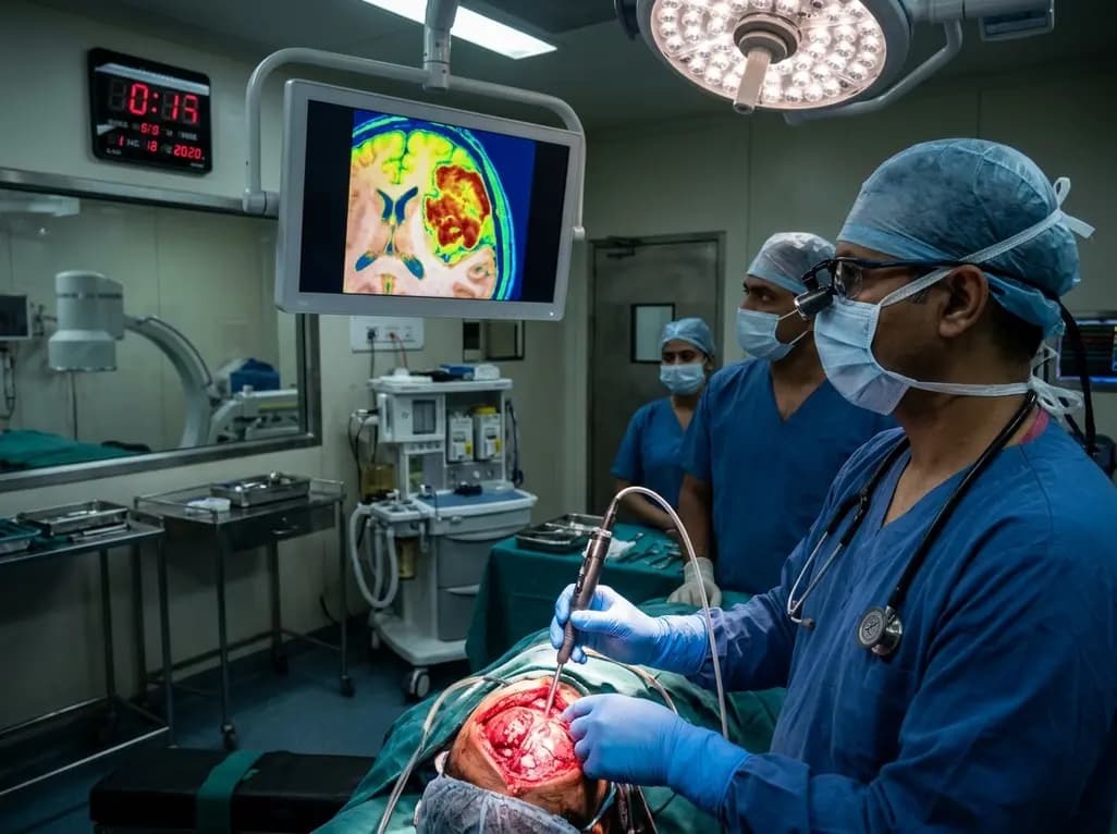
Surgical resection remains the definitive cornerstone for managing intracranial meningiomas, which represent the most common primary brain tumors encountered in neurosurgical practice. Achieving maximal safe resection while strictly preserving functional neurovascular architecture dictates long-term progression-free survival. However, visual differentiation between tumor tissue, adjacent dura mater, and compressed cerebral cortex remains notoriously challenging under conventional white-light microscopy. Recently, intraoperative hyperspectral imaging has emerged as an objective, non-contact, and label-free diagnostic modality that captures quantitative optical data across continuous wavelength spectra. When coupled with advanced computational algorithms, this technology provides real-time guidance that could soon enhance intraoperative decision-making across global neurosurgical operating theaters.
Meningiomas arise from arachnoid cap cells and typically exhibit an extra-axial growth pattern. Although benign in most cases, these neoplasms frequently encase critical cerebral vessels, compress eloquent cortical regions, and infiltrate contiguous dural leaflets. Consequently, neurosurgeons must balance aggressive tumor removal against the disastrous risk of causing new neurological deficits. The classical Simpson grading system highlights that complete excision of the tumor and its dural attachment drastically reduces recurrence rates. Nevertheless, distinguishing microscopic tumor invasion from reactive dural hypervascularity or edematous cerebral cortex often eludes conventional microscopic inspection.
Currently, surgeons occasionally rely on fluorescence guidance using agents such as sodium fluorescein or 5-aminolevulinic acid. Although these fluorophores can assist tumor demarcation, they require intravenous administration, cause systemic side effects, and exhibit variable uptake patterns across heterogeneous meningioma subtypes. Furthermore, optical tissue contrast frequently degrades due to photobleaching and background ambient light contamination. Therefore, neurosurgeons have actively sought label-free, non-invasive optical platforms that reliably characterize intracranial tissue properties without requiring exogenous fluorescent tracers.
To overcome the limitations of conventional operative visualization, intraoperative hyperspectral imaging captures hundreds of contiguous, narrow spectral bands beyond the visible spectrum. Standard human vision captures only red, green, and blue wavelengths, whereas hyperspectral sensors record light reflectance from the ultraviolet through the near-infrared spectrum. Because biological tissues exhibit unique molecular compositions, light absorption, and scattering behaviors, each intracranial structure produces a distinct biochemical signature.
Specifically, this optical methodology measures fundamental tissue constituents, including oxyhemoglobin, deoxyhemoglobin, water content, and structural lipids. For instance, cerebral cortex demonstrates intense microvascular perfusion with prominent oxyhemoglobin absorption bands. In contrast, neoplastic meningioma tissue displays altered metabolic activity, disrupted microarchitecture, and altered vascular patterns. Consequently, cranial meningiomas exhibit marked attenuation of the classical oxyhemoglobin double absorption peak alongside significantly lower tissue oxygenation levels compared to normal cortex and dura mater. Therefore, capturing these subtle spectral shifts enables automated systems to identify neoplastic tissue borders with outstanding precision.
Transforming complex multi-dimensional spectral cubes into actionable surgical maps requires sophisticated algorithmic processing. In an ongoing prospective observational study at a tertiary neurosurgical center, investigators captured high-resolution hyperspectral images across elective cranial meningioma cases. Surgical experts systematically annotated verified regions of interest representing meningioma, healthy cerebral cortex, and dura mater on calibrated color reconstructions. This comprehensive protocol yielded hundreds of thousands of individual pixel-wise reflectance spectra for quantitative computational assessment.
Subsequently, data scientists evaluated four distinct supervised machine learning classifiers: linear discriminant analysis, support vector machines, logistic regression, and multilayer perceptrons. To eliminate baseline optical drift and ambient illumination irregularities, researchers tested raw reflectance alongside standard normal variate normalization and higher-order spectral derivatives. Furthermore, they instituted a strict leave-one-patient-out cross-validation framework. This rigorous validation technique ensures that the algorithms evaluate test data from unseen patients, thereby providing an unbiased assessment of algorithmic generalizability.
The interim analysis evaluated 70 surgical patients, incorporating 291,104 meningioma pixels, 141,515 cortex pixels, and 333,244 dura pixels. In the binary classification task comparing cranial meningioma against healthy cerebral cortex, the multilayer perceptron coupled with standard normal variate-normalized spectra demonstrated superior performance. Specifically, this neural network achieved an overall accuracy of 0.928, a precision of 0.960, a recall of 0.932, an F1 score of 0.946, and a Matthews correlation coefficient of 0.840. Consequently, these metrics indicate exceptional reliability in separating neoplastic tissue from viable neural parenchyma.
However, multiclass discrimination involving meningioma, adjacent cortex, and dura mater presented greater analytical difficulty. In this three-tier classification task, the multilayer perceptron maintained viable performance, demonstrating an accuracy of 0.773, a macro F1 score of 0.782, and a macro Matthews correlation coefficient of 0.662. The slight reduction in multiclass performance largely stems from overlapping fibrovascular signatures between meningioma and adjacent dura mater. Nonetheless, these interim findings robustly prove that automated optical classification can reliably support intraoperative surgical navigation.
Despite these promising interim results, several substantial hurdles remain before clinicians can routinely deploy hyperspectral imaging in daily surgical workflows. First, current hyperspectral image acquisition and processing pipelines require dedicated cameras and off-line computational processing. To assist real-time decision-making during microsurgical dissection, engineering teams must streamline data acquisition to generate instantaneous augmented-reality overlays within the surgical microscope eyepiece.
Second, researchers must conduct multicenter external validation studies across broader patient cohorts and diverse meningioma grades. Future investigations should determine whether hyperspectral reflectance signatures correlate with World Health Organization histological grading or aggressive molecular profiles. Moreover, investigators must establish rigorous, spatially matched histopathological confirmation along resection margins to guarantee diagnostic ground truth. Addressing these translational challenges will enable neurosurgeons in India and worldwide to harness label-free optical guidance, ultimately improving surgical safety and oncological outcomes.
Conventional neurosurgical fluorescence requires intravenous administration of exogenous dyes such as 5-aminolevulinic acid or sodium fluorescein, which can cause patient hypersensitivity and phototoxicity. In contrast, intraoperative hyperspectral imaging operates as an entirely label-free, non-contact optical modality. It measures intrinsic biochemical reflectance, tissue oxygenation, and hemoglobin absorption across continuous wavelengths without injecting chemical contrast agents into the patient.
Meningioma tissue exhibits distinct optical characteristics compared with adjacent normal cerebral cortex and dura mater. Neoplastic tissue demonstrates significant attenuation of the characteristic oxyhemoglobin-associated double absorption peak and displays lower baseline tissue oxygenation. These distinct biochemical differences allow machine learning algorithms to accurately separate pathological tumor tissue from functional cortical parenchyma during surgical resection.
Currently, processing massive hyperspectral data cubes requires several seconds to minutes of dedicated computational power, which limits instantaneous intraoperative feedback. However, researchers are actively optimizing hardware accelerators, edge computing, and streamlined neural network architectures. These technological advancements aim to deliver instantaneous, real-time augmented reality visual overlays directly into neurosurgical operating microscopes in the near future.
Disclaimer: This content is for informational and educational purposes only. It is not intended to provide medical advice, diagnosis, or treatment. Always seek the advice of your physician or other qualified health provider with any questions you may have regarding a medical condition. Never disregard professional medical advice or delay in seeking it because of something you have read here. Clinical judgment must guide therapeutic decisions. Refer to the latest local and national guidelines for clinical practice.
References

Read summarized clinical updates, watch expert medical content, and earn CME certifications right from your smartphone.
Open your smartphone camera to scan and install.





Interim findings from a prospective clinical study demonstrate that intraoperative hyperspectral imaging combined with machine learning accurately discriminates cranial meningioma from adjacent brain cortex and dura mater without requiring exogenous fluorescent dyes.
Today

Delhi civic authorities have cleared 1,066 contractual public health posts and a Rs 923.6-crore road-sweeping initiative to tackle vector-borne illnesses and air pollution. Learn how enhanced entomological surveillance, anti-larval drives, and dust control bolster community preventive medicine and clinical outcomes.
Today

Medical experts caution that GLP-1 receptor agonists offer metabolic support rather than a curative answer for polyendocrine metabolic ovarian syndrome. Clinicians must evaluate glycemic markers, reproductive plans, and lifestyle interventions while discouraging unsupervised medication use among female patients.
Today

A retrospective CAHAL study comparing Amplatzer and Occlutech devices in 413 patients found no significant difference in moderate-to-large residual shunt rates at 6 months follow-up after percutaneous PFO closure, supporting clinical equivalence between both platforms.
Today

Age-related spinal kyphosis is driven by vertebral fragility, disc degeneration, and paraspinal muscle dysfunction. Coexisting with locomotive syndrome and frailty, its management requires combined anti-osteoporotic therapy, spinal extensor rehabilitation, and comprehensive nutritional optimization.
Today

A longitudinal feasibility pilot study highlights that combining polygenic risk score disclosure with in-person lifestyle counseling significantly enhances genomic literacy in individuals at risk for type 2 diabetes, overcoming educational disparities and reinforcing patient motivation.
Today
