

Loading, please wait...
Loading, please wait...

Physical aging brings progressive structural alterations to the human skeleton. In clinical geriatrics, age-related spinal kyphosis represents an increasingly recognized postural deformity that threatens mobility. While clinicians often dismiss it as benign aging, the deformity can precipitate severe functional impairment, balance instability, and restrictive pulmonary compromise. Consequently, understanding its pathophysiological ties to skeletal, muscular, and systemic decline is essential for clinical management.
Age-related spinal kyphosis develops through a complex cascade of mechanical and degenerative factors. The thoracic spine naturally carries a gentle convex curvature. However, degenerative changes in intervertebral discs reduce anterior column height over time. Furthermore, chronic disc desiccation shifts compressive spinal loading anteriorly. This forward load transfer markedly amplifies bending moments across the thoracic and thoracolumbar segments. As a result, the anterior vertebral margins endure disproportionate compressive stresses during daily weight-bearing activities.
In addition, degenerative changes in facet joints and posterior ligamentous laxity worsen sagittal imbalance. When passive stabilizers deteriorate, the muscular envelope must generate higher counter-forces to maintain an erect posture. Unfortunately, aged muscle tissue often fails to compensate adequately. Therefore, the spine settles progressively into an anteriorly displaced center of gravity. This mechanical displacement initiates a vicious cycle of increasing kyphosis and localized tissue strain. In severe cases, marked thoracic curvature restricts rib cage expansion, which diminishes vital capacity and impairs diaphragmatic excursion. Clinicians must recognize that spinal sagittal malalignment is not merely a static structural flaw. Instead, it serves as an active biomechanical stressor that accelerates musculoskeletal degradation across the entire axial skeleton.
Osteoporosis plays a central structural role in progressive spinal deformity. In elderly individuals, trabecular thinning and cortical bone loss severely compromise vertebral compressive strength. Consequently, even low-energy axial loads can produce microfractures within the anterior vertebral cortex. These structural failures frequently manifest as anterior wedge deformities. Studies demonstrate that multiple wedge fractures drastically increase local thoracic curvature. Moreover, a substantial proportion of osteoporotic vertebral fractures occur asymptomatically without acute pain episodes. As a result, patients may accumulate silent deformities over several years before seeking medical evaluation.
Furthermore, untreated skeletal fragility accelerates adjacent segment breakdown. Once an anterior wedge collapse occurs, the local mechanical axis tilts forward, dramatically increasing stress on neighboring vertebral bodies. Therefore, single vertebral fragility fractures frequently trigger subsequent fracture cascades across adjacent levels. In clinical practice, bone mineral density alone does not fully predict fracture vulnerability because microarchitectural deterioration also impairs mechanical toughness. For this reason, clinicians must actively screen for hidden morphological fractures using dedicated spinal radiographs or vertebral fracture assessment tools. Addressing skeletal fragility early with potent anti-osteoporotic therapies remains paramount to arrest deformity progression and preserve spinal alignment.
The muscular contribution to spinal alignment has generated significant debate among musculoskeletal specialists. Historically, researchers assumed that generalized sarcopenia drove age-related thoracic deformities. However, recent clinical investigations reveal inconsistent associations between whole-body muscle wasting and kyphotic progression. Generalized reductions in appendicular lean mass do not reliably correlate with spinal sagittal deviation. Instead, local paraspinal muscle dysfunction appears to exert a far more decisive influence.
Specifically, pathological degeneration of deep intrinsic trunk extensors undermines active postural stabilization. Muscle biopsy and cross-sectional magnetic resonance imaging confirm that fatty infiltration and myosteatosis frequently replace functional muscle fibers within the erector spinae and multifidus muscles. This local myopathy markedly diminishes isometric extension strength. Consequently, the posterior muscular tension band fails to counterbalance the forward gravitational moment arm. Although systemic sarcopenia and nutritional decline can exacerbate muscle weakness, local trunk extensor quality represents the critical mechanical determinant of posture. Therefore, therapeutic interventions must prioritize targeted thoracic extensor strengthening rather than relying solely on general physical conditioning. Restoring dynamic extensor capacity provides the biomechanical resistance needed to prevent sagittal collapse.
Age-related spinal deformity rarely exists in clinical isolation. Instead, it frequently coexists with locomotive syndrome and physical frailty, forming an interconnected geriatric triad. Locomotive syndrome reflects progressive functional mobility impairment caused by underlying disorders of bones, joints, muscles, and nerves. When thoracic kyphosis advances, the anterior displacement of the body center of mass impairs dynamic balance. Consequently, affected individuals walk with a slower gait velocity, shortened stride lengths, and widened bases of support.
Moreover, postural instability drastically increases the risk of accidental falls and subsequent catastrophic fractures. To compensate for forward trunk inclination, patients frequently hyperextend their hips and flex their knees. However, these compensatory mechanisms require excessive energy expenditure, which induces chronic physical fatigue and muscular exhaustion. This heightened physiological demand rapidly accelerates the transition into physical frailty. Frail individuals experience reduced physical activity, social withdrawal, and depressive symptoms, which further worsen musculoskeletal deconditioning. Although observational data highlight strong associations between kyphosis, locomotive syndrome, and frailty, clinicians should recognize that these relationships are complex and reciprocal. Therefore, interventions must target mobility preservation to break this self-reinforcing downward spiral.
Effective management of spinal deformity requires a comprehensive multidisciplinary approach. Clinicians must initiate evaluation with dual-energy X-ray absorptiometry to quantify bone mineral density. In addition, standing full-spine radiographs are indispensable to assess regional thoracic kyphosis and global sagittal vertical axis. Physicians should also evaluate trunk extensor endurance, gait speed, and five-times sit-to-stand performance to detect locomotive impairment early.
From a therapeutic perspective, medical management should integrate pharmacological and non-pharmacological modalities. First, patients with confirmed bone loss require anti-resorptive or osteoanabolic agents to prevent subsequent vertebral fractures. Concurrently, rehabilitation specialists must prescribe targeted spinal extension exercises, core stabilization protocols, and postural biofeedback training. These specific exercises strengthen paraspinal muscles and enhance postural awareness without imposing excessive flexion moments on vulnerable vertebral bodies. Furthermore, optimizing daily nutritional intake is crucial. Clinicians should ensure adequate high-quality dietary protein along with vitamin D and calcium repletion to support muscle protein synthesis and bone mineralization. By combining structural bone protection with functional neuromuscular rehabilitation, healthcare providers can mitigate kyphotic progression, restore functional mobility, and safeguard independence in older adults.
Osteoporosis significantly reduces trabecular microarchitecture and bone mineral density within vertebral bodies. As a result, the anterior vertebral cortex becomes vulnerable to compressive loads during ordinary daily activities. Repetitive microtrauma causes anterior cortical collapse and wedge-shaped fractures. When multiple thoracic vertebrae collapse anteriorly, the spinal column tilts forward into progressive thoracic kyphosis. Therefore, aggressive anti-osteoporotic therapy is crucial to halt further wedge deformity progression and preserve spinal posture.
While generalized sarcopenia reduces whole-body muscle mass, spinal alignment depends primarily on local dynamic stabilization. Intrinsic paraspinal extensors, including the erector spinae and multifidus, actively counterbalance the forward bending torque of gravity. When fat infiltrates these paraspinal muscles, local contractile strength diminishes drastically despite normal appendicular muscle measurements. Consequently, localized trunk myosteatosis and muscular atrophy compromise active sagittal balance far more directly than generalized reductions in peripheral limb musculature.
Non-pharmacological management relies on targeted spinal extensor exercises, core muscle conditioning, and postural retraining. Focused thoracic extension routines strengthen the posterior paraspinal musculature to counteract anterior gravitational collapse. Furthermore, balance training and gait re-education help mitigate fall risks associated with locomotive syndrome. Clinicians should also combine physical exercise with personalized nutritional support, ensuring adequate dietary protein and vitamin D supplementation to optimize musculoskeletal healing and maintain overall functional independence.
Disclaimer: This content is for informational and educational purposes only... Refer to the latest local and national guidelines for clinical practice.
References

Read summarized clinical updates, watch expert medical content, and earn CME certifications right from your smartphone.
Open your smartphone camera to scan and install.





Age-related spinal kyphosis is driven by vertebral fragility, disc degeneration, and paraspinal muscle dysfunction. Coexisting with locomotive syndrome and frailty, its management requires combined anti-osteoporotic therapy, spinal extensor rehabilitation, and comprehensive nutritional optimization.
Today
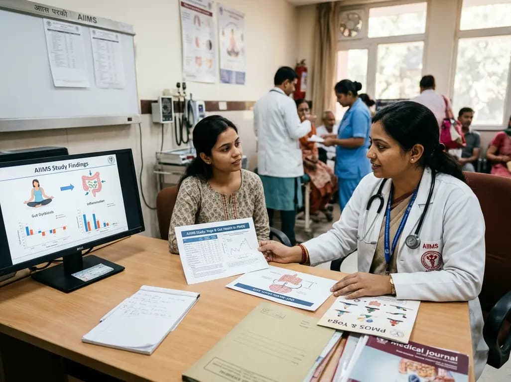
AIIMS researchers suggest yoga may alleviate gut dysbiosis in Polyendocrine Metabolic Ovarian Syndrome (PMOS) by modulating the gut-brain axis, lowering chronic inflammation, and enhancing parasympathetic tone to support metabolic and hormonal equilibrium.
Today

A prospective cohort study demonstrates that feeding tubes, formula exposure, aortic cross-clamp duration, and socioeconomic deprivation disrupt the neonatal gut microbiome in congenital heart disease, emphasizing the need for targeted nutritional and perioperative gut-protective strategies.
Today
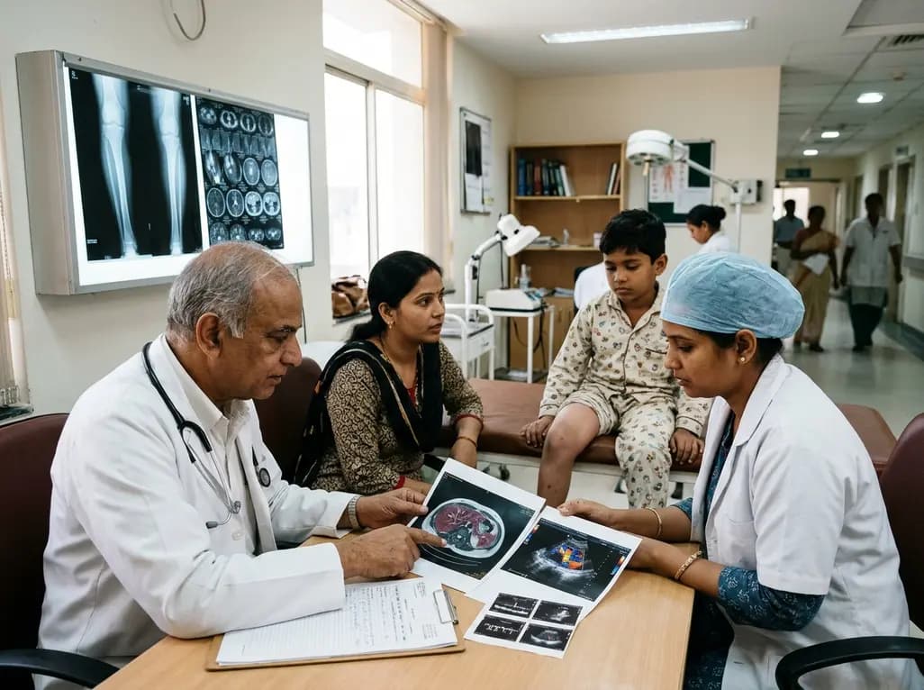
Deep intramuscular vascular malformations in pediatric Klippel-Trenaunay syndrome can mimic Maffucci syndrome. Multimodality imaging with Doppler ultrasound and MRI, along with histopathology, ensures prompt differential diagnosis, targeted surgical debulking, and optimized conservative surveillance.
Yesterday

A longitudinal feasibility pilot study highlights that combining polygenic risk score disclosure with in-person lifestyle counseling significantly enhances genomic literacy in individuals at risk for type 2 diabetes, overcoming educational disparities and reinforcing patient motivation.
Today
