.webp&w=384&q=75)

Loading, please wait...
Loading, please wait...
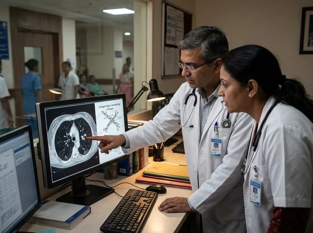
Recent breakthroughs in molecular imaging offer unprecedented diagnostic avenues for modern thoracic oncology. Specifically, precision lung cancer MRI detection represents a transformative approach for spotting early malignant lesions. Today, clinicians diagnose pulmonary carcinomas primarily after regional extension or distant organ metastasis occurs. Consequently, curative therapeutic windows narrow significantly, while patient morbidity escalates. Conventional imaging modalities, including chest radiographs and computed tomography scans, often fail to characterize lesions smaller than five millimeters accurately. Moreover, repetitive computed tomography exposure raises concerns regarding cumulative ionizing radiation, particularly during long-term surveillance regimens. In response to these persistent clinical hurdles, investigators developed a novel protein-based contrast agent known as hProCA32.Collagen. Preclinical trials in Science Advances demonstrate that this specialized agent resolves primary tumors as small as one millimeter. Additionally, it visualizes micro-metastatic deposits within distant organs with exceptional clarity. Therefore, this molecular platform bridges a major diagnostic gap in early disease identification. Furthermore, the technology avoids radioactive exposure entirely while delivering superior soft-tissue contrast. As a result, oncologists may soon identify localized neoplasms when curative surgical resection remains entirely feasible.
The novel contrast agent operates through a unique biochemical mechanism that exploits tumor microenvironment biology. Specifically, hProCA32.Collagen targets type I collagen, a dominant structural scaffold protein within lung parenchyma. Healthy pulmonary tissues maintain organized, stable extracellular matrix frameworks. In contrast, aggressive neoplastic cells dramatically accelerate extracellular matrix turnover and aberrant remodeling. Stromal fibroblasts deposit dense, disorganized type I collagen fibers directly along invasive tumor boundaries. Consequently, this dense fibrous matrix creates an identifiable molecular signature for progressive malignancy. Researchers engineered the protein agent to exhibit exceptional thermodynamic stability and high binding affinity for human collagen fibers. Furthermore, the molecule demonstrates roughly ten-fold higher relaxivity than conventional gadolinium chelates at clinical magnetic field strengths. This pronounced relaxation enhancement yields brilliant signal contrast over surrounding normal lung structures. As a result, radiologic scans illuminate previously imperceptible microscopic infiltrates. In addition, the targeted agent binds persistently to tumor margins without displacing vital physiological metals. Therefore, this biological targeting mechanism converts matrix remodeling into a bright visual beacon for radiologists.
Low-dose computed tomography currently represents the global standard for high-risk lung cancer screening programs. However, this established radiological technique presents noticeable clinical challenges and technical constraints. For example, computed tomography frequently detects benign non-calcified pulmonary nodules that mimic early malignant lesions. Consequently, false-positive findings trigger substantial patient anxiety, repeated radiation exposure, and invasive diagnostic procedures. Furthermore, transthoracic needle biopsies carry tangible risks of pneumothorax, pulmonary hemorrhage, and pleural seeding. In contrast, the collagen-targeted magnetic resonance protocol provides both structural and biochemical information simultaneously. Preclinical evaluations showed that this targeted magnetic resonance technique achieved threefold higher contrast than high-resolution computed tomography. Thus, physicians can distinguish actively remodeling malignant stroma from indolent inflammatory granulomas with greater diagnostic confidence. Moreover, magnetic resonance imaging does not expose patients to ionizing radiation. This feature provides immense safety benefits during mandatory serial follow-up scans. Ultimately, adopting targeted magnetic resonance protocols could substantially reduce unnecessary tissue biopsies in vulnerable populations. Additionally, whole-body screening protocols allow comprehensive surveillance without raising cumulative radiation burdens. Therefore, thoracic clinicians can safely execute repeated follow-ups across prolonged surveillance intervals for high-risk individuals.
Beyond straightforward lesion detection, this molecular imaging platform delivers pivotal prognostic data regarding tumor biology. Specifically, aggressive lung adenocarcinomas often harbor mutations in key tumor suppressor genes like liver kinase B1. These aggressive genetic variants produce profound extracellular matrix alterations and exhibit high resistance to standard chemo-immunotherapy. Fortunately, the collagen-targeted contrast agent accurately maps heterogeneous collagen densities at invasive margins. Consequently, clinicians can directly visualize microscopic invasion before overt structural disruption becomes visible on standard cross-sectional imaging. Furthermore, the agent readily detects distant occult metastases in high-risk sanctuary organs such as adrenal glands and kidneys. In animal models, precision imaging identified secondary deposits as small as one millimeter across multiple organ systems. Thus, oncologists can stage systemic disease comprehensively in a single imaging session. Meanwhile, tracking collagen remodeling over time provides early insight into therapeutic responsiveness. If a cytotoxic or targeted drug halts matrix remodeling, signal intensity decreases rapidly. Conversely, persistent collagen signal highlights treatment failure long before dimensional tumor shrinkage manifests. Additionally, radiomic scoring models synthesized from these magnetic resonance images accurately predict long-term clinical survival. Therefore, physicians gain actionable prognostic intelligence that directly shapes personalized oncologic interventions.
The emergence of radiation-free, precision molecular imaging holds substantial significance for oncology practice across India. Currently, Indian clinicians encounter a high proportion of lung cancer patients presenting at advanced stages. Furthermore, non-smoking related lung adenocarcinoma, often driven by distinct genetic mutations, occurs frequently across urban demographics. Consequently, accurate, radiation-free detection tools could markedly improve survival metrics by catching aggressive disease early. Additionally, benign pulmonary infections such as endemic tuberculosis frequently produce confounding granulomatous nodules on routine computed tomography scans. These benign lesions frequently trigger false-positive diagnoses and unwarranted invasive lung biopsies. In contrast, molecular magnetic resonance imaging targeting active collagen remodeling may help differentiate active malignant stroma from healed tubercular scars. Moreover, Indian tertiary medical centers already possess widespread 1.5 Tesla and 3.0 Tesla magnetic resonance infrastructure. Therefore, clinical translation would primarily require contrast agent procurement rather than massive capital investments in novel imaging hardware. Ultimately, incorporating targeted contrast agents could optimize resource allocation and enhance early diagnostic yields throughout regional healthcare networks. As a result, timely therapeutic intervention could dramatically curb cancer-related mortality in diverse clinical settings. Similarly, early noninvasive staging preserves valuable surgical windows for operable candidates.
Q1: What is hProCA32.Collagen and how does it work?
The agent hProCA32.Collagen is an engineered protein-based magnetic resonance contrast dye designed to bind human type I collagen. Aggressive pulmonary tumors remodel extracellular matrix structures, depositing dense collagen fibers along their margins. When administered intravenously, this agent binds to remodeled fibers, enhancing local relaxivity. Consequently, standard magnetic resonance imaging visualizes malignant nodules and micro-metastases as small as one millimeter without using ionizing radiation.
Q2: How does this agent compare with low-dose computed tomography?
Low-dose computed tomography reliably identifies small lung nodules but frequently detects harmless benign granulomas, causing high false-positive rates. Furthermore, repeated computed tomography exposes individuals to cumulative ionizing radiation. In contrast, this collagen-targeted magnetic resonance protocol provides high diagnostic specificity without radiation exposure. By highlighting active collagen turnover rather than mere tissue density, this imaging agent accurately distinguishes aggressive carcinomas from indolent lesions while delivering threefold higher contrast.
Q3: Can collagen-targeted MRI monitor cancer treatment response?
Yes, collagen-targeted magnetic resonance imaging offers valuable prognostic monitoring during anti-cancer therapy. Because tumor cells stimulate collagen synthesis during disease progression, reductions in matrix turnover reflect effective therapeutic response. If systemic therapies successfully suppress malignant activity, collagen remodeling diminishes, leading to decreased contrast enhancement on sequential scans. Conversely, persistent signal intensity warns clinicians of treatment resistance. Consequently, oncologists can adjust regimens earlier without waiting for late macroscopic changes.
Disclaimer: This content is for informational and educational purposes only. It does not constitute medical advice or replace professional judgment. Refer to the latest local and national guidelines for clinical practice.
References

Read summarized clinical updates, watch expert medical content, and earn CME certifications right from your smartphone.
Open your smartphone camera to scan and install.





Preclinical findings demonstrate that hProCA32.Collagen, an innovative protein-based contrast agent, detects micro-metastases and aggressive lung adenocarcinomas down to 1 mm on MRI. By targeting collagen type I remodeling, this technology provides radiation-free diagnostic and prognostic evaluation for clinicians.
Today

Gestational diabetes doubles the future risk of type 2 diabetes for mothers and offspring. Adopting a life-course approach centered on family meals and brief, supportive nutrition messaging empowers households to reduce metabolic risks and establish sustainable dietary habits across generations.
Today

A comprehensive meta-analysis evaluates the association between Body Mass Index and outcomes following abdominal aortic aneurysm repair. Discover how overweight and obesity correlate with lower mortality while underweight status confers heightened risk, challenging conventional preoperative risk stratification.
Today

The SOLARIO trial shows that a short course (≤7 days) of systemic antibiotic therapy is noninferior to long courses (≥4 weeks) in orthopedic infections treated with local antibiotic carriers, with significantly fewer adverse events.
Today
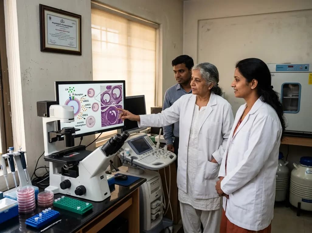
New research reveals that preovulatory follicles communicate via LH-induced paracrine signaling. Diffusible EGF ligands epiregulin and amphiregulin coordinate oocyte meiotic resumption across neighboring follicles, challenging traditional concepts of follicular autonomy.
Today
