.webp&w=384&q=75)

Loading, please wait...
Loading, please wait...

Surgeons frequently encounter immense challenges when managing massive posterosuperior rotator cuff tears in younger, active individuals. In these demanding scenarios, lower trapezius transfer serves as an effective joint-preserving reconstruction instead of early reverse shoulder arthroplasty. Traditional tendon transfers, such as latissimus dorsi reconstruction, often pull along non-anatomical axes. Consequently, orthopedic surgeons increasingly favor arthroscopic-assisted lower trapezius transfer to recreate the native vector of the infraspinatus tendon. This modern approach bridges the lower trapezius muscle to the greater tuberosity using a soft-tissue interposition graft. Furthermore, arthroscopic visualization minimizes surgical trauma while protecting deltoid attachments. Patients presenting with persistent pain and functional limitations frequently demonstrate severe infraspinatus atrophy and fatty infiltration. Because conventional tendon repair predictably fails under these compromised biological conditions, restoring the transverse force couple represents the primary surgical goal. Clinical investigations have therefore focused on identifying baseline predictors of recovery. Specifically, surgeons evaluate whether a severe preoperative deficit, such as an external rotation lag sign, compromises long-term clinical outcomes.
Biomechanical stability in the glenohumeral joint depends upon coordinated force couples across the transverse and coronal planes. When irreparable tears disrupt the posterior cuff, unopposed forces from the subscapularis pull the humeral head anteriorly and superiorly. Latissimus dorsi transfer historically counteracted this destabilization; however, its vector tends to induce internal rotation during active elevation. In contrast, the lower trapezius muscle fibers share an almost identical line of pull with the native infraspinatus. Therefore, transferring the lower trapezius restores physiological external rotation moments while dynamically depressing the humeral head. During surgery, clinicians harvest the lower trapezius tendon along the medial scapular spine. Surgeons must carefully protect the nearby spinal accessory nerve and dorsal scapular vasculature throughout dissection. Next, the surgical team securely sutures an Achilles allograft or hamstring autograft to the mobilized tendon. Arthroscopic instruments then guide the extended graft underneath the posterior deltoid into the subacromial space. Finally, surgeons anchor the graft securely onto the infraspinatus footprint, successfully re-establishing glenohumeral balance.
Preoperative clinical evaluation provides indispensable diagnostic and prognostic information when assessing posterosuperior cuff deficiency. Specifically, clinicians evaluate active external rotation at the patient's side, termed ER1, and at ninety degrees of abduction, termed ER2. When an examiner rotates the arm externally and the patient cannot maintain that position, an external rotation lag sign occurs. This drop confirms structural incompetence of the infraspinatus and suggests significant involvement of the adjacent teres minor. Orthopedic surgeons historically considered an active lag sign an indicator of poor outcomes following tendon reconstruction. Nevertheless, contemporary clinical evidence disproves this traditional assumption. Reconstructive lower trapezius surgery reliably resolves the dropping limb in patients who present with severe baseline lag signs. Although patients with advanced atrophy face significant functional deficits initially, the transferred tendon establishes critical active posterior restraint. In addition, dynamic tendon transfers encourage functional neurological retraining over time. The transposed muscle gradually adapts during shoulder motion, allowing patients to regain purposeful, stable arm control during daily activities.
Recent clinical cohort studies deliver valuable insights into patient recovery following lower trapezius reconstruction. In a two-year study of fifty-three patients, researchers stratified clinical outcomes by preoperative ER1 lag sign status. Forty percent of the cohort demonstrated a positive lag sign preoperatively. Importantly, both patient groups achieved remarkable, statistically significant improvements in range of motion and functional scores by twenty-four months. Patients without a preoperative lag achieved fifty-eight degrees of external rotation compared to thirty-two degrees in the lag-positive cohort. Similarly, external rotation at ninety degrees abduction reached higher absolute values in patients without baseline lag signs. However, both cohorts reported equivalent improvements in subjective shoulder values, satisfaction ratings, and pain reduction. Furthermore, longitudinal evaluations demonstrated ongoing, progressive recovery between six and twenty-four months postoperatively. These findings clearly confirm that a preoperative lag sign should never rule out surgical reconstruction. While patients with preserved muscle tone attain superior motion, individuals with severe lag signs gain meaningful functional independence.
Achieving successful joint preservation mandates precise candidate selection and dedicated postoperative rehabilitation. Surgeons predominantly select patients under sixty-five years who maintain high physical demands and suffer from irreparable posterosuperior cuff tears without severe glenohumeral osteoarthritis. Crucially, the subscapularis tendon must remain intact or fully repairable. If anterior cuff deficiency persists, the reconstructed shoulder loses stability despite successful posterior transfer. Following surgery, clinicians immobilize the operative limb in a specialized external rotation abduction brace for six weeks. This immobilization protects the healing tendon-graft interface against mechanical tension and tissue failure. During this initial phase, patients perform passive elbow and hand exercises while strictly avoiding active internal rotation. After six weeks, therapists introduce supervised passive and active-assisted shoulder movements. Consequently, clinicians initiate light isometric strengthening around twelve weeks postoperatively, gradually advancing toward active functional exercises. Because the lower trapezius originally serves as a scapular retractor, patients require targeted biofeedback to retrain the transposed muscle during glenohumeral rotation. Full recovery requires twelve to twenty-four months of structured rehabilitation.
The lower trapezius possesses an anatomical trajectory that directly mirrors the physiological vector of the infraspinatus muscle. When surgeons transfer this tendon to the greater tuberosity using an interposition graft, the muscle dynamically depresses the humeral head and produces external rotation torque. This reconstruction restores the transverse force couple against the subscapularis, stabilizing the glenohumeral fulcrum and enabling pain-free active shoulder movement during overhead arm elevation.
A preoperative external rotation lag sign indicates advanced muscle wasting, but it does not prevent surgical success. Clinical evidence confirms that patients with a baseline lag sign achieve marked pain relief, functional recovery, and resolution of the lag sign after surgery. Although their absolute degrees of external rotation remain lower than those in lag-negative patients, both groups report comparable patient satisfaction and substantial improvements in overall shoulder function.
Surgeons frequently utilize an Achilles tendon allograft because it offers robust tensile strength, substantial length, and broad bone-to-tendon contact area. Alternatively, surgeons can harvest an autologous hamstring tendon, such as the semitendinosus, when allografts are unavailable or when autologous tissue is preferred. Both graft options effectively span the anatomical distance between the harvested lower trapezius tendon edge and the greater tuberosity footprint, facilitating solid biological incorporation.
Disclaimer: This content is for informational and educational purposes only and should not be considered as medical advice or used for self-diagnosis or treatment. Comprehensive evaluation by a qualified healthcare professional is necessary for proper assessment, diagnosis, and management of any health condition or concern. The content here should not replace consultation with a physician, surgeon, or other medical specialist. Use of any diagnostic tools, risk calculators, clinical criteria, or scoring systems mentioned in the text must be guided by appropriate clinical training and applied with sound professional judgment. Always consult with a registered medical practitioner before adopting new clinical practices or altering current therapeutic strategies. Refer to the latest local and national guidelines for clinical practice.
References

Read summarized clinical updates, watch expert medical content, and earn CME certifications right from your smartphone.
Open your smartphone camera to scan and install.





A landmark study reveals that arthroscopic-assisted lower trapezius transfer provides durable functional restoration and pain relief in patients with irreparable posterosuperior rotator cuff tears, successfully reversing deficits even in those presenting with a severe preoperative external rotation lag sign.
Today
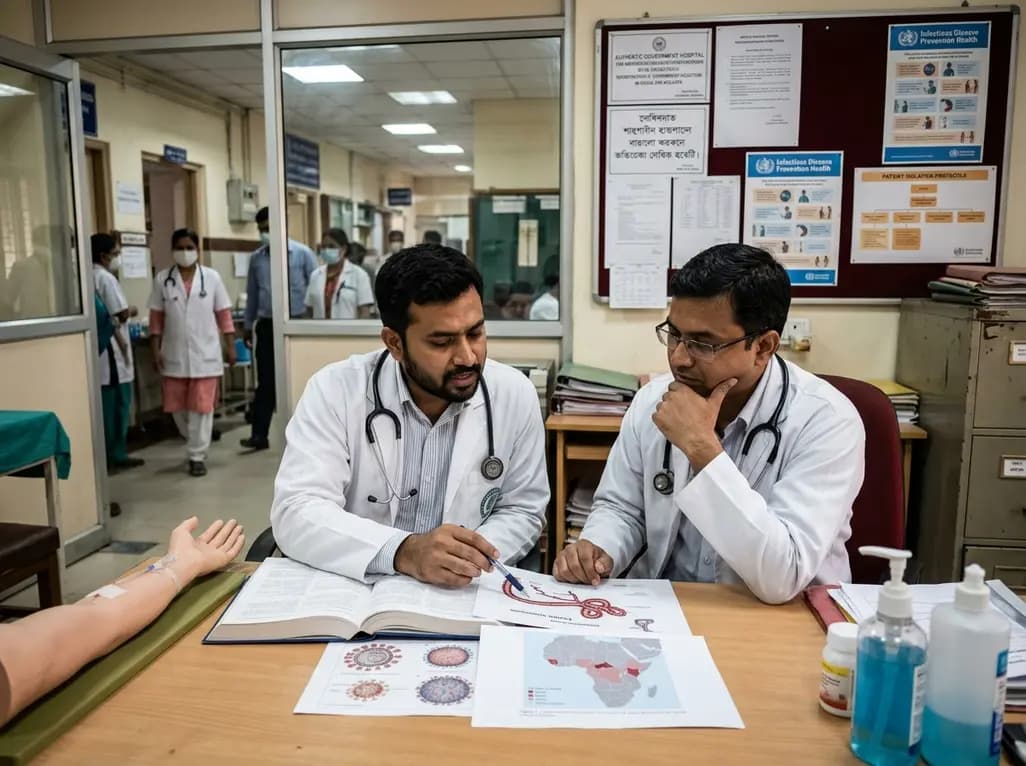
Health authorities in the Democratic Republic of Congo confirmed the spread of Ebola to North Ubangi province. With cross-border infections and high case counts, healthcare professionals worldwide must review early clinical recognition, differential diagnosis, infection control measures, and isolation protocols.
Today
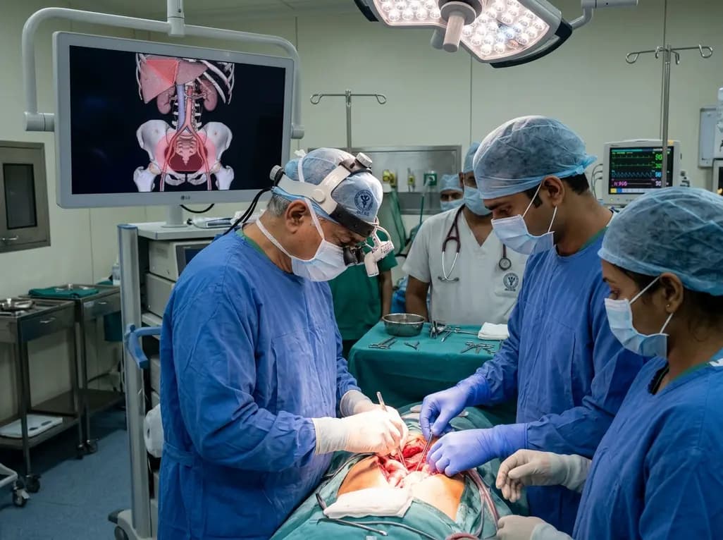
Surgical oncologists at AIIMS Delhi have mapped two critical anatomical landmarks: Ray's Zone in the deep pelvis and Ray's Hepatocaval Triangle near the liver. These predefined hazard boundaries guide dissection, dramatically reduce severe intraoperative bleeding, and preserve vital nerves and visceral organs.
Today
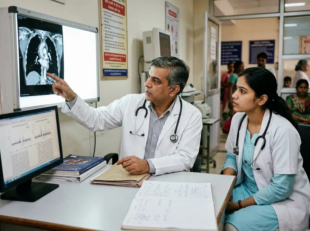
A study reveals that the MYLK c.4079G>A, p.(Gly1360Asp) variant is linked to high-penetrance heritable thoracic aortic disease and early-onset aortic dissection, showing a founder effect in Finnish cohorts. Findings emphasize early cascade screening and vigilant surveillance.
Today
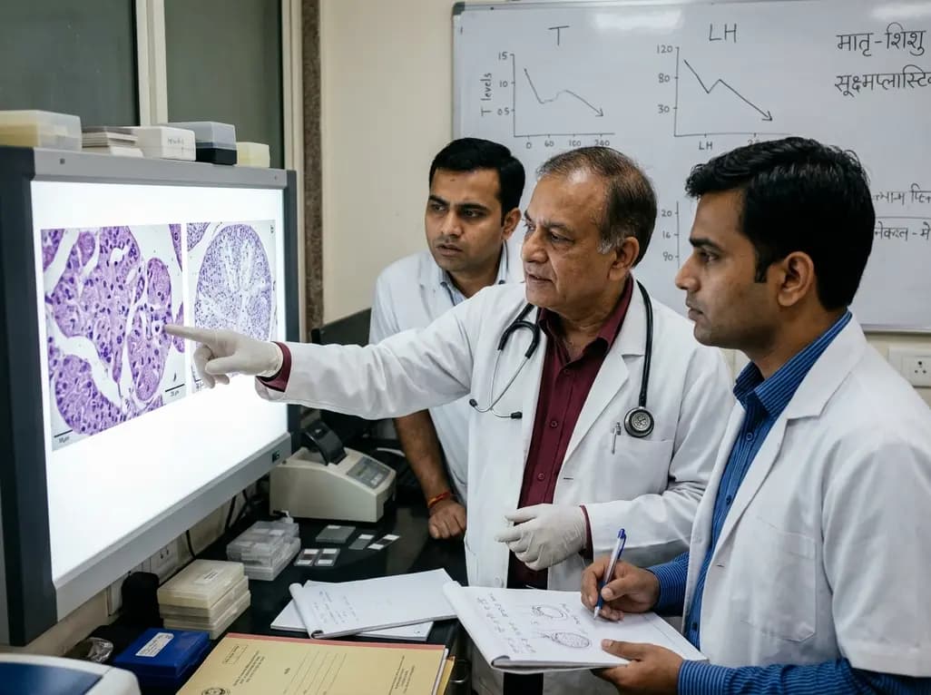
An animal model study reveals that gestational exposure to polystyrene microplastics causes persistent intrauterine growth restriction, testicular hypoplasia, Leydig cell depletion, and an atypical low-testosterone, low-LH endocrine profile in male offspring from birth into adulthood.
Today
