.webp&w=384&q=75)

Loading, please wait...
Loading, please wait...
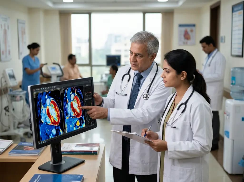
Atrial fibrillation represents one of the most common sustained cardiac arrhythmias encountered in adult medical practice. Clinicians have traditionally focused on left atrial dilatation and systemic vascular resistance when assessing arrhythmic vulnerability. However, advancing magnetic resonance imaging modalities now illuminate the critical contribution of ventricular tissue remodeling to atrial pathology. Specifically, non-invasive assessment of left ventricular fibrosis provides objective insight into microscopic myocardial disease. A landmark prospective investigation from the Multi-Ethnic Study of Atherosclerosis demonstrates that diffuse left ventricular fibrosis independently predicts incident atrial fibrillation across diverse community-dwelling populations.
Cardiac magnetic resonance imaging has transformed our clinical approach to myocardial tissue characterization. Standard late gadolinium enhancement accurately detects localized replacement scarring from past ischemic events. However, conventional techniques frequently overlook diffuse microscopic collagen deposition within the interstitium. In contrast, T1 mapping and extracellular volume fraction calculation quantify diffuse matrix expansion with high histological fidelity. Clinicians calculate extracellular volume by measuring pre- and post-contrast longitudinal relaxation times alongside patient hematocrit levels. Consequently, this parametric mapping method provides an objective metric of diffuse fibrotic remodeling throughout the entire ventricular myocardium.
The Multi-Ethnic Study of Atherosclerosis prospectively followed 1,261 participants who were free of baseline atrial fibrillation between 2010 and 2012. Over a median follow-up of 7.82 years, 156 individuals developed verified incident atrial fibrillation. Multivariable Cox proportional hazards regression demonstrated that extracellular volume expansion independently predicted new-onset arrhythmia after rigorous adjustment for CHARGE-AF risk factors. Specifically, each 1% absolute increase in extracellular volume conferred a 10.5% higher risk of incident atrial fibrillation. Furthermore, participants in the highest extracellular volume quartile experienced nearly a two-fold greater risk than those in the lowest quartile.
Why does ventricular interstitial expansion predispose individuals to supraventricular rhythm disturbances? Diffuse interstitial collagen accumulation increases ventricular myocardial stiffness and significantly impairs active and passive diastolic filling. As diastolic compliance deteriorates, left ventricular filling pressures rise steadily throughout the cardiac cycle. Consequently, these elevated diastolic pressures transmit retrograde mechanical stress directly into the thin-walled left atrium. Chronic hemodynamic overload subsequently drives left atrial structural stretch, electrical dispersion, and cellular remodeling. Notably, the Multi-Ethnic Study of Atherosclerosis confirmed that higher extracellular volume strongly correlated with increased atrial volume and reduced emptying function.
A crucial insight from this prospective investigation is the persistence of risk across comprehensive statistical adjustments. Left ventricular extracellular volume remained a robust predictor of incident atrial fibrillation even after accounting for focal myocardial scar, left ventricular ejection fraction, and detailed left atrial parameters. Therefore, diffuse ventricular interstitial expansion represents more than a mere bystander to macroscopic atrial enlargement. Instead, microscopic ventricular remodeling serves as an early, independent substrate that promotes the cascade toward sustained electrophysiological instability.
Conventional clinical scores such as CHARGE-AF rely primarily on demographic features, blood pressure measurements, and baseline electrocardiograms. While these tools offer valuable population-level stratification, they cannot evaluate ongoing subclinical tissue remodeling. Integrating extracellular volume quantification into diagnostic pathways helps clinicians identify vulnerable individuals before overt atrial dilatation occurs. Furthermore, early detection of ventricular fibrosis encourages timely lifestyle optimization and targeted pharmacological therapy. Prescribing neurohormonal antagonists and modern metabolic agents can reduce fibrotic signaling, maintain chamber compliance, and mitigate future arrhythmia risk.
Left ventricular fibrosis increases ventricular stiffness and impairs normal diastolic relaxation. As a consequence, left ventricular filling pressures rise and transmit retrograde stress into the left atrium. This persistent hemodynamic burden induces left atrial enlargement, structural stretch, and electrophysiological heterogeneity. Over time, these combined pathological changes create an optimal arrhythmogenic substrate that facilitates the spontaneous initiation and maintenance of incident atrial fibrillation.
T1 mapping and extracellular volume calculation provide quantitative non-invasive assessments of diffuse interstitial myocardial fibrosis. Unlike late gadolinium enhancement, which only highlights localized macroscopic scar tissue, extracellular volume mapping accurately detects early diffuse microscopic collagen expansion. Therefore, this imaging modality allows clinicians to identify subclinical structural cardiac disease, track therapeutic responses, and refine cardiovascular risk prediction across diverse patient populations.
Emerging clinical evidence indicates that targeted medical management can decelerate or potentially reverse diffuse myocardial fibrosis. Specifically, neurohormonal antagonists, including angiotensin-converting enzyme inhibitors, angiotensin receptor blockers, and mineralocorticoid receptor antagonists, suppress pro-fibrotic signaling cascades. In addition, sodium-glucose cotransporter-2 inhibitors and optimal blood pressure control reduce mechanical wall tension, thereby mitigating ongoing extracellular matrix accumulation and preserving long-term chamber compliance.
Disclaimer: This content is for informational and educational purposes only and does not constitute medical advice. Treatment decisions must rely on individualized clinical evaluations by licensed healthcare professionals. Refer to the latest local and national guidelines for clinical practice.
References

Read summarized clinical updates, watch expert medical content, and earn CME certifications right from your smartphone.
Open your smartphone camera to scan and install.





A prospective MESA study of 1,261 adults reveals that diffuse left ventricular fibrosis measured via T1 mapping and ECV independently predicts incident atrial fibrillation risk, highlighting ventricular remodeling as an early substrate for arrhythmogenesis.
last month

Emerging neuroenergetic research reveals that cognitive fatigue stems from local metabolic constraints and regional aerobic glycolysis rather than global resource depletion. This paradigm shift offers valuable clinical insights for managing chronic mental exhaustion, post-viral syndromes, and neurodegenerative fatigue.
Today

A scoping review reveals how pregnancy affects phonation through laryngeal edema and reduced phonation time rather than major acoustic pitch alterations. Learn how to identify, evaluate, and manage vocal symptoms in expectant mothers.
Today

According to WHO data, average lifespan reached 73.3 years in 2023, recovering from pandemic lows. However, healthy life expectancy lags at 62.8 years due to surging noncommunicable conditions like cardiovascular disease and dementia, demanding proactive primary screening and sustained chronic care.
Today

Researchers have engineered an artificial sensory-pain integrated receptor using a multi-threshold organic synaptic transistor. The system distinguishes benign touch from noxious stimuli, enabling safer, more responsive prosthetic limbs with built-in memory and simplified hardware architecture.
Today

Medical robotics is transitioning beyond operative suites into diagnostics, cardiology, and endocrinology. Explore how innovations in tele-robotics, AI integration, and nanomedicine are overcoming geographical barriers and transforming precision healthcare delivery across India's evolving disease landscape.
Today
