.webp&w=384&q=75)

Loading, please wait...
Loading, please wait...

Standardizing Knee Bone Mechanics Analysis through advanced imaging represents a major leap in orthopedic diagnostics. High-resolution peripheral quantitative CT (HR-pQCT) now allows clinicians to assess bone strength at a microstructural level in vivo. This technology, previously limited to the wrist and ankle, now addresses the complex geometry of the knee joint. Consequently, researchers can noninvasively evaluate how injuries like ACL tears alter the mechanical environment of the femur and tibia.
The integration of microfinite element (μFE) analysis into HR-pQCT imaging transforms static images into dynamic mechanical models. However, modeling the knee joint remains computationally demanding due to its large size and intricate load-bearing surfaces. A recent study by Stirling et al. addresses these hurdles by introducing standardized support layers. Specifically, the researchers found that an anatomically shaped layer with approximately 2500MPa stiffness ensures high reproducibility across tibial scans. Furthermore, this advancement is vital for longitudinal studies that monitor post-traumatic osteoarthritis progression.
The study utilized sensitivity analyses to determine that support layer stiffness significantly impacts strain energy density (SED). In contrast, the geometry and length of the support layer had minimal influence on the outcomes. Therefore, adopting a 3mm extruded PMMA-like support layer provides a computationally efficient and stable solution for researchers. This protocol facilitates better comparability between different clinical studies and patient populations.
Moreover, the reproducibility of these models is excellent, particularly in peri-articular trabecular regions. Stable results were observed in areas 5-7.5mm below the articular surface. Such precision allows clinicians to detect subtle mechanical changes following reconstructive surgeries. Additionally, these standardized models could eventually help in personalizing rehabilitation protocols for athletes returning to sport after major knee trauma.
μFE analysis provides a noninvasive way to measure bone mechanical properties like stiffness and strain energy density. This helps in understanding how an ACL injury or surgery affects the long-term structural integrity of the femur and tibia.
Standardization ensures that results are reproducible across different scans and patients. Because knee geometry is complex, using consistent support layer parameters allows researchers to accurately compare data in longitudinal studies.
While not a direct crystal ball, these mechanical assessments identify high-stress regions that are prone to bone remodeling and cartilage degradation. Identifying these areas early may help in predicting the risk of post-traumatic osteoarthritis.
Disclaimer: This content is for informational and educational purposes only and does not constitute medical advice or a professional relationship. The information provided is based on recent research findings and should be interpreted by qualified healthcare professionals. Refer to the latest local and national guidelines for clinical practice.
References
Stirling CE et al. Finite Element Analysis for the Load-Bearing Femur and Tibia in the Human Knee Using an In Vivo HR-pQCT Protocol. J Biomech Eng. 2026 Mar 27. doi: 10.1115/1.4071535. PMID: 41893870.
Manske SL et al. HR-pQCT measurements of changes in periarticular bone density and microarchitecture one year after acute knee injury and after reconstructive surgery. Bone. 2025 Mar;192:117376.
Chapurlat R et al. The clinical application of high-resolution peripheral computed tomography (HR-pQCT) in adults: state of the art and future directions. Osteoporos Int. 2021 Jun;32(6):1033-1048.

Read summarized clinical updates, watch expert medical content, and earn CME certifications right from your smartphone.
Open your smartphone camera to scan and install.





Researchers have established a standardized HR-pQCT and micro-FE modeling protocol for the human knee to improve reproducibility in longitudinal bone studie...
6 months ago
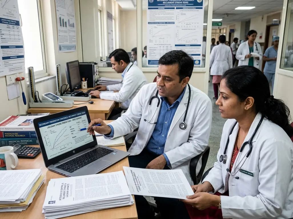
Explore vital recurrent stroke predictors in lacunar stroke patients managed with intensive blood pressure lowering. This analysis examines trial findings from SPS3 to uncover residual vascular risks, metabolic factors, and targeted secondary prevention strategies for high-risk neurological patients.
Today

Odisha has established India's first state-level clinical protocol specifically tailored for pregnant women with sickle cell disease. Developed with NHM and UNICEF, the framework provides structured antenatal screening, crisis mitigation, standardized referral pathways, and specialized interdisciplinary care.
Today
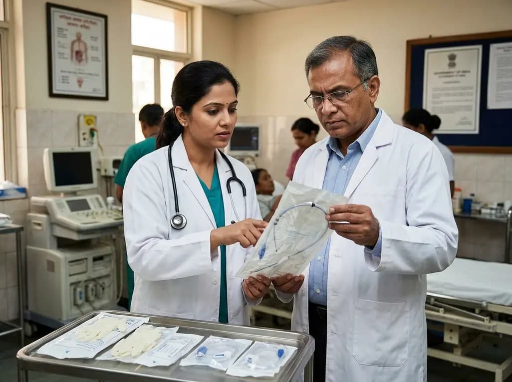
The Directorate General of Health Services directs Indian states to halt the hazardous reuse and reprocessing of single-use medical devices. Discover regulatory implications under the Drugs and Cosmetics Act, critical infection risks, legal liabilities, and operational changes required across clinical specialties.
Today

Mitochondria-targeted nanoparticles self-assembled from EGCG and L-glutamic acid effectively restore mitochondrial homeostasis, suppress synovial inflammation, and attenuate chondrocyte senescence in osteoarthritis, offering a promising disease-modifying nanotherapy.
Today

India is advancing preventive healthcare by integrating genomics, artificial intelligence, and quantum technologies. With the SANGAM programme sequencing one million Indian genomes, clinicians can unlock personalized therapies, detect cardiometabolic risks early, and curb the burden of diabetes and heart disease.
Today
