.webp&w=384&q=75)

Loading, please wait...
Loading, please wait...
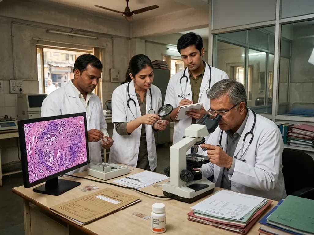
Kikuchi-Fujimoto disease, or histiocytic necrotizing lymphadenitis, remains a clinical enigma that frequently puzzles even seasoned internists and hematologists. Traditionally characterized as a benign and self-limiting condition, it primarily manifests as fever and localized cervical lymphadenopathy. While it has a well-documented predilection for young women of East Asian descent, recent clinical observations have highlighted its occurrence across diverse demographics, including Western populations. However, the diagnostic journey is often complicated because the condition lack pathognomonic clinical features. Consequently, a formal Kikuchi-Fujimoto disease diagnosis necessitates a high index of clinical suspicion and rigorous pathological confirmation. In some instances, the disease presents with severe systemic inflammatory markers that closely mirror much more aggressive syndromes. This overlap can lead to significant diagnostic uncertainty, potentially resulting in the initiation of unnecessarily intensive immunosuppressive therapies. Therefore, clinicians must remain vigilant, especially when encountering cases that deviate from the classic epidemiologic profile, such as the 23-year-old white male described in recent medical literature. This patient presented with a two-week history of intermittent high-grade fevers and significant weight loss, raising immediate concerns for malignancy or severe systemic infection.
One of the most daunting challenges in the clinical landscape is distinguishing Kikuchi-Fujimoto disease from hemophagocytic lymphohistiocytosis (HLH). HLH is a life-threatening hyperinflammatory state characterized by an uncontrolled immune response and cytokine storm. Interestingly, KFD can occasionally trigger secondary HLH-like features, including profound cytopenias, transaminitis, and extreme hyperferritinemia. In the reported case, the patient displayed a ferritin level of 8757 µg/L, which is significantly higher than what is typically observed in self-limiting lymphadenitis. Furthermore, a bone marrow biopsy revealed evidence of hemophagocytosis, which is a hallmark of HLH. However, a structured evaluation using the HLH-2004 criteria revealed that the patient only met four out of the eight necessary indicators. Specifically, while he exhibited fever, cytopenias, hyperferritinemia, and hemophagocytosis, he lacked splenomegaly, hypofibrinogenemia, and other metabolic derangements required for a definitive HLH diagnosis. This borderline presentation underscores the critical importance of integrating multiple diagnostic modalities. Relying solely on laboratory trends might lead to a misdiagnosis of true HLH, which requires aggressive chemotherapy and even stem cell transplantation. In contrast, managing KFD with systemic features often requires a far more conservative approach, focusing on symptom control and targeted anti-inflammatory measures.
Although KFD is often described as a disease of Asian females, the medical community is increasingly recognizing its presence in other groups. This particular case involving a young white male in the United Kingdom serves as a potent reminder that geographic and ethnic boundaries are not absolute in clinical medicine. In India, for example, several case series from tertiary care centers in South and Eastern India have documented KFD across various age groups and genders. Many of these patients initially present with tender cervical lymphadenopathy that is frequently misdiagnosed as extrapulmonary tuberculosis, a much more prevalent condition in the region. Additionally, the presence of systemic symptoms like weight loss and night sweats can mimic lymphoma, leading to extensive and sometimes invasive diagnostic workups. The case highlighted here was further complicated by the patient’s poor dentition, which initially led clinicians to consider odontogenic infections or deep neck space abscesses as potential etiologies. Nevertheless, when initial infectious and autoimmune screenings return negative results, clinicians should pivot toward less common inflammatory conditions. Recognizing these atypical demographic presentations is essential for ensuring that patients receive timely care without undergoing redundant or harmful diagnostic procedures that stem from an narrow clinical focus.
The cornerstone of a definitive Kikuchi-Fujimoto disease diagnosis remains the excisional biopsy of an involved lymph node. While fine-needle aspiration cytology is often performed as an initial step, it frequently provides insufficient detail to distinguish KFD from other necrotizing processes. Histologically, KFD is characterized by well-circumscribed areas of necrosis within the paracortical regions of the lymph node. These areas typically contain abundant karyorrhectic debris, which resembles "nuclear dust," along with a distinct population of crescentic histiocytes. Crucially, a hallmark feature of KFD is the conspicuous absence of neutrophils and eosinophils within these necrotic foci. This pathological finding helps exclude bacterial infections and other suppurative processes. Additionally, immunohistochemical staining often reveals a predominance of CD8-positive T cells and CD68-positive histiocytes, providing further support for the diagnosis. In the case of the 23-year-old patient, the biopsy results were pivotal. They provided the definitive evidence needed to shift the clinical focus away from the potentially fatal diagnosis of HLH and toward the management of KFD. Without this histopathological clarity, the patient might have been subjected to the HLH-94 or HLH-2004 treatment protocols, which involve potent cytotoxic agents like etoposide. This highlights why surgical biopsy should never be delayed when systemic symptoms persist despite negative infectious workups.
Management of KFD is predominantly supportive, as the majority of cases resolve spontaneously within a few months. Standard treatment typically involves the use of non-steroidal anti-inflammatory drugs (NSAIDs) for analgesia and fever control. However, in patients presenting with severe systemic features, marked hyperferritinemia, or HLH-like complications, a more proactive approach is required. Corticosteroids have emerged as a vital therapeutic option for these complex cases. Specifically, a short course of oral prednisolone or dexamethasone can lead to rapid clinical improvement and biochemical normalization. In the reported case, the patient was treated with oral corticosteroids following a multidisciplinary team discussion involving hematologists and infectious disease specialists. This intervention resulted in a swift resolution of his fevers and a significant downward trend in his inflammatory markers. Furthermore, the use of steroids in KFD is generally associated with a low risk of relapse, provided the tapering is managed appropriately. It is important to note that while steroids are effective, they should only be initiated once malignancy and infections have been thoroughly excluded, as immunosuppression can exacerbate underlying infectious processes like tuberculosis. This case demonstrates that a tailored, multidisciplinary approach can successfully manage even the most severe presentations of KFD without resorting to aggressive chemotherapy.
The long-term outlook for patients with Kikuchi-Fujimoto disease is exceptionally favorable, with a recurrence rate of approximately 3% to 4%. Most patients experience complete recovery without any residual complications. However, because KFD shares certain immunological pathways with systemic lupus erythematosus (SLE), long-term surveillance is often recommended. Some studies suggest that KFD may precede or occur concurrently with SLE, particularly in patients who exhibit positive antinuclear antibodies at the time of diagnosis. Consequently, follow-up should involve periodic clinical assessments to monitor for any emerging signs of connective tissue disease. In the case of the young white male, follow-up imaging and laboratory tests confirmed continued remission without evidence of relapse. His case serves as a successful model for managing atypical KFD, emphasizing that early biopsy and conservative inflammation control are key. By avoiding the toxicities associated with HLH-directed therapies, the medical team ensured a smooth recovery for the patient. For clinicians in non-endemic areas or regions where tuberculosis is prevalent, the primary takeaway is the importance of histopathology in the diagnostic algorithm. A structured approach ensures that rare but benign mimics are identified, preventing the physical and psychological burden of a misdiagnosis of more severe hematological disorders.
Kikuchi-Fujimoto disease can mimic HLH because both conditions involve significant immune system activation, leading to systemic symptoms. Patients with KFD may exhibit high fevers, cytopenias (leukopenia or neutropenia), and extreme elevations in serum ferritin. In rare, severe cases, hemophagocytosis can also be seen in the bone marrow. These shared features often lead to diagnostic confusion, as they overlap significantly with the established HLH-2004 clinical criteria used by hematologists globally.
A definitive diagnosis of KFD is made through a lymph node biopsy. The characteristic findings include paracortical areas of necrosis containing significant karyorrhectic debris, often referred to as nuclear dust. Additionally, the presence of crescentic histiocytes and a notable absence of neutrophils are key diagnostic indicators. Immunohistochemistry typically shows a high concentration of CD8-positive T-cells and plasmacytoid dendritic cells, which helps differentiate KFD from other conditions like lymphoma or tuberculosis.
While most KFD cases are self-limiting and managed with NSAIDs, corticosteroids are indicated for patients with severe systemic involvement or those who do not respond to initial supportive care. Specifically, steroids are used when a patient presents with high, persistent fevers, significant weight loss, or laboratory evidence of hyperinflammation, such as very high ferritin levels. A short, tapered course usually results in rapid clinical improvement and helps prevent the progression toward more severe hyperinflammatory syndromes.
Disclaimer: This content is for informational and educational purposes only. It is not intended to provide specific medical advice or to substitute for the professional judgment of a healthcare provider. Always seek the advice of a physician or other qualified health provider with any questions you may have regarding a medical condition. Refer to the latest local and national guidelines for clinical practice.
References
Chan WK et al. Kikuchi-Fujimoto disease mimicking hemophagocytic lymphohistiocytosis in a young white male: a case report. J Med Case Rep. 2026 Jul 11. doi: 10.1186/s13256-026-06247-w. PMID: 42436523.
Kucukardali Y, Solmazgul E, Kunter E, Oncul O, Yildirim S, Kaplan M. Kikuchi-Fujimoto Disease: analysis of 244 cases. Clin Rheumatol. 2007 Jan;26(1):50-54.
Henter JI, Horne A, Aricó M, et al. HLH-2004: Diagnostic and therapeutic guidelines for hemophagocytic lymphohistiocytosis. Pediatr Blood Cancer. 2007 Feb;48(2):124-31.
Mohite RS, Kaur T, MN V, et al. Kikuchi-Fujimoto Disease: An Experience from a Tertiary Care Center in South India. Pediatr Inf Dis. 2022;4(2):38-42.

Read summarized clinical updates, watch expert medical content, and earn CME certifications right from your smartphone.
Open your smartphone camera to scan and install.





Kikuchi-Fujimoto disease (KFD) often mimics aggressive conditions like hemophagocytic lymphohistiocytosis (HLH). This case report of a 23-year-old male highlights the diagnostic challenges, the importance of lymph node biopsy, and the role of corticosteroids in managing severe systemic KFD presentations.
3 months ago
An ICMR study reveals that nearly 25% of tribal adults in Odisha suffer from renal decline. Driven by untreated hypertension, diabetes, and lifestyle risks, this hidden burden highlights the critical need for point-of-care diagnostics, proactive screening, and targeted non-communicable disease interventions.
Today

A study in Ultrasound in Obstetrics & Gynecology shows that quantitative vascularization of the anterior fornix/cervix with transvaginal ultrasound reliably identifies placenta accreta spectrum and predicts severe intraoperative hemorrhage, offering vital objective risk stratification.
Today
A landmark study details how an enterprise pediatric remote patient management model expanded from a single cardiology pilot to 16 diverse specialty programs by combining centralized digital operations with specialty clinical leadership.
Yesterday
Peripheral nerve injuries present clinical challenges due to regenerative mismatches between sensory and motor neurons. This review explores cutting-edge tissue-engineered nerve grafts designed as central-peripheral signal relays to overcome denervation atrophy and restore functional motor-sensory recovery.
Today
Protein O-GlcNAcylation couples cellular nutrient status to vital neuronal functions. This review explores how disrupted O-GlcNAc cycling drives both neurodevelopmental delays and neurodegenerative cascades, positioning metabolic post-translational modifications as therapeutic targets in clinical neurology.
Today
