

Loading, please wait...
Loading, please wait...

Mild traumatic brain injury poses significant diagnostic challenges in clinical neurology. Standard neuroimaging often fails to detect subtle microstructural and microvascular disruptions. Consequently, patients frequently experience persistent cognitive deficits without visible structural damage on conventional computed tomography or magnetic resonance imaging. Recent advancements have identified advanced diffusion techniques as promising solutions. In particular, the clinical implementation of IVIM MRI in mTBI allows clinicians to measure tissue diffusivity and microvascular perfusion simultaneously. This breakthrough study evaluates the distinct utility of bi-exponential and tri-exponential intravoxel incoherent motion models. Ultimately, these advanced biomarkers offer deeper insight into the complex pathophysiological mechanisms underlying subacute traumatic brain trauma.
Mild traumatic brain injury accounts for the vast majority of all head injuries globally. In acute care facilities, emergency clinicians encounter numerous patients presenting with concussive trauma. However, conventional computed tomography and standard structural MRI scans frequently yield completely normal findings. Therefore, physicians struggle to validate persistent patient complaints, including cognitive fatigue, memory deficits, and executive dysfunction. In addition, routine diffusion-weighted imaging captures only gross molecular water displacement. This fundamental limitation leaves microscopic cellular injury and subtle microvascular compromise entirely undetected. Consequently, objective prognostic biomarkers remain scarce in routine clinical practice. When biomechanical forces disrupt brain tissue, microcirculatory disturbances and axonal shearing develop simultaneously. These pathological cascades impair cellular metabolism without triggering macroscopic parenchymal hemorrhage or visible contusions. As a result, physicians require more sensitive non-invasive imaging tools to identify occult neurovascular alterations. Developing precise imaging biomarkers will significantly improve initial diagnostic accuracy. Moreover, objective metrics will help clinicians identify individuals at high risk for protracted post-concussive recovery. Reliable physiological quantification ultimately enables tailored neurorehabilitation protocols and informed clinical decision-making.
Intravoxel incoherent motion imaging represents a significant evolution in quantitative magnetic resonance technology. Originally introduced by Denis Le Bihan, this technique evaluates the microscopic translational motions occurring within each imaging voxel. Specifically, IVIM differentiates between thermal Brownian diffusion of interstitial water and pseudodiffusion within capillary microcirculation. Traditional diffusion tensor imaging combines these distinct biological compartments into a single apparent diffusion coefficient. In contrast, IVIM uses multiple low and high b-values to uncouple cellular diffusivity from microvascular blood flow. Thus, clinicians acquire quantitative information regarding both microstructural parenchymal integrity and microvascular perfusion density in a single acquisition. In addition, the method operates entirely without intravenous gadolinium contrast agents. This feature eliminates contrast-associated risks such as nephrogenic systemic fibrosis or tissue gadolinium retention. Consequently, applying IVIM MRI in mTBI provides an ideal non-invasive framework for delicate neurovascular monitoring. Furthermore, the resulting parametric maps generate reproducible indices of tissue health. Clinicians can track longitudinal alterations in perfusion fractions alongside true interstitial diffusivity changes across vulnerable anatomical tracts.
Historically, researchers applied a bi-exponential mathematical model to fit intravoxel incoherent motion data. This standard bi-exponential fit divides tissue water into two primary compartments. These compartments comprise true slow extravascular molecular diffusion and fast intravascular microvascular pseudodiffusion. However, cerebral tissue architecture exhibits far greater physiological complexity. Recent computational neuroimaging advances have introduced a tri-exponential IVIM model to capture this heterogeneity. Specifically, the tri-exponential formulation separates the water signal into three distinct compartments. It resolves slow parenchymal diffusion, intermediate microvascular perfusion, and very fast flowing fluid components. In this pilot comparative study, investigators analyzed 20 subacute mTBI patients and 20 healthy controls. Furthermore, eleven control participants underwent repeated scans to evaluate test-retest reproducibility. Notably, the tri-exponential model demonstrated superior statistical fitting performance and heightened sensitivity compared to the bi-exponential approach. The tri-exponential framework also delivered greater parametric reproducibility across critical gray and white matter regions. Therefore, tri-exponential modeling offers a refined mathematical architecture for resolving delicate microcirculatory disturbances in traumatized brain tissue.
The study revealed widespread parametric alterations across multiple cerebral regions in subacute patients. Voxel-wise analytical comparisons demonstrated pronounced reductions in the apparent slow diffusion coefficient among injured participants. Concurrently, patients exhibited significant elevations in perfusion-related volume fractions across affected areas. These neurovascular anomalies appeared prominently within the frontal white matter, corpus callosum, and deep subcortical structures. Interestingly, these specific tracts represent neuroanatomical pathways highly susceptible to mechanical shear forces during traumatic impact. The reduction in slow diffusion likely reflects cytotoxic edema, neurofilament compaction, or localized astrocytic swelling. Conversely, elevated perfusion fractions may indicate post-traumatic hyperemia, neuroinflammatory vasodilation, or compensatory microvascular recruitment. Thus, the tri-exponential framework successfully captures both microstructural axonal compromise and active vascular responses. Standard clinical sequences routinely overlook these subtle neurovascular interactions. In addition, the robust detection of localized perfusion abnormalities highlights the persistent vascular dysfunction accompanying subacute concussive injury. Consequently, these multi-compartment parametric signatures provide an unprecedented window into post-traumatic brain pathophysiology.
Establishing clinical relevance requires linking quantitative imaging metrics directly to functional patient performance. In this investigation, researchers administered the Montreal Cognitive Assessment to all participants during the subacute phase. The findings demonstrated robust, statistically significant correlations between altered IVIM parameters and lower cognitive scores. Specifically, reductions in slow tissue diffusion and elevated perfusion fractions correlated strongly with impaired executive functioning and memory performance. These findings confirm that microstructural disruption directly impairs neural processing efficiency across distributed cognitive networks. Interestingly, the associations between IVIM parameters and Glasgow Outcome Scale-Extended scores appeared much more modest. This disparity highlights the fundamental differences between global functional recovery scales and fine-grained cognitive batteries. The GOS-E measures broad disability and social autonomy, which often mask subtle cognitive deficits in mildly injured individuals. Conversely, the MoCA sensitively captures targeted executive impairments caused by frontal and corpus callosum microstructural damage. Therefore, multi-exponential IVIM metrics serve as reliable objective surrogates for patient cognitive health following concussions.
Advanced diffusion modeling holds tremendous potential for the future of clinical neurotrauma management. Currently, clinicians evaluate mild traumatic brain injuries relying predominantly on subjective symptom checklists. However, subjective scoring frequently complicates return-to-play decisions in sports medicine and return-to-work clearances in occupational health. Implementing tri-exponential IVIM provides an objective, contrast-free imaging biomarker to resolve these diagnostic ambiguities. Furthermore, this imaging approach could facilitate the clinical trial stratification of neuroprotective compounds. By quantifying both cellular integrity and capillary perfusion, clinicians can objectively track therapeutic responses over time. Nevertheless, several translational hurdles remain before widespread routine adoption can occur. Standardizing acquisition protocols across different MRI scanner manufacturers represents an essential prerequisite. Additionally, computational processing times must decrease to allow automated bed-side interpretation in high-volume radiological centers. Larger multicenter prospective cohorts will also help establish normative reference intervals across diverse age demographics. Ultimately, integrating advanced IVIM modeling into clinical workflows will transform the diagnostic precision and prognostic management of mild head injuries.
Standard diffusion MRI calculates a single apparent diffusion coefficient combining all fluid movement. In contrast, tri-exponential IVIM applies sophisticated mathematical fitting across multiple b-values. This approach cleanly isolates true tissue diffusion, intermediate microvascular capillary perfusion, and rapid fluid shifts without needing intravenous contrast agents.
Subacute mTBI patients exhibit reduced slow cellular diffusion and elevated microvascular perfusion fractions in the frontal white matter and corpus callosum. Importantly, these specific microstructural and hemodynamic changes correlate strongly with reduced Montreal Cognitive Assessment scores, providing objective radiological evidence of cognitive impairment.
Currently, tri-exponential IVIM complements rather than replaces emergent computed tomography or conventional MRI. While emergency CT rapidly excludes acute intracranial hemorrhages and surgical emergencies, advanced IVIM serves as a specialized subacute biomarker that detects subtle neurovascular damage and predicts cognitive recovery trajectories.
Disclaimer: This content is for informational and educational purposes only and should not be considered medical advice. Healthcare professionals should exercise their independent clinical judgment. Refer to the latest local and national guidelines for clinical practice.
References

Read summarized clinical updates, watch expert medical content, and earn CME certifications right from your smartphone.
Open your smartphone camera to scan and install.





A pilot study demonstrates that tri-exponential IVIM MRI outperforms bi-exponential models in detecting occult microstructural and microvascular brain alterations in subacute mild traumatic brain injury, correlating robustly with cognitive performance.
Today
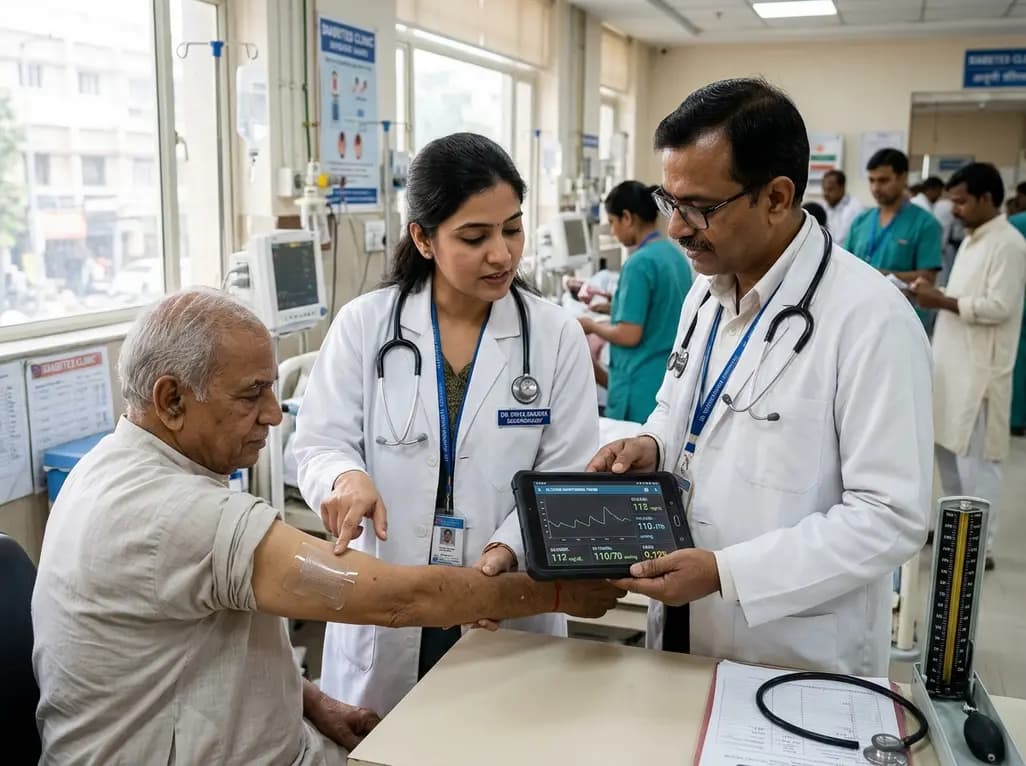
A breakthrough ultrathin multimodal sensor decouples physical hemodynamic monitoring from disposable electrochemical calibration, achieving long-term wearable glucose prediction with 81.36% accuracy and a 9.12% MARD. This offers a robust, drift-free solution for outpatient metabolic surveillance.
Today

A clinical review of menopause and neurology highlights the neurobiological impacts of midlife hormonal shifts on cognition, epilepsy, migraine, multiple sclerosis, and stroke, emphasizing safe hormone therapy prescribing.
Today

Co-administering antiseizure medications and DOACs frequently causes pharmacokinetic interactions, subtherapeutic drug levels, and thromboembolic failures. This review highlights enzyme induction risks, unpredictable outcomes with levetiracetam, and the vital role of therapeutic drug monitoring.
Today

A comparative review of tension band wiring and intramedullary screw fixation in thumb metacarpophalangeal arthrodesis, evaluating union rates, hardware complications, and clinical outcomes.
Today
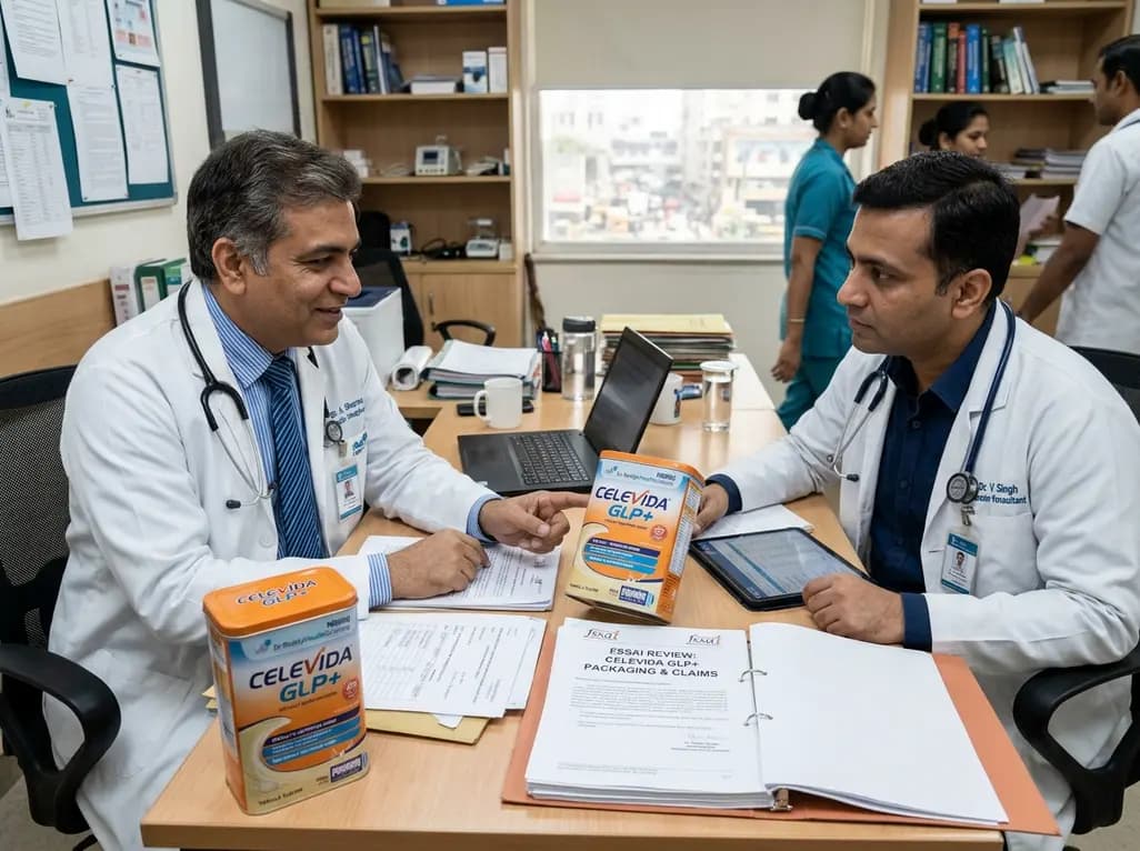
Following regulatory scrutiny from FSSAI regarding promotional representations and lean muscle preservation claims, Dr. Reddy's Laboratories and Nestlé Health Science are currently reviewing the branding and marketing strategy for their joint nutritional supplement Celevida GLP+.
Today
