.webp&w=384&q=75)

Loading, please wait...
Loading, please wait...
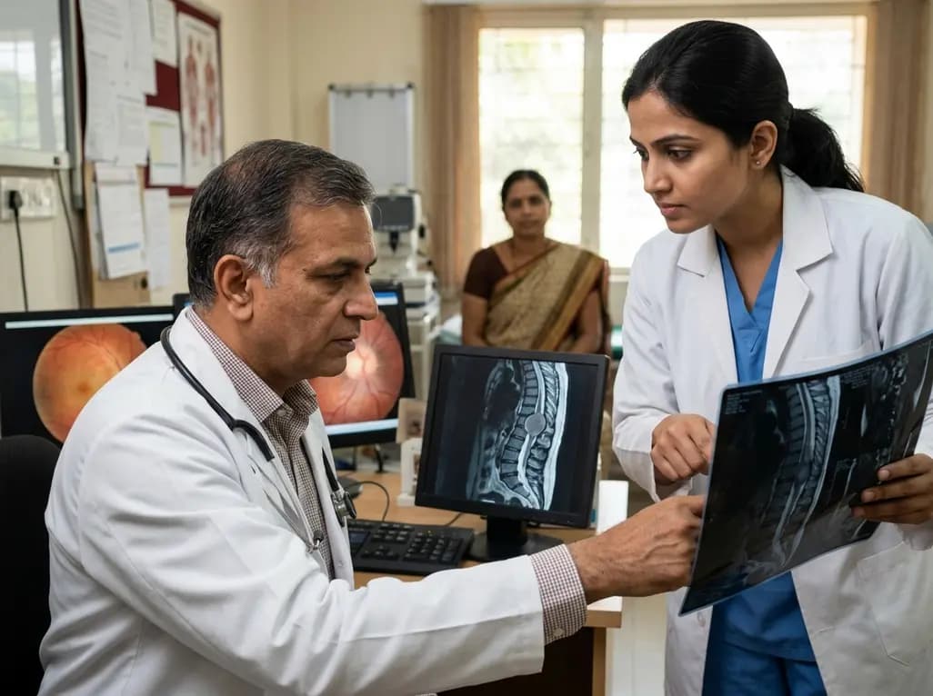
Physicians typically associate papilloedema with intracranial pathologies such as brain tumors, venous sinus thrombosis, or idiopathic intracranial hypertension. However, recent clinical evidence confirms that spinal tumor papilloedema represents a rare but critical diagnostic challenge for modern practitioners. This phenomenon occurs when intradural spinal masses trigger raised intracranial pressure, often in the total absence of ventriculomegaly or hydrocephalus. Because these patients frequently lack typical limb neurology, such as weakness or sensory loss, initial diagnostic efforts often focus exclusively on the brain. Consequently, this narrow focus can lead to severe diagnostic delays, misdiagnosis, and potentially permanent vision loss. Recognizing that the cause of optic nerve swelling may lie far below the skull is essential for every neurologist and ophthalmologist. Furthermore, timely intervention can transform the clinical course, shifting the prognosis from certain blindness to full visual recovery. Therefore, clinicians must maintain a high index of suspicion when intracranial imaging fails to explain significant disc swelling. Specifically, understanding the subtle links between spinal pathology and cranial pressure is the first step toward effective management.
The exact mechanism behind how a spinal mass influences cranial pressure remains a subject of intense medical discussion. Notably, several complex dynamics involving cerebrospinal fluid (CSF) appear to be at play. Many researchers suggest that high CSF protein levels secreted by the spinal tumor significantly increase fluid viscosity. This thickened fluid subsequently obstructs the arachnoid villi, which are responsible for CSF absorption into the venous system. When absorption fails, intracranial pressure rises despite a lack of physical obstruction within the brain itself. Additionally, malignant cells from certain tumors may directly infiltrate subarachnoid pathways, causing increased resistance to flow. Some cases also point toward venous stasis caused by tumor compression of spinal venous plexuses. This compression produces unfavorable hydrostatic pressure, further hindering the natural drainage of CSF. Moreover, the spinal canal acts as an elastic reservoir for CSF; when a tumor occupies this space, the system loses its compliance. Consequently, even minor changes in fluid volume can lead to dramatic spikes in pressure. This complex interplay explains why a lumbar or thoracic mass can manifest as a primary ophthalmic emergency.
One of the most confounding aspects of this condition is the frequent absence of classic spinal symptoms. Specifically, many patients present with isolated visual disturbances such as blurring, transient obscurations, or persistent headaches. Because they do not report back pain, radiculopathy, or leg weakness, the spine is rarely considered the primary site of pathology. However, the presence of bilateral papilloedema should always trigger a comprehensive search for the underlying cause of hypertension. In the cases described, patients often underwent multiple brain scans that appeared normal or showed only subtle signs of high pressure. This lack of ventriculomegaly often leads clinicians down the path of diagnosing idiopathic intracranial hypertension. Nevertheless, a failure to respond to standard medical treatments like acetazolamide should serve as a major red flag. Furthermore, clinicians should ask detailed questions about subtle postural changes in vision or very mild back discomfort that the patient might have dismissed. Ultimately, the lack of limb neurology does not rule out the spine. Instead, it highlights the need for a broader diagnostic perspective to prevent permanent optic nerve atrophy.
When a patient presents with unexplained intracranial hypertension, the diagnostic workup must be systematic and thorough. Initially, most protocols mandate high-resolution MRI of the brain and orbits to rule out mass lesions or venous sinus thrombosis. If these results are inconclusive, many doctors proceed to a lumbar puncture to confirm elevated opening pressure. However, if the CSF analysis reveals significantly elevated protein levels without pleocytosis, the clinical suspicion must shift toward the spine. Consequently, clinicians should order a contrast-enhanced MRI of the entire neuroaxis, including the cervical, thoracic, and lumbosacral regions. This step is vital because tumors at any level of the spinal cord can trigger the protein-driven mechanism of hypertension. Furthermore, optical coherence tomography (OCT) and formal visual field testing are essential to quantify the severity of the papilloedema. These tools help monitor the progression of optic nerve damage and guide the urgency of surgical intervention. By "thinking outside the box" and imaging the spine early, practitioners can avoid the pitfall of repetitive cranial imaging. Therefore, whole-spine imaging should be a standard consideration in any case of atypical or refractory papilloedema.
Managing cases where a spinal tumor threatens vision requires a dual approach focusing on both the cause and the symptoms. The definitive treatment is usually the surgical resection of the intradural spinal mass. Successfully removing the tumor often leads to a rapid normalization of CSF protein levels and a subsequent decrease in intracranial pressure. However, the acuity of visual deterioration often dictates the immediate management strategy. If a patient is rapidly losing sight, the surgical team must prioritize emergency CSF diversion. This may involve the placement of a temporary external ventricular drain or a more permanent shunt before addressing the spinal lesion. Conversely, in more stable cases, direct tumor excision might suffice to resolve the ophthalmic symptoms. Notably, the choice between different operative strategies depends on the tumor's location and histology. In many instances, once the physical or biochemical obstruction is removed, the papilloedema regresses significantly over several weeks. Furthermore, postoperative follow-up with neuro-ophthalmology is crucial to ensure that the optic nerve head returns to its normal state. Ultimately, a coordinated effort between neurosurgeons and eye specialists ensures the best possible patient journey.
The relationship between intradural spinal tumors and intracranial hypertension serves as a powerful reminder of the interconnected nature of the central nervous system. Although rare, the potential for permanent blindness makes this a diagnosis that no clinician can afford to miss. By recognizing that spinal tumor papilloedema can occur without hydrocephalus or leg weakness, doctors can significantly reduce diagnostic delays. Furthermore, the use of comprehensive neuroaxis imaging in atypical cases of raised pressure is a lifesaving practice. We must emphasize the priority of vision preservation through timely CSF management and tumor removal. Most patients experience a significant improvement in symptoms and a reversal of disc swelling following successful intervention. However, the key to these positive outcomes remains early identification and a high index of clinical suspicion. As medical educators, we urge our colleagues to look beyond the cranium when the diagnostic puzzle does not fit. By integrating spinal pathology into the differential diagnosis of papilloedema, we can protect our patients from the devastating consequences of misdiagnosis. Thus, vigilance and a broad clinical perspective remain our best tools in the fight against vision loss.
Yes, intradural spinal tumors can occasionally present with isolated intracranial hypertension and papilloedema. In such cases, patients may not exhibit any motor or sensory deficits in their limbs. This absence of typical spinal symptoms often leads to diagnostic confusion and necessitates a high index of clinical suspicion for the spine.
The primary mechanism involves elevated CSF protein levels secreted by the tumor, which increases fluid viscosity and obstructs absorption at the arachnoid villi. Additionally, the tumor may physically occupy space in the spinal canal, reducing the system's ability to compensate for fluid volume changes, leading to hypertension.
The immediate priority is the preservation of vision. If papilloedema is severe or vision is deteriorating rapidly, emergency CSF diversion, such as a shunt or drain, may be required. Following stabilization, the surgical team typically performs a resection of the spinal tumor to address the underlying cause of the pressure.
Disclaimer: This content is for informational and educational purposes only. It does not constitute medical advice or establish a doctor-patient relationship. Refer to the latest local and national guidelines for clinical practice.
References
Sun R et al. Intra-cranial hypertension and vision-threatening papilloedema caused by intradural spinal tumours: a case series of three. Br J Neurosurg. 2025 Aug. doi: 10.1080/02688697.2023.2263087. PMID: 37807636.
Al-Houshimy A, et al. Hydrocephalus and Papilledema in Spinal Cord Tumors: A Report of Two Cases. PMC. 2023.
Xie JS, et al. Papilledema: A review of etiology, pathophysiology, diagnosis, and management. Surv Ophthalmol. 2022;67(4):935-959.

Read summarized clinical updates, watch expert medical content, and earn CME certifications right from your smartphone.
Open your smartphone camera to scan and install.





Spinal tumors can unexpectedly cause intracranial hypertension and papilloedema without ventriculomegaly. This case-based review explores why early spinal imaging and vision preservation are critical when patients present with isolated eye symptoms and no limb neurology.
3 months ago

Review how tailored exercise programs benefit frail older patients with heart failure. Explore pathophysiological mechanisms, individualized training prescriptions, and strategies to overcome adherence barriers in clinical practice.
Today

Leucine Rich Bio has introduced South Asia's first comprehensive vaginal microbiome test, BugSpeaks. By profiling microbial dysbiosis, pathogens, and antimicrobial resistance, this next-generation diagnostic platform provides clinicians with deeper insights into recurrent infections, fertility, and reproductive health.
Today

In high-risk pulmonary embolism complicated by severe anemia and active uterine bleeding, systemic thrombolysis is contraindicated. Large-bore aspiration thrombectomy provides rapid hemodynamic rescue without fibrinolysis, supported by inferior vena cava filter placement and multidisciplinary critical care.
Today

This comparative study evaluates posterior labral tears with and without glenohumeral instability, highlighting distinct demographic patterns, MRI underestimation in stable shoulders, and successful arthroscopic repair outcomes.
Today

A clinical study demonstrates that early intervention with tocilizumab, either as monotherapy or bridged with corticosteroids, achieves significant proptosis reduction and resolves active inflammation in treatment-naive thyroid eye disease.
Today
