.webp&w=384&q=75)

Loading, please wait...
Loading, please wait...

Early identification of malignant liver nodules dramatically alters patient survival. Clinicians face significant diagnostic hurdles when differentiating early malignancies from benign lesions in chronic liver disease. Consequently, accurate small HCC diagnosis remains a challenging endeavor when nodules measure two centimeters or less. Traditional imaging protocols often fail to capture subtle microvascular alterations in tiny lesions. Moreover, benign regenerative nodules, dysplastic nodules, and hemangiomas frequently mimic early malignancy on standard contrast computed tomography. Therefore, radiologists need advanced, noninvasive imaging biomarkers to establish definitive diagnoses without risky biopsy procedures.
Gadolinium ethoxybenzyl diethylenetriamine pentaacetic acid, commonly known as Gd-EOB-DTPA, represents a major advance in functional hepatobiliary imaging. This hepatocyte-specific agent offers dual extracellular vascular and functional hepatobiliary properties. Consequently, functioning hepatocytes clear the contrast through biliary excretion, creating stark contrast against malignant lesions that lack functioning organic anion transporters. However, interpreting these subtle signals requires extensive clinical experience. Junior radiologists frequently struggle to distinguish atypical benign lesions from well-differentiated hepatocellular carcinoma. Therefore, developing automated, highly interpretable artificial intelligence systems provides an invaluable clinical solution. By decoding complex image patterns beyond human visual perception, artificial intelligence can transform hepatology workflows and streamline clinical decision-making worldwide.
To overcome diagnostic variability across institutions, a multicenter investigation evaluated machine learning algorithms across multiple tertiary academic hospitals. The researchers assembled a retrospective cohort comprising 296 hepatic lesions across three distinct clinical centers. Specifically, Center A provided 209 lesions, which the investigators divided into 146 training cases and 63 internal test cases. Additionally, Centers B and C provided 87 external validation cases to confirm broad clinical generalizability.
The investigative team methodically extracted three comprehensive feature categories from each observation. First, they extracted high-dimensional radiomics features from Gd-EOB-DTPA-enhanced MRI scans across multiple imaging phases. Second, they recorded standardized qualitative MRI features routinely assessed by abdominal radiologists, such as arterial hyperenhancement and capsule appearance. Third, they gathered vital clinical indicators, including serological markers and underlying viral etiology. Furthermore, the researchers utilized a strict multistep feature selection pipeline exclusively within the training cohort. They implemented nested cross-validation protocols to prevent data leakage and avoid model overfitting. Subsequently, the investigators systematically compared seven machine learning classifiers to determine optimal predictive performance. This meticulous methodological architecture ensured robust algorithmic training before evaluating real-world validation data.
The resulting multimodal fusion model demonstrated remarkable discriminative capability during rigorous evaluation. In the internal testing set, the fusion model achieved an impressive area under the receiver operating characteristic curve of 0.913. In contrast, the standalone radiomics-only model yielded an area under the curve of only 0.791. This noticeable difference underscores the immense diagnostic value of integrating clinical parameters and qualitative imaging signs with computational radiomics. Furthermore, the fusion model preserved its robust diagnostic precision within the independent external validation cohort, yielding an area under the curve of 0.824.
Interscanner variability often degrades artificial intelligence performance across different commercial hospital imaging platforms. To address this technical challenge, the investigators applied ComBat harmonization across the multicenter datasets. Consequently, the harmonized external model maintained stable discriminative power with an area under the curve of 0.806. Readers also observed that the computational model achieved numerically superior accuracy compared to junior radiologists. Simultaneously, the algorithmic platform matched the diagnostic precision of senior gastrointestinal imaging specialists. Therefore, the multimodal system demonstrates substantial clinical reliability across disparate imaging environments.
Black-box artificial intelligence models frequently meet resistance from skeptical medical practitioners in oncology. Therefore, establishing transparent algorithmic reasoning represents an essential prerequisite for routine clinical implementation. To provide actionable transparency, the investigators integrated SHapley Additive exPlanations, commonly designated as SHAP. This mathematical framework precisely calculates the individual contribution of each predictive variable to the final diagnostic classification.
Notably, the SHAP analysis illuminated three dominant drivers of malignancy risk in small nodules. First, positive hepatitis B virus status emerged as a paramount clinical indicator of hepatocellular malignancy. Second, elevated serum alpha-fetoprotein levels strongly reinforced the probability of primary liver cancer. Third, specific radiomic textures extracted from hepatobiliary phase images contributed profound diagnostic weight. During the hepatobiliary phase, malignant hepatocytes exhibit decreased organic anion-transporting polypeptide expression. Consequently, early carcinomas fail to absorb Gd-EOB-DTPA, causing conspicuous signal loss. The quantitative texture features captured microscopic architectural heterogeneity within these hypointense zones. Thus, SHAP successfully bridged advanced computer science and physiological hepatocarcinogenesis, instilling clinical confidence in the model's automated recommendations.
Integrating artificial intelligence into hepatology workflows offers substantial promise for global healthcare delivery. In high-prevalence regions like India and East Asia, chronic viral hepatitis and metabolic dysfunction drive substantial liver cancer morbidity. However, many peripheral diagnostic centers lack dedicated subspecialty abdominal radiologists. Consequently, junior clinicians frequently encounter indeterminate nodules that require definitive characterization. Deploying an interpretable algorithmic tool provides dependable diagnostic assistance in resource-constrained environments.
Although statistical significance versus junior readers was limited by external sample size, numerical trends strongly favored algorithmic triage. Moreover, implementing dynamic threshold optimization in future software versions will allow clinicians to adjust sensitivity according to specific institutional protocols. For instance, screening programs could maximize diagnostic sensitivity to ensure early detection, whereas surgical planning suites could prioritize specificity to avoid unwarranted resections. Furthermore, avoiding invasive biopsy prevents potential complications such as tumor seeding, localized hemorrhage, or procedural delay. Ultimately, this validated artificial intelligence platform paves the way for accessible, standardized, and precise liver cancer diagnosis worldwide.
Gd-EOB-DTPA functions as a dual-action contrast agent that provides vascular dynamic imaging alongside targeted hepatobiliary phase evaluation. Healthy hepatocytes take up this agent through organic anion-transporting polypeptides and eliminate it into the bile ducts. In contrast, dedifferentiated malignant hepatocytes downregulate these transporters, resulting in distinct hypointensity during delayed hepatobiliary phases. Consequently, this functional contrast agent reveals microscopic hepatocarcinogenesis far earlier than conventional extracellular agents can achieve.
Interpretability builds clinical confidence by transforming opaque algorithmic predictions into biologically plausible diagnostic explanations. Oncologists and radiologists must understand why an artificial intelligence platform flags a specific hepatic lesion as malignant. Frameworks like SHapley Additive exPlanations quantify the exact importance of individual clinical indicators and radiomic features. Therefore, transparent reasoning empowers clinicians to verify algorithmic outputs, prevent catastrophic diagnostic errors, and defend personalized therapeutic strategies effectively.
ComBat harmonization effectively eliminates technical batch effects caused by diverse MRI scanners, acquisition parameters, and institutional protocols across multicenter studies. Without harmonization, machine learning algorithms frequently latch onto scanner-specific noise rather than genuine disease biomarkers. Consequently, predictive accuracy drops significantly when testing external hospital data. Harmonization standardizes feature distributions while preserving essential biological variation. Thus, it ensures reproducible diagnostic performance across varied tertiary healthcare environments.
Disclaimer: This content is for informational and educational purposes only and should not be taken as professional medical advice. Always consult a qualified healthcare provider for personalized clinical diagnosis and treatment. Refer to the latest local and national guidelines for clinical practice.
References

Read summarized clinical updates, watch expert medical content, and earn CME certifications right from your smartphone.
Open your smartphone camera to scan and install.





A multicenter study validates an interpretable machine learning model integrating Gd-EOB-DTPA MRI radiomics and clinical factors to identify small hepatocellular carcinomas (≤2 cm), achieving high diagnostic accuracy comparable to senior radiologists.
Today
A comprehensive meta-analysis of over 4.4 million out-of-hospital cardiac arrest patients reveals a global bystander CPR rate of only 32.1%, showing marked disparities between high-income and developing nations. Healthcare professionals must advocate for community resuscitation training.
Today
A recent interventional study assessed whether adding a single 150 IU dose of follitropin alfa before or after clomiphene citrate improves clinical outcomes in unexplained infertility. The findings demonstrate that sequential gonadotropin therapy does not increase ovulation or pregnancy rates.
Today
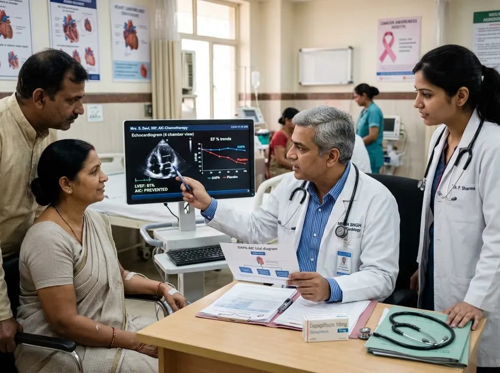
The phase II DAPA-AIC trial demonstrates that dapagliflozin 10 mg daily significantly preserves left ventricular ejection fraction compared to placebo over four months in adults undergoing anthracycline chemotherapy, highlighting a potential preventive strategy in cardio-oncology.
Today
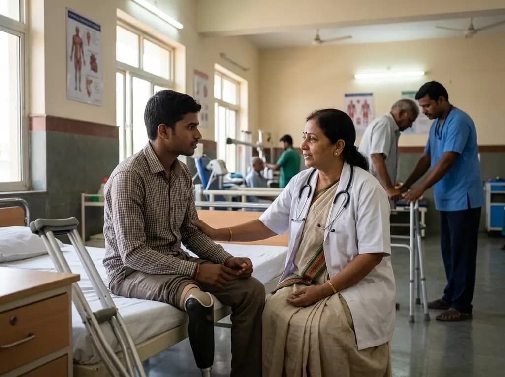
A scoping review reveals severe gaps in long-term assistive technology provision following humanitarian emergencies. Addressing post-disaster rehabilitation requires systematic follow-up, workforce training, and integration into national health systems to sustain mobility and recovery.
Today
A new study reveals that high-fat, cholesterol, and fructose intake drives MASH and hepatic fibrosis in a sex- and hepatocyte PPARγ-dependent manner. Male mice showed severe fibrosis and downregulated methionine metabolism, while hepatocyte PPARγ knockout protected against progression.
Today
