

Loading, please wait...
Loading, please wait...

Estimating biological senescence through structural neuroimaging offers profound insights into individual neural vulnerability. Consequently, brain age prediction has emerged as a transformative quantitative biomarker across modern neurobiology and clinical neurology. Chronological age frequently fails to capture subtle variations in individual organ health. In contrast, machine learning models estimate biological brain age directly from magnetic resonance imaging data. When a patient exhibits a brain age that markedly exceeds chronological age, physicians recognize potential early neurodegenerative risk. However, conventional deep learning models frequently operate as opaque black boxes, preventing clinicians from understanding specific anatomical drivers.
Brain age prediction provides an objective metric to quantify deviations from healthy neuroanatomical aging trajectories. Specifically, the difference between predicted biological brain age and chronological age represents the brain age gap. A positive gap suggests accelerated cerebral aging, which strongly associates with conditions such as Alzheimer's disease, vascular dementia, and major depression. Furthermore, elevated gaps correlate with subtle cognitive decline, lifestyle risk factors, and elevated mortality. Clinicians traditionally evaluate structural neuroimaging through qualitative inspection of generalized atrophy or regional ventricular enlargement. However, manual radiologic review often misses subtle, diffuse microstructural changes occurring in preclinical disease stages. Automated neural networks solve this challenge by evaluating complex, non-linear morphological relationships across three-dimensional brain scans. For example, deep convolutional architectures examine voxel-level cortical thickness, sulcal widening, and subcortical volume reductions simultaneously. As a result, automated models capture early neurodegenerative pathology well before overt functional impairment emerges. Nevertheless, healthcare providers cannot comfortably adopt these algorithmic scores without clear biological justification. Neurologists and radiologists require verifiable anatomical evidence to confirm that predictions reflect genuine neuropathology rather than technical imaging artifacts. Therefore, establishing transparent interpretability serves as the critical prerequisite for integrating neuroimaging artificial intelligence into real-world geriatric and neurological care.
Explainable artificial intelligence organizes deep learning methodologies into two primary paradigms: post-hoc explanation techniques and intrinsically interpretable models. Post-hoc methods inspect established networks after training completes. Common approaches include gradient-weighted class activation mapping, integrated gradients, and perturbation-based occlusion maps. These tools generate visual saliency maps highlighting the voxels that drove network output. In addition, localized attribution methods like Shapley additive explanations identify how individual features modify final numerical estimates. However, post-hoc methods often demonstrate considerable instability. Small image perturbations or scanner noise can drastically alter saliency maps without changing the model's numerical output. Furthermore, gradient-based heatmaps frequently display diffuse, anatomically non-specific highlights that fail to isolate discrete anatomical nuclei. In response, artificial intelligence researchers increasingly prioritize intrinsically interpretable architectures. These self-explaining frameworks incorporate transparency directly into network layers. For instance, vision transformers employ multi-head self-attention mechanisms to map dependencies across distant anatomical regions. Similarly, disentangled representation learning isolates distinct biological factors, such as tissue atrophy and ventriculomegaly, into separate latent variables. Moreover, generative adversarial networks visualize counterfactual aging trajectories, showing how an individual brain scan would appear at varying ages. Consequently, these modern intrinsic architectures align machine reasoning far more closely with human neuroanatomy.
To bridge the gap between algorithmic engineering and diagnostic neurology, researchers propose a robust four-dimensional evaluation framework. The first dimension, Algorithm Reliability, measures fundamental predictive performance using metrics such as mean absolute error and explained variance. Models must demonstrate consistent mathematical convergence alongside high predictive fidelity across diverse cohorts. The second dimension, Robustness and Reproducibility, assesses stability across scanner variations, image noise, and subtle spatial transformations. If minor head rotation or altered contrast ruins interpretability, clinicians cannot trust the algorithm during routine workflows. The third dimension, Biological Validation, tests whether algorithmic explanations align with established neuropathology. For example, explainability heatmaps in elderly populations should pinpoint hippocampal atrophy, temporal lobe thinning, and ventricular enlargement. When explanations target irrelevant extracranial structures or image background, algorithms clearly lack biological credibility. Finally, the fourth dimension, Clinical Utility, examines whether brain age outputs improve patient management. Clinicians must determine whether predicted gaps meaningfully differentiate normal aging from mild cognitive impairment or dementia. Furthermore, utility metrics evaluate whether models assist risk stratification, predict longitudinal decline, and guide therapeutic decision-making. Therefore, adopting this multi-dimensional framework ensures that neuroimaging algorithms satisfy strict medical standards before routine deployment in memory clinics.
Despite encouraging technological advancements, substantial methodological obstacles hinder the broad clinical adoption of brain age models. A prominent challenge involves age dependency bias, often called regression toward the mean. Specifically, standard regression algorithms systematically overestimate biological age in younger participants and underestimate age in older subjects. Consequently, researchers must implement post-processing statistical calibration to adjust for this baseline dependency before interpreting the gap. In addition, the demographic composition of reference training populations profoundly influences model generalizability. Many existing models rely primarily on Western neuroimaging repositories, such as the UK Biobank. However, cerebral morphology, skull thickness, and vascular risk profiles vary considerably across global and Indian populations. Applying Western-trained algorithms directly to Indian clinical cohorts without local recalibration introduces significant demographic bias. Furthermore, multicenter site heterogeneity creates substantial technical noise. Imaging centers employ magnetic resonance scanners from different manufacturers featuring distinct field strengths, coil designs, and acquisition parameters. These technical discrepancies produce spurious inter-site variations that corrupt biological age predictions. To mitigate this hurdle, development teams must integrate rigorous image harmonization algorithms like ComBat. Ultimately, establishing standardized normalization pipelines across local centers ensures fair, reproducible, and ethnically inclusive neuroimaging analytics.
Integrating explainable neuroimaging systems into clinical memory workflows offers substantial diagnostic advantages for multidisciplinary teams. First, transparent models provide objective quantitative evidence that complements traditional cognitive assessments, such as the Mini-Mental State Examination. When a clinician detects borderline cognitive impairment, an elevated brain age gap confirmed by regional temporal lobe atrophy provides compelling evidence for early intervention. Moreover, interpretable brain age metrics help physicians monitor disease progression and evaluate therapeutic efficacy during clinical drug trials. For instance, novel disease-modifying therapies for Alzheimer's disease target amyloid clearance and slow neurodegeneration. By tracking whether an investigational drug reduces the biological brain age gap over time, neurologists obtain measurable evidence of neuroprotection. In addition, transparent explanations enhance clinical communication between healthcare professionals, patients, and family caregivers. Visualizing counterfactual images or anatomical attention maps helps families comprehend the biological reality of cognitive decline. Furthermore, clear visual explanations foster greater trust in algorithmic recommendations among attending physicians. Nevertheless, successful clinical translation demands prospective longitudinal trials rather than purely retrospective evaluations. Clinicians must validate whether acting upon algorithmic predictions genuinely improves long-term patient outcomes. As multidisciplinary collaboration expands, explainable artificial intelligence will undoubtedly redefine personalized neurodegenerative care.
The brain age gap quantifies accelerated tissue loss exceeding normal chronological aging patterns. In neurodegenerative conditions like Alzheimer's disease, pathological processes trigger widespread cortical thinning and hippocampal atrophy. Consequently, deep learning models trained on healthy scans detect these structural anomalies as advanced biological age. Identifying this difference provides clinicians with an early, sensitive metric for disease vulnerability before overt cognitive decline appears.
Post-hoc explanation methods, including saliency maps and SHAP, analyze neural networks after complete training to highlight influential input voxels. However, these techniques often produce noisy heatmaps that lack mathematical stability. In contrast, intrinsically interpretable architectures integrate transparency directly into the model design. For instance, vision transformers explicitly display attention weights across brain regions during inference. Therefore, intrinsic methods provide clinicians with clearer, more dependable biological explanations for predictions.
Site heterogeneity occurs because different imaging facilities utilize diverse magnetic resonance scanner manufacturers, magnetic field strengths, and scanning protocols. These variations introduce non-biological noise and systematic domain shifts into imaging data. Consequently, a deep learning algorithm trained at one hospital frequently experiences performance drops when tested elsewhere. Applying statistical harmonization, standardizing acquisition parameters, and validating algorithms across multi-institutional external cohorts are necessary steps to ensure robust real-world clinical performance.
Disclaimer: This content is for informational and educational purposes only... Refer to the latest local and national guidelines for clinical practice.
References

Read summarized clinical updates, watch expert medical content, and earn CME certifications right from your smartphone.
Open your smartphone camera to scan and install.





Interpretable deep learning bridges predictive accuracy and clinical trust in brain age prediction. This review examines post-hoc and intrinsic explainable AI methods, introducing a four-dimensional framework to evaluate algorithmic reliability, robustness, biological validity, and clinical utility in neuroimaging.
Today
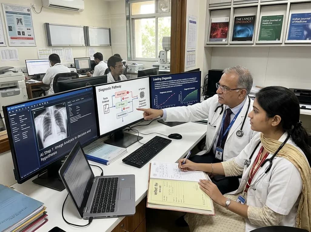
A breakthrough peer-reviewed study demonstrates that a sequential AI diagnosis system can mirror real-world clinical reasoning. By ordering stepwise tests and updating hypotheses dynamically, this multi-agent orchestrator achieved superior diagnostic accuracy while reducing investigative costs.
Today

A prenatal case highlights severe glutaric acidemia type II presenting as bilateral hyperechoic cystic kidneys at 18 weeks. Whole-exome sequencing revealed a novel homozygous ETFDH frameshift variant, demonstrating the power of rapid genetic testing in fetal renal anomalies.
Today

Review how tailored exercise programs benefit frail older patients with heart failure. Explore pathophysiological mechanisms, individualized training prescriptions, and strategies to overcome adherence barriers in clinical practice.
Yesterday

This comparative study evaluates posterior labral tears with and without glenohumeral instability, highlighting distinct demographic patterns, MRI underestimation in stable shoulders, and successful arthroscopic repair outcomes.
Yesterday

A clinical study demonstrates that early intervention with tocilizumab, either as monotherapy or bridged with corticosteroids, achieves significant proptosis reduction and resolves active inflammation in treatment-naive thyroid eye disease.
Yesterday
