.webp&w=384&q=75)

Loading, please wait...
Loading, please wait...

Decades after refractive surgery, corneal surgeons still encounter rare but visually devastating complications in post-LASIK eyes. Among these entities, interface fluid syndrome remains one of the most diagnostically challenging conditions in clinical practice. The accumulation of transudative fluid within the latent interlamellar space compromises visual acuity and distorts standard clinical measurements. Furthermore, recent evidence synthesis reveals that while elevated intraocular pressure triggers most presentations, endothelial pump decompensation also generates this distinctive clinical pathology.
The creation of a lamellar corneal flap permanently alters the biomechanics of the anterior stroma. Histological studies show that the LASIK interface never achieves the tensile strength of virgin tissue. Instead, non-fibrillar proteoglycans fill this microscopic potential space, creating a hydrophilic cleft between the flap and stromal bed. Under physiological conditions, healthy endothelial cells constantly dehydrate the cornea, keeping this potential plane tightly apposed.
However, whenever corneal stromal hydration increases beyond physiologic limits, fluid preferentially dissects into this weak lamellar boundary. This phenomenon explains why interface fluid syndrome acts as a final common pathway rather than a single isolated disease. When hydraulic hydrostatic pressure drives fluid forward across a compromised endothelial barrier, fluid accumulates in the flap pocket. Consequently, both increased anterior chamber pressure and defective endothelial fluid clearance produce identical anatomical pooling.
Clinicians frequently encounter immense diagnostic confusion when evaluating post-LASIK patients with unexplained visual blurring. Patients typically present with painless vision reduction, variable stromal haze, and marked central corneal thickening. Slit-lamp biomicroscopy often reveals an optically clear space between the flap and underlying stroma, but subtle presentations easily escape detection without specialized imaging. High-resolution anterior segment optical coherence tomography provides vital confirmation by directly imaging the fluid-filled interface cleft.
Crucially, central Goldmann applanation tonometry generates severe diagnostic errors in this setting. Because the interface fluid acts like a soft cushion, the tonometer indents the anterior flap without transmitting force to the anterior chamber. As a result, central applanation tonometry yields artificially low or deceptively normal pressure values, despite perilous anterior chamber hypertension. Therefore, ophthalmologists must measure intraocular pressure over the peripheral cornea or sclera using dynamic contour tonometry or pneumotonometry to reveal the true underlying pressure.
A recent comprehensive synthesis of 83 studies analyzing 225 eyes demonstrates that elevated intraocular pressure causes 81.33% of interface fluid syndrome cases. Prolonged topical corticosteroid therapy represents the primary culprit in most instances. Many patients initially receive corticosteroids following refractive surgery, cataract extraction, or minor ocular trauma. In steroid-responsive individuals, trabecular outflow resistance rises sharply, driving fluid forward into the corneal stroma.
Tragically, interface fluid syndrome closely mimics diffuse lamellar keratitis on initial slit-lamp inspection. If treating clinicians mistakenly diagnose sterile inflammation, they typically intensify topical corticosteroid frequency. This therapeutic misstep accelerates intraocular pressure elevation and expands the fluid pocket, initiating a dangerous clinical spiral. Once clinicians recognize the true pressure-dependent etiology, management requires immediate steroid cessation combined with aggressive pressure-lowering agents. In most cases, controlling intraocular pressure promptly resolves the interlamellar fluid and restores baseline corneal clarity.
Although ocular hypertension causes most presentations, primary endothelial dysfunction accounts for approximately 12% of reported cases. In these patients, interface fluid develops in the presence of completely normal intraocular pressure. Conditions such as Fuchs endothelial corneal dystrophy, toxic anterior segment syndrome, and surgical trauma destroy endothelial pump capacity. When the endothelium fails to pump fluid out of the stroma, fluid collects inside the latent LASIK interface.
Historically, endothelial-driven fluid accumulation required invasive surgical intervention, such as endothelial keratoplasty. However, groundbreaking evidence reveals a novel presentation occurring after Descemet stripping only for Fuchs dystrophy. In two newly reported cases, stripping central guttata created transient endothelial pump failure, precipitating early postoperative interface fluid syndrome despite normotension. Remarkably, these cases resolved spontaneously without corneal transplantation. As peripheral endothelial cells migrated centrally and established functional coverage, fluid cleared completely, confirming the self-limiting nature of this unique post-stripping manifestation.
Successful management of interlamellar fluid accumulation requires clinicians to adopt a rigorous, mechanism-based evaluation protocol. First, ophthalmologists must review surgical history to identify remote refractive procedures, which may date back over three decades. Second, clinicians must verify pressure levels using peripheral tonometry to circumvent misleading central applanation values. In addition, anterior segment optical coherence tomography should serve as the primary objective modality to measure fluid thickness and track resolution.
Once clinicians establish the underlying mechanism, targeted intervention must follow immediately. For pressure-driven presentations, providers should taper topical steroids rapidly and initiate topical aqueous suppressants. Conversely, in patients with confirmed endothelial failure, treatment strategy depends on graft integrity and potential for cellular repopulation. While chronic endothelial decompensation mandates Descemet membrane endothelial keratoplasty, transient dysfunction following Descemet stripping only justifies watchful observation. Through precise etiology-specific treatment, clinicians can prevent permanent glaucomatous neuropathy and preserve long-term corneal clarity.
Central applanation tonometry measures resistance across the anterior cornea. When interface fluid syndrome occurs, the fluid pocket acts as a hydraulic cushion. Consequently, the tonometer indents only the thin anterior flap rather than assessing true anterior chamber tension. This phenomenon causes falsely low or normal central readings. Therefore, clinicians must measure pressure over the peripheral cornea or sclera using dynamic contour or pneumotonometry to accurately detect elevated intraocular pressure.
Diffuse lamellar keratitis represents an inflammatory leukocyte infiltration localized strictly within the stromal interface. Conversely, interface fluid syndrome involves transudative serous fluid accumulation without genuine cellular infiltration. While inflammatory keratitis requires aggressive topical corticosteroid therapy, steroids exacerbate pressure-driven interface fluid accumulation by increasing intraocular pressure. High-resolution anterior segment optical coherence tomography readily separates these entities by demonstrating a clear, hypo-reflective fluid pocket rather than hyper-reflective inflammatory granular debris.
Clinicians should manage post-stripping interface fluid conservatively rather than rushing toward corneal transplantation. Descemet stripping only temporarily disrupts central endothelial pumping capacity, which permits fluid transudation into the pre-existing lamellar flap space. Because intraocular pressure remains normal in these eyes, clinicians should monitor corneal clearance using serial optical coherence tomography. As peripheral endothelial cells migrate centrally and repopulate the bare stroma, endothelial pump activity recovers spontaneously, clearing interface fluid without graft placement.
Disclaimer: This content is for informational and educational purposes only. It is not intended to provide medical advice, diagnosis, or treatment and should not be relied upon as a substitute for professional clinical judgment. Medical knowledge is constantly evolving, and readers are strongly encouraged to independently verify all information, consult senior colleagues, and refer to current primary literature, institution-specific protocols, and up-to-date regional guidelines before making clinical decisions. Clinical discretion is paramount. Refer to the latest local and national guidelines for clinical practice.
References

Read summarized clinical updates, watch expert medical content, and earn CME certifications right from your smartphone.
Open your smartphone camera to scan and install.





A systematic review of 225 eyes identifies intraocular hypertension as the main cause of interface fluid syndrome, while also revealing endothelial dysfunction after Descemet stripping only as a novel etiology that resolves with endothelial repopulation.
Today
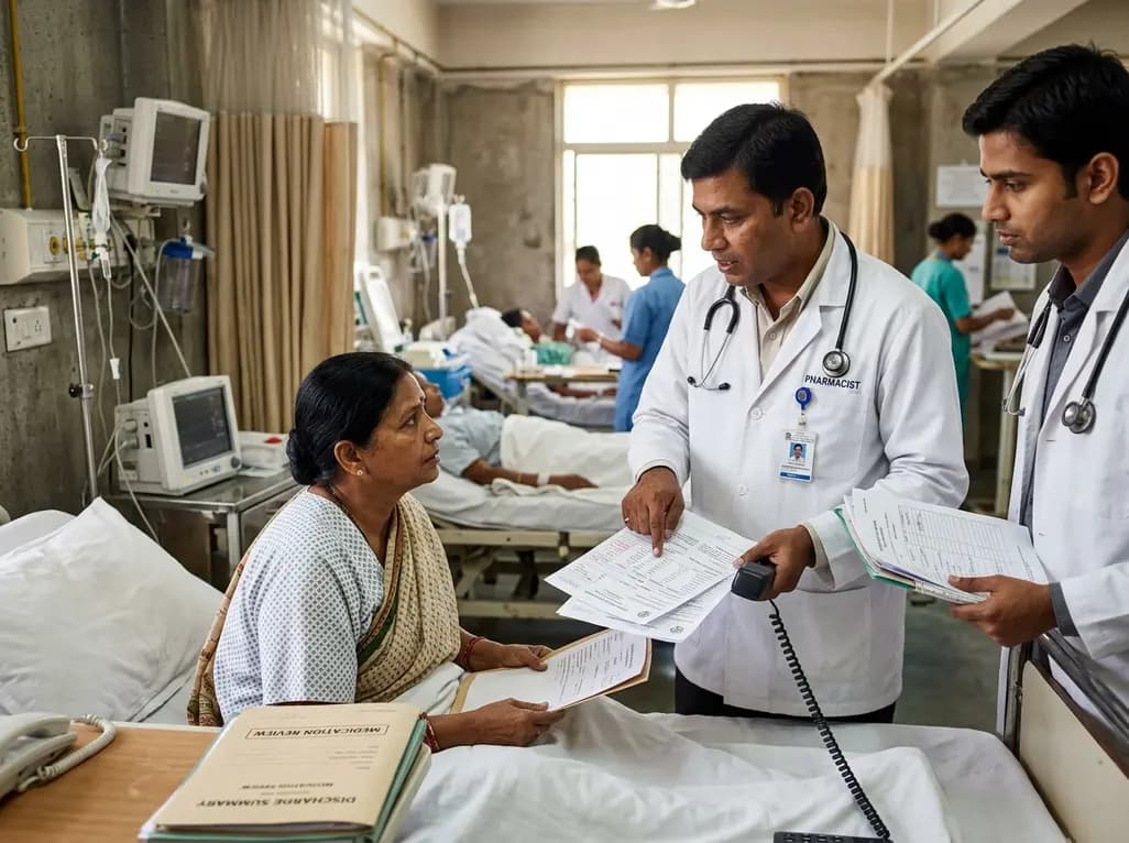
A retrospective cohort study demonstrates that comprehensive pharmacy transitions of care—including admission medication history, discharge review, and 72-hour telephone follow-up—significantly reduced 30-day readmission rates in acute care patients, highlighting the value of pharmacist-led clinical interventions.
Today

Meghalaya recorded a historic 55% reduction in maternal deaths, dropping from 244 to 107 over five years. Through the MOTHER app, dedicated transit homes, and expanded infrastructure like the 150-bed Tura Maternity Hospital with its first public NICU, the state demonstrates an effective public health model.
Today

Explore modern perioperative management protocols for clinical xenotransplantation. Learn how costimulation blockade, anti-inflammatory perfusion, and coagulopathy rescue overcome interspecies hurdles.
Today
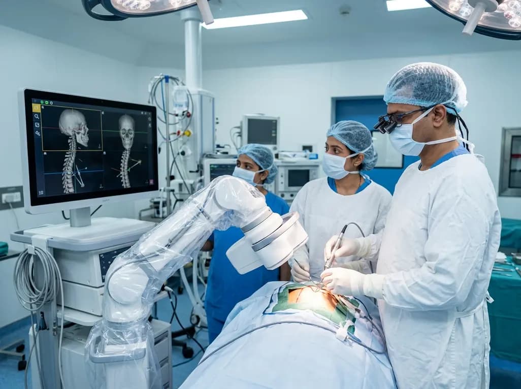
North India's first Centre of Excellence for Brain and Spine at Amrita Hospital Faridabad integrates Brainlab's Loop-X mobile robot. This landmark facility combines cone-beam computed tomography with surgical navigation to elevate precision, optimize operative workflows, and enhance specialized clinician training.
Today

At Homa CME 2026 at Hyderabad's T-Hub, healthcare leaders emphasized moving beyond routine metrics through digital calculators and precision stratification. Discover why characterizing individual insulin secretion, resistance patterns, and vascular risks transforms chronic metabolic care in India.
Today
