.webp&w=384&q=75)

Loading, please wait...
Loading, please wait...

Multiparametric magnetic resonance imaging has revolutionized modern prostate cancer diagnosis, biopsy targeting, and active surveillance. However, achieving diagnostic precision requires consistent prostate MRI quality assessment across imaging centers. High-quality scans allow clinicians to differentiate indolent lesions from clinically significant neoplasms with high confidence. Conversely, poor image quality obscures anatomy, produces artificial signal alterations, and inflates indeterminate category assignments. When images exhibit blur or noise, radiologists frequently face diagnostic dilemmas that impair clinical staging.
To overcome technical variations, the European Society of Urogenital Radiology introduced standardized quality criteria. The Prostate Imaging Quality version 2 system refines these standards by evaluating both multiparametric and biparametric acquisitions. This system focuses heavily on structural delineation on T2-weighted imaging and functional clarity on diffusion-weighted sequences. Although standardized criteria establish essential baselines, image interpretation inherently retains human subjectivity. Interpreting radiologists often disagree on whether subtle image flaws truly invalidate diagnostic reporting. Consequently, evaluating how controlled image degradation impacts observer agreement provides vital insights into diagnostic reliability in routine urological practice.
To assess how technical flaws alter professional judgment, researchers developed an experimental paradigm using controlled image degradation. The study utilized ten single-slice prostate image sets and introduced seven distinct levels of synthetic distortion. Specifically, investigators degraded T2-weighted images by progressively adding Gaussian noise and spatial blurring. For diffusion-weighted imaging, the team applied controlled noise, spatial blur, and non-linear elastic deformation. Additionally, researchers created twelve datasets featuring progressive anatomical mismatch between apparent diffusion coefficient maps and T2-weighted structural images.
Ten expert radiologists independently reviewed these randomized cases using criteria aligned with Prostate Imaging Quality version 2. The readers categorized image adequacy while remaining blinded to the degradation parameters. The researchers evaluated inter-reader agreement using proportion agreement and Gwet's agreement coefficient 1. Gwet's statistical approach provides dependable concordance estimates because it remains robust against marginal distribution imbalances. By isolating specific artifact types through synthetic alteration, the authors established precise perceptual boundaries. Unlike retrospective clinical audits with uncontrolled confounders, this experimental model revealed exactly how each degradation parameter influences specialist visual evaluation.
The experimental findings demonstrated a fascinating relationship between degradation severity and reader concordance. For T2-weighted acquisitions, perceived image adequacy steadily decreased as noise and blurring intensified. However, inter-reader agreement followed a pronounced U-shaped curve across degradation tiers. Radiologists exhibited nearly perfect agreement when assessing pristine baseline examinations or severely degraded scans. Under those extreme conditions, Gwet's agreement coefficient reached 0.98. In sharp contrast, agreement collapsed entirely at intermediate degradation levels, dropping to a coefficient of -0.03.
This striking drop exposes a critical diagnostic gray zone in radiological practice. When images are flawless, experts effortlessly confirm diagnostic adequacy. Likewise, when severe distortion obliterates anatomical landmarks, radiologists unanimously reject the scan. However, moderate degradation forces readers to make subjective judgments regarding diagnostic safety. One specialist might judge a moderately blurred scan acceptable for identifying organ-confined tumors. Meanwhile, another expert may classify that exact examination as clinically uninterpretable. Consequently, this wide variability proves that visual thresholds differ substantially among qualified specialists, creating clinical uncertainty in borderline examinations.
Functional diffusion-weighted imaging displayed distinct vulnerability characteristics compared to structural T2-weighted sequences. As blur and elastic warping increased, radiologists reported progressive declines in diffusion sequence adequacy. Interestingly, agreement regarding noise degradation in diffusion imaging followed an unexpected pattern. Concordance actually rose from moderate levels at baseline to substantial agreement at severe noise levels. This pattern suggests that severe noise creates unmistakable visual artifacts that clinicians easily recognize and penalize.
Nevertheless, diffusion imaging overall demonstrated greater inter-reader variability than structural sequences. Diffusion acquisitions inherently suffer from lower signal-to-noise ratios and vulnerability to susceptibility artifacts from rectal air. Furthermore, the analysis revealed striking findings regarding apparent diffusion coefficient alignment. When anatomical mismatch between functional maps and T2 images measured 3 millimeters or less, reader agreement remained almost perfect. Conversely, when mismatch exceeded 5 millimeters, agreement dropped precipitously to a negative coefficient of -0.08. This pronounced disagreement highlights that geometric distortion and spatial misalignment present major hurdles for consistent image quality interpretation.
These observed perceptual variations carry substantial clinical significance for urologists, oncologists, and radiologists. When image quality assessment fluctuates, patient pathways can experience undesirable disruption. For example, if one reader rejects a borderline examination, the patient faces rescanning delays and unnecessary anxiety. Conversely, if another reader accepts a compromised scan, the team risks missing aggressive tumors or underestimating extraprostatic extension. Therefore, establishing reproducible quality standards directly impacts biopsy targeting accuracy and subsequent oncological decision-making.
To minimize subjective discrepancies, multidisciplinary teams must develop institutional consensus guidelines and audit protocols. Centers should implement regular peer-review sessions where radiologists calibrate their interpretive thresholds against standardized benchmark cases. Furthermore, facilities must leverage automated software solutions that evaluate signal-to-noise ratios and motion artifacts during image acquisition. Providing immediate feedback to MRI technologists allows on-table sequence repetition before patient discharge. Additionally, scanner vendors should implement improved distortion-correction algorithms to reduce spatial mismatch. Combining objective computational metrics with calibrated human oversight will ensure dependable prostate imaging across clinical environments.
The updated system simplifies prostate MRI quality scoring by replacing the original five-point scale with an intuitive three-tier framework. It categorizes examinations as inadequate, acceptable, or optimal. Furthermore, version 2 evaluates both multiparametric and biparametric scans, removing strict dependence on dynamic contrast enhancement. It focuses primarily on T2-weighted and diffusion-weighted acquisitions, providing clear visual criteria for technical parameters and anatomical structures to improve practical workflow and reproducibility across varied imaging centers.
Radiologists readily achieve consensus at the extremes of image quality because pristine scans and ruined scans present unmistakable visual evidence. However, intermediate degradation introduces subjective diagnostic thresholds. Readers must weigh whether subtle blurring or moderate noise impedes tumor identification and staging accuracy. Because individual radiologists maintain different personal risk tolerances and clinical experience, their interpretations diverge widely in this gray zone, resulting in poor statistical agreement coefficients despite standardized scoring criteria.
Anatomical mismatch occurs when geometric distortion or patient motion shifts apparent diffusion coefficient maps relative to structural T2-weighted images. When spatial shifts remain under 3 millimeters, expert readers reliably agree on image adequacy. However, when misalignment exceeds 5 millimeters, lesion localization becomes highly uncertain, causing reader agreement to drop sharply. Severe mismatch prevents accurate correlation between suspicious diffusion signals and anatomical prostate zones, undermining overall diagnostic staging precision.
Disclaimer: This content is for informational and educational purposes only... Refer to the latest local and national guidelines for clinical practice.
References

Read summarized clinical updates, watch expert medical content, and earn CME certifications right from your smartphone.
Open your smartphone camera to scan and install.





Evaluating reader variability in prostate MRI quality assessment under PI-QUAL v2 reveals high agreement at extreme quality levels but notable discordance at intermediate degradation. Understanding these perceptual thresholds is crucial for optimizing imaging quality and diagnostic consistency in clinical practice.
Today

Cardiovascular disease remains a primary health threat in India, where heart attacks strike a decade earlier than in Western nations. Through multidisciplinary heart teams, expanded diagnostic access, and breakthrough interventions like pulse field ablation, modern cardiology offers renewed survival and clinical recovery.
Today

The FEBE study investigates the prevalence of post-traumatic stress disorder symptoms in women receiving high-dose-rate brachytherapy for cervical cancer, highlighting the critical role of procedural sedation and trauma-informed clinical care.
Today

Aortic root aneurysms often develop silently, yet timely intervention provides a critical window to salvage the native aortic valve. Preserving the patient's own anatomy avoids lifelong anticoagulation, provided clinicians identify root dilatation before secondary leaflet distortion and ventricular failure occur.
Today

A Level I trauma center study shows a brief bedside mental health intervention boosts 30-day follow-up screening adherence by 84% in nonviolent injury patients. However, outcomes for violence survivors reveal critical care gaps, underscoring the urgent need for specialized community violence intervention programs.
Today
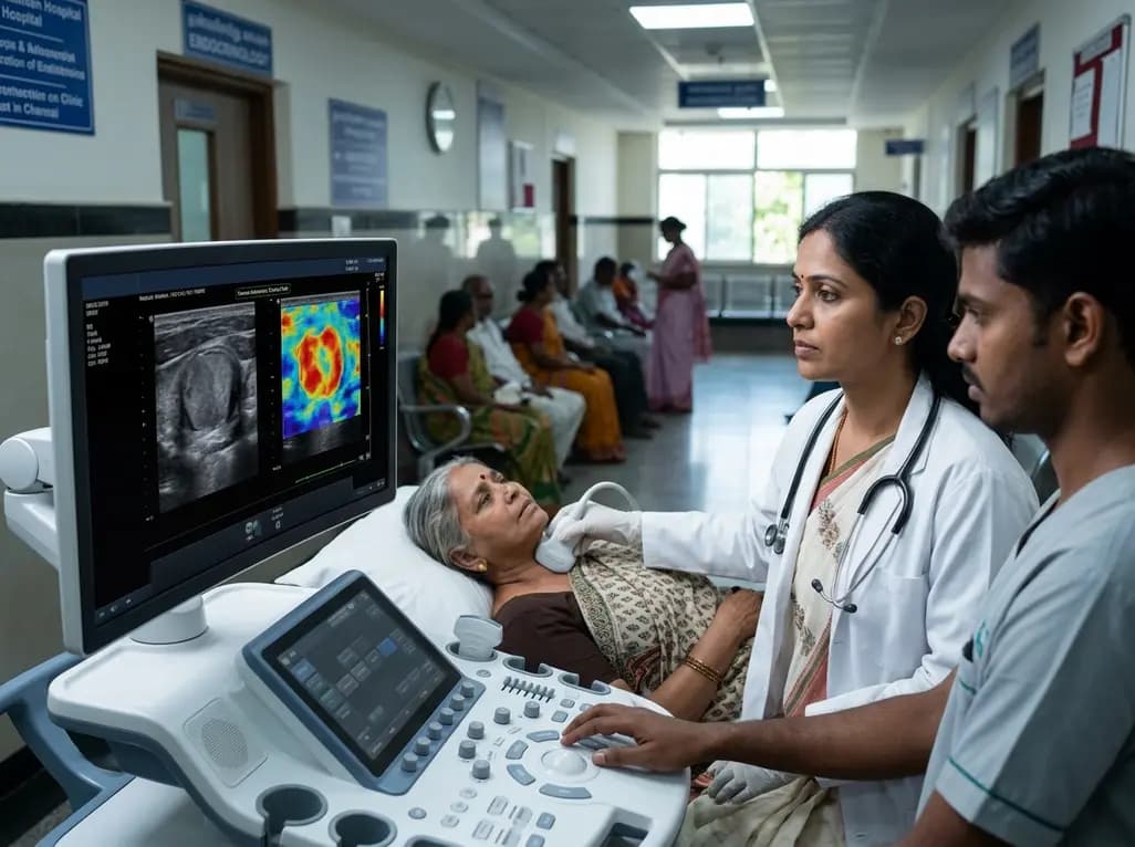
A study introduces a dual-discriminator generative adversarial network (DD-GAN) capable of generating virtual elastography ultrasound from standard B-mode images. The tool matches real elastography performance and improves thyroid nodule risk stratification when combined with TI-RADS.
Today
