.webp&w=384&q=75)

Loading, please wait...
Loading, please wait...
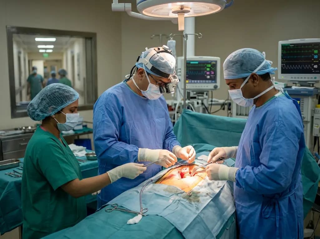
Aortic root aneurysms often expand silently over several years without causing overt cardiopulmonary symptoms. Consequently, many patients present only after substantial aortic regurgitation or ventricular dysfunction has already developed. The David procedure offers a reconstructive surgical strategy that replaces the dilated aortic root while preserving the patient's native aortic valve. However, clinical success depends entirely on surgical timing. When clinicians identify aneurysmal enlargement early, surgeons can salvage pliable valve cusps before irreversible secondary damage takes place.
Thoracic aortic root aneurysms develop insidiously due to progressive medial degeneration and loss of elastic fibers. Therefore, patients rarely experience dyspnea, angina, or functional decline in the initial stages. During this latency period, the aortic root gradually widens, but the valve cusps frequently remain thin and pliable. This hemodynamic stability creates a deceptive sense of safety for both patients and clinicians.
However, continuous mechanical wall stress inevitably disrupts the delicate geometry of the aortic root. Progressive annular dilatation separates the leaflet commissures, which impairs central coaptation. Consequently, secondary aortic regurgitation develops, imposing severe volume and pressure overload on the left ventricle. If clinicians delay intervention until congestive heart failure manifests, ventricular remodeling may become irreversible. Furthermore, severe leaflet stretching undermines the durability of subsequent reparative techniques. Therefore, regular surveillance with transthoracic echocardiography and gated computed tomography angiography remains essential. Timely imaging detects pathological root expansion before secondary valvular deterioration compromises surgical feasibility.
The David procedure, recognized formally as valve-sparing aortic root reimplantation, requires meticulous reconstruction of complex annular geometry. During this operation, the surgical team completely resects the diseased aneurysmal sinuses while carefully mobilizing the coronary buttons. Subsequently, the surgeon secures the intact aortic valve inside a tailored Dacron vascular graft.
Moreover, the surgeon anchors the prosthetic conduit below the anatomic annulus using circumferentially placed subannular sutures. This precise anchoring stabilizes the fibrous annulus and prevents future annular dilatation. Next, the surgeon reimplants the native aortic valve complex inside the graft lumen. Precise resuspension ensures symmetrical leaflet coaptation height and restores physiological cusp geometry. In addition, the operator mobilizes and anastomoses the left and right coronary ostia directly into the neoroot wall. Because even minute geometric misalignments can cause eccentric regurgitant jets, the procedure demands advanced surgical expertise. Operating teams in high-volume aortic centers consistently achieve superior long-term hemodynamics through standardized intraoperative transesophageal echocardiographic assessment.
Traditional surgical management of aortic root aneurysms typically involves composite graft replacement, known as the Bentall procedure. In younger populations, this approach generally necessitates a mechanical valve prosthesis, requiring lifelong oral anticoagulation with warfarin. Consequently, patients face persistent risks of major gastrointestinal hemorrhage, intracranial bleeding, and thromboembolic stroke. Furthermore, systemic anticoagulation creates severe lifestyle restrictions for physically active individuals and introduces complex teratogenic risks for women contemplating pregnancy.
Alternatively, bioprosthetic valves eliminate anticoagulant requirements but undergo rapid structural valve deterioration in younger hosts. Therefore, young recipients of biological prostheses face inevitable structural reoperations within ten to fifteen years. In contrast, the David procedure preserves native endothelialized tissue, ensuring physiological transvalvular flow and superior hemodynamics. Patients retain normal exercise capacity without needing routine international normalized ratio monitoring. In addition, preserving native cusps significantly reduces the incidence of prosthetic valve endocarditis. Clinical evidence confirms that successful reimplantation grants patients decades of normal functional life without prosthetic hardware.
Careful patient selection determines the ultimate durability of valve-sparing root reimplantation. Specifically, candidates must possess structurally pliable valve leaflets free from extensive calcification, fenestrations, or profound retraction. Therefore, younger adults presenting with trileaflet aortic valve anatomy and isolated root dilatation represent the ideal clinical demographic. However, experienced centers also treat carefully selected patients with bicuspid aortic valves, provided cusp tissue exhibits adequate mobility.
Furthermore, patients diagnosed with connective tissue disorders require heightened vigilance. Genetically triggered conditions, such as Marfan syndrome and Loeys-Dietz syndrome, induce aggressive vascular wall remodeling at early ages. International consensus guidelines strongly recommend earlier surgical thresholds for these heritable aortopathies, typically at root diameters of 45 to 50 millimeters. Conversely, elderly patients over seventy years usually gain greater benefit from composite bioprosthetic replacement. Extensive tissue friability and preexisting cusp degeneration make valve preservation less predictable in older cohorts. Therefore, multidisciplinary aortic teams must evaluate annular diameter, effective cusp height, and ventricular dimensions before finalizing the operative strategy.
Despite exceptional initial surgical outcomes, long-term clinical registries in India face persistent operational challenges. Many patients travel across state borders to access advanced quaternary centers but fail to return for routine follow-up. Consequently, surgical teams encounter significant difficulty when tracking valve durability beyond five years. Historical institutional audits often reveal invalid contact details, disconnected telephone numbers, and substantial geographic attrition.
Additionally, echocardiographic examinations obtained from rural or non-standardized peripheral facilities frequently exhibit variable image quality. Such inconsistencies hamper the objective detection of progressive aortic regurgitation or graft-related complications. To address these systemic hurdles, premier Indian cardiac centers are adopting integrated electronic health records and dedicated outreach registries. Moreover, structured digital telemedicine platforms allow remote clinicians to transmit standardized Doppler studies directly to surgical experts. Strengthening this regional referral and surveillance network ensures early detection of recurrent valvular incompetence, ultimately protecting patient survival across the country.
Q1: What are the primary indications for performing the David procedure?
The David procedure is primarily indicated for patients with aortic root or ascending aortic aneurysms who maintain structurally preserved valve leaflets. Clinicians strongly favor this approach in younger individuals, physically active patients, and females of childbearing potential. In addition, patients suffering from connective tissue disorders such as Marfan syndrome qualify for early intervention before extensive leaflet stretching or acute aortic dissection develops.
Q2: How long does a preserved native aortic valve remain durable after surgery?
Extensive international data demonstrate that a successfully reimplanted native aortic valve provides excellent durability, with freedom from significant regurgitation exceeding eighty percent at twenty years. However, long-term longevity depends closely on baseline leaflet quality, precise surgical geometry, and meticulous blood pressure control. Patients require periodic Doppler echocardiography to monitor ongoing coaptation and detect late degenerative changes early.
Q3: Why is early surgical referral necessary before aortic symptoms manifest?
Aortic aneurysms frequently expand without pain or breathlessness, leaving patients entirely unaware of underlying structural danger. If surgery is delayed until severe aortic regurgitation develops, chronic volume overload causes irreversible left ventricular remodeling. Furthermore, prolonged cusp stretching reduces the likelihood of achieving a durable repair, ultimately forcing surgeons to replace rather than spare the native valve.
Disclaimer: This content is for informational and educational purposes only. It does not constitute medical advice or replace professional judgment. Refer to the latest local and national guidelines for clinical practice.
References

Read summarized clinical updates, watch expert medical content, and earn CME certifications right from your smartphone.
Open your smartphone camera to scan and install.





Aortic root aneurysms often develop silently, yet timely intervention provides a critical window to salvage the native aortic valve. Preserving the patient's own anatomy avoids lifelong anticoagulation, provided clinicians identify root dilatation before secondary leaflet distortion and ventricular failure occur.
Today
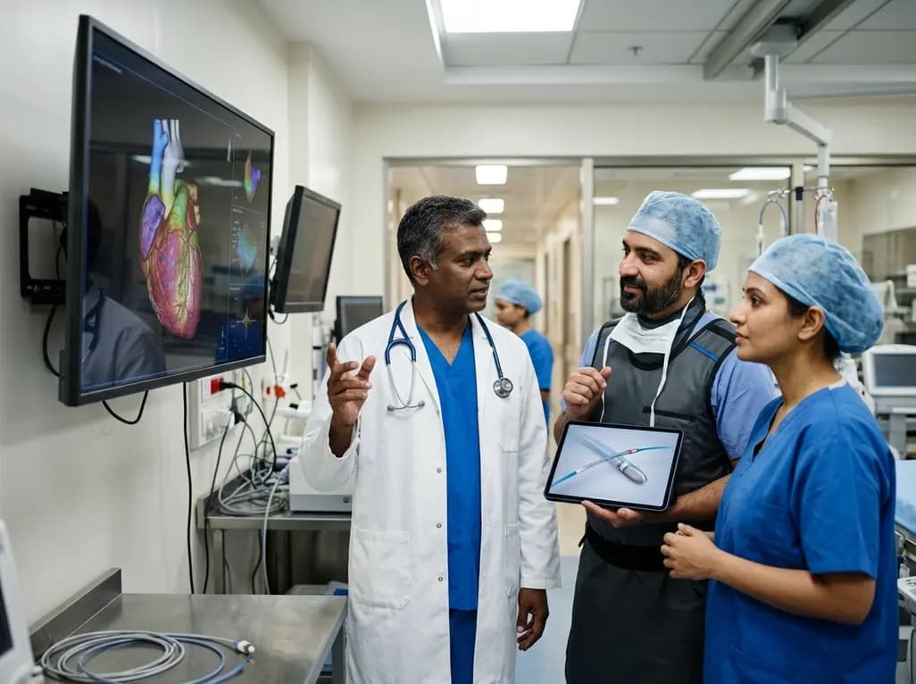
Cardiovascular disease remains a primary health threat in India, where heart attacks strike a decade earlier than in Western nations. Through multidisciplinary heart teams, expanded diagnostic access, and breakthrough interventions like pulse field ablation, modern cardiology offers renewed survival and clinical recovery.
Today
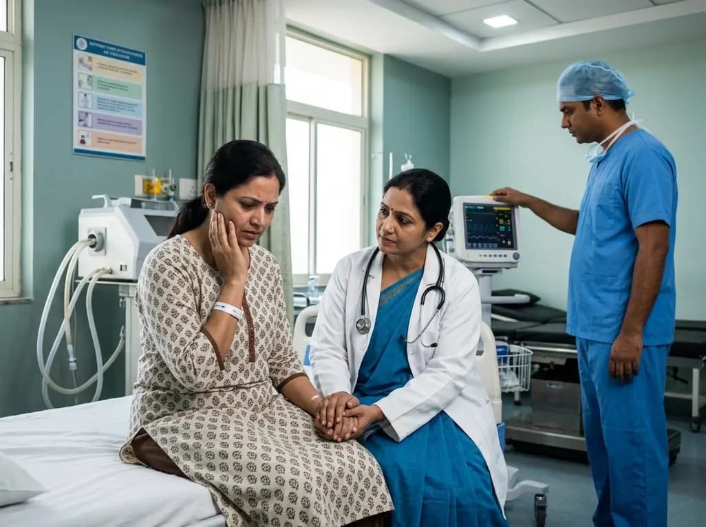
The FEBE study investigates the prevalence of post-traumatic stress disorder symptoms in women receiving high-dose-rate brachytherapy for cervical cancer, highlighting the critical role of procedural sedation and trauma-informed clinical care.
Today
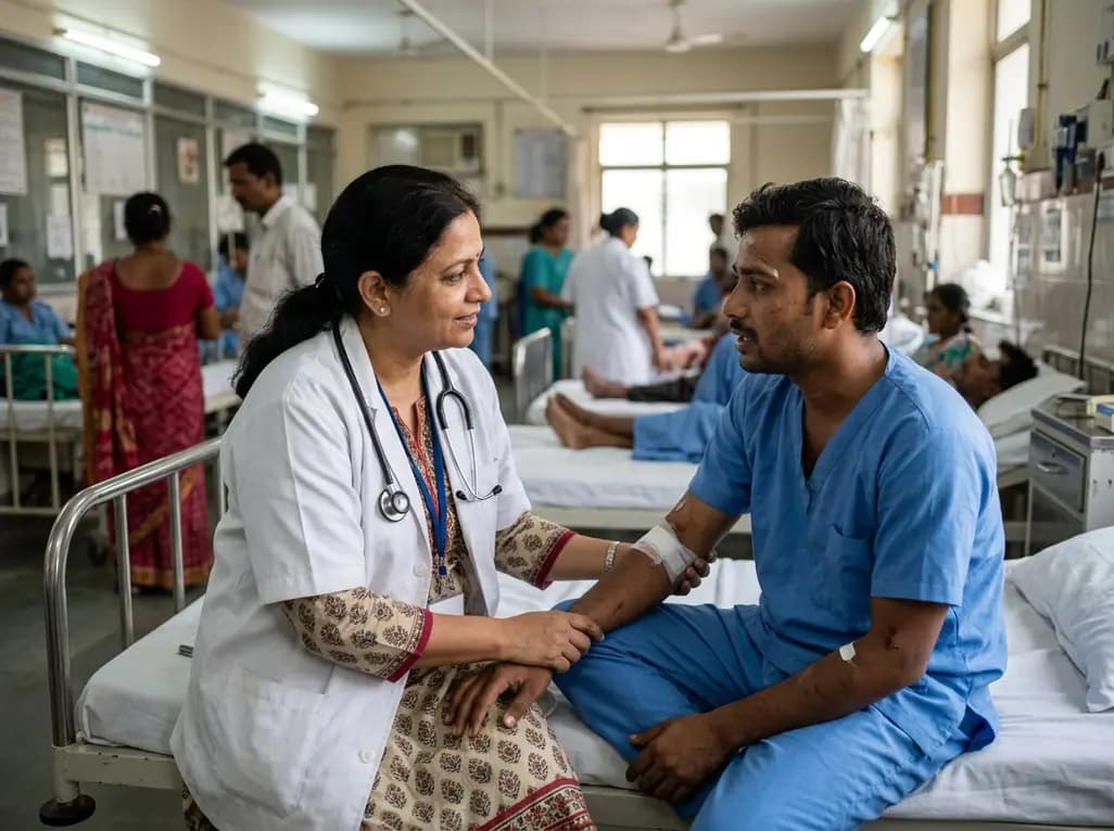
A Level I trauma center study shows a brief bedside mental health intervention boosts 30-day follow-up screening adherence by 84% in nonviolent injury patients. However, outcomes for violence survivors reveal critical care gaps, underscoring the urgent need for specialized community violence intervention programs.
Today
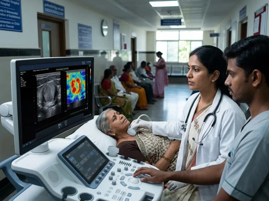
A study introduces a dual-discriminator generative adversarial network (DD-GAN) capable of generating virtual elastography ultrasound from standard B-mode images. The tool matches real elastography performance and improves thyroid nodule risk stratification when combined with TI-RADS.
Today
