

Loading, please wait...
Loading, please wait...

Acute intracerebral hemorrhage represents a devastating medical emergency with significant morbidity and mortality worldwide. For decades, emergency physicians and neurointensivists have actively debated the optimal hemodynamic targets during the hyperacute phase of bleeding. Several landmark clinical trials investigated whether intensive blood pressure reduction limits secondary neurological damage. However, emerging evidence suggests that therapeutic responses vary markedly depending on the anatomical location of the hematoma. A recent exploratory analysis of the ATACH-2 trial offers critical insights into this clinical question. Specifically, the study compares how deep and lobar intracerebral hemorrhages respond to aggressive blood pressure lowering. Understanding these pathophysiological differences is essential for optimizing neurocritical care protocols.
Spontaneous intracerebral hemorrhage arises from fundamentally distinct vascular pathologies depending on whether bleeding occurs in deep or lobar brain regions. Deep cerebral hematomas typically originate from hypertensive arteriolosclerosis that affects the small penetrating arteries of the basal ganglia and thalamus. In contrast, lobar hemorrhages frequently result from cerebral amyloid angiopathy. This distinct disease process promotes fragile beta-amyloid peptide deposition within cortical and leptomeningeal arterial walls.
Consequently, these distinct underlying microangiopathies alter vessel compliance and local autoregulatory capacity differently. In deep subcortical structures, severe hypertensive surges transmit intense hydrostatic pressure against chronically damaged arteriole walls. Rapidly lowering systemic arterial pressure reduces this transmural bursting force, which effectively halts active bleeding. Conversely, cortical vessels damaged by amyloid angiopathy exhibit severe structural brittleness, recurrent microaneurysms, and chronic wall remodeling. As a result, sudden reductions in blood pressure may fail to seal these frail cortical channels. Furthermore, aggressive systemic hypotension risks creating focal hypoperfusion in vulnerable surrounding brain parenchyma. Recognizing these distinct anatomical responses allows clinicians to personalize acute resuscitation strategies.
The Antihypertensive Treatment of Acute Cerebral Hemorrhage II trial was an international, randomized clinical trial evaluating hyperacute blood pressure lowering. The study enrolled adult patients presenting within four and a half hours after symptom onset with spontaneous intracerebral hemorrhage. Eligible participants demonstrated baseline systolic blood pressure readings of at least 180 mm Hg upon initial presentation. Investigators randomly allocated participants to either intensive blood pressure management or standard hemodynamic control.
The intensive treatment protocol aimed for a target systolic range of 110 to 139 mm Hg. Meanwhile, the standard treatment protocol pursued a target systolic range of 140 to 179 mm Hg. Physicians administered intravenous nicardipine infusions to achieve and sustain these predefined hemodynamic parameters. This exploratory post hoc analysis examined 875 trial participants with complete baseline and 24-hour neuroimaging data. Deep hematomas comprised 89 percent of the evaluated population, representing 778 individuals. Conversely, lobar hematomas represented 11 percent, comprising 97 patients. Researchers assessed hematoma expansion, defined as an increase greater than six milliliters within 24 hours, along with 90-day functional recovery and adverse renal events.
The multivariable regression analysis unveiled a clear divergence in radiographic responses between anatomical cohorts. Among patients suffering from deep intracerebral hemorrhage, intensive blood pressure reduction significantly decreased the risk of hematoma expansion. Specifically, the intensive intervention produced an adjusted odds ratio of 0.60, with a 95 percent confidence interval of 0.38 to 0.93. This confirmed a statistically significant 40 percent risk reduction in hematoma growth for deep lesions.
In contrast, patients presenting with lobar hemorrhage achieved no significant radiographic benefit from aggressive blood pressure reduction. In the lobar subgroup, the adjusted odds ratio for hematoma expansion was 0.91, with a broad 95 percent confidence interval spanning 0.34 to 2.41. Consequently, aggressive hemodynamic lowering did not confer meaningful protection against hematoma expansion in cortical bleeding. However, this notable reduction in hematoma enlargement among deep hemorrhage patients did not improve 90-day functional recovery. Unfavorable functional outcomes remained practically identical between the treatment arms for both deep and lobar hematomas. Deep subcortical bleeds destroy dense sensorimotor tracts immediately, which limits the clinical utility of hematoma restriction alone.
Although intensive hemodynamic control limited hematoma expansion in deep bleeds, it simultaneously generated significant systemic safety concerns. Patients in the intensive reduction arm suffered a marked increase in acute renal adverse events. In the deep hemorrhage subgroup, intensive therapy more than doubled the odds of renal complications, yielding an odds ratio of 2.42 with a 95 percent confidence interval of 1.29 to 4.72.
This heightened incidence of acute renal injury directly reflects impaired perfusion dynamics. Rapid and steep reductions in mean arterial pressure compromise renal autoregulation, particularly in chronically hypertensive individuals whose autoregulatory curves are shifted rightward. When systemic pressure falls precipitously, glomerular filtration rates decline, leading to acute tubular dysfunction. Furthermore, acute kidney injury complicates neurocritical care management by impairing fluid balance, electrolyte stability, and pharmacological clearance. Clinicians must therefore weigh the modest radiographic benefit of preventing hematoma expansion against the immediate risk of iatrogenic renal hypoperfusion. Navigating this clinical trade-off requires vigilant monitoring and measured hemodynamic targets rather than unconstrained pressure lowering.
These findings carry profound clinical implications for emergency physicians and neurointensivists managing spontaneous intracerebral hemorrhage. Current international guidelines generally endorse acute systolic blood pressure reduction to roughly 140 mm Hg while cautioning against excessive drops below 130 mm Hg. The post hoc ATACH-2 data strongly support avoiding systolic targets beneath 130 mm Hg, given the heightened risk of acute nephrotoxicity.
For deep intracerebral hemorrhages, clinicians should pursue steady, moderate blood pressure control to mitigate hematoma growth without inducing systemic hypoperfusion. Continuous invasive blood pressure monitoring and smooth intravenous drug titration help eliminate hazardous hemodynamic fluctuations. In contrast, clinicians evaluating lobar hematomas should recognize that intensive blood pressure reduction offers limited efficacy in preventing hematoma expansion. In these cortical cases, treatment teams must prioritize rapid diagnostic neuroimaging, reversal of underlying coagulopathies, and neurosurgical evaluation. Overall, neurocritical care protocols must evolve beyond a generic approach to acute intracerebral hemorrhage. Integrating neuroanatomical localization into emergency algorithms enables tailored, patient-centered hemodynamic resuscitation.
Future randomized clinical trials should stratify acute intracerebral hemorrhage cohorts prospectively according to neuroanatomical location. Because lobar hemorrhages constituted only eleven percent of the ATACH-2 trial cohort, dedicated clinical investigations are needed to definitively evaluate cortical responses. Such prospective studies will clarify whether distinct target thresholds improve outcomes in specific patient subsets.
Moreover, contemporary neurocritical research increasingly emphasizes real-time physiological monitoring over rigid population-wide targets. Incorporating continuous cerebral autoregulation monitoring and noninvasive intracranial pressure assessments may help neurointensivists identify individualized hemodynamic limits. Maintaining adequate cerebral perfusion pressure while avoiding hypertensive surges represents the premier clinical objective during hyperacute resuscitation. Additionally, researchers must investigate multimodal neuroprotective strategies that stabilize the vascular endothelium alongside blood pressure management. Until prospective trials validate location-tailored goals, clinicians should adopt prudent systolic targets between 130 and 140 mm Hg. This balanced approach protects fragile cerebral parenchyma while preserving renal function during critical care admissions.
Intensive blood pressure reduction significantly curtails hematoma expansion in deep intracerebral hemorrhage by reducing hydrostatic force on ruptured arterioles. However, lobar hemorrhages do not experience similar radiographic benefits. This discrepancy likely reflects different underlying vascular diseases, as lobar hemorrhages typically stem from brittle amyloid angiopathy rather than hypertensive arteriosclerosis.
Deep intracerebral hemorrhages directly involve eloquent subcortical architecture, particularly the internal capsule and thalamus. Initial mechanical tissue destruction inflicts severe baseline axonal disruption that limiting hematoma expansion cannot reverse. Additionally, therapy-induced renal adverse events and potential perihematomal hypoperfusion may counteract the clinical advantages of smaller hematoma volumes over long-term follow-up.
Current guidelines advise rapidly lowering systolic blood pressure to approximately 140 mm Hg while avoiding drops below 130 mm Hg. ATACH-2 demonstrated that reducing systolic pressure below 130 mm Hg increases renal injury without improving functional recovery. Clinicians should prioritize smooth, steady control and avoid extreme hemodynamic fluctuations.
Disclaimer: This content is for informational and educational purposes only and should not be taken as professional medical advice. Healthcare professionals should always exercise their own independent clinical judgment. Patients should consult their physician regarding any health questions or conditions. Refer to the latest local and national guidelines for clinical practice.
References

Read summarized clinical updates, watch expert medical content, and earn CME certifications right from your smartphone.
Open your smartphone camera to scan and install.





A post hoc analysis of the ATACH-2 trial demonstrates that intensive blood pressure reduction significantly curtails hematoma expansion in deep intracerebral hemorrhage but not lobar ICH. However, aggressive lowering also raises renal adverse events, highlighting the need for tailored, anatomically guided treatment.
Today

Review how tailored exercise programs benefit frail older patients with heart failure. Explore pathophysiological mechanisms, individualized training prescriptions, and strategies to overcome adherence barriers in clinical practice.
Today
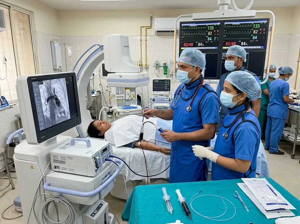
In high-risk pulmonary embolism complicated by severe anemia and active uterine bleeding, systemic thrombolysis is contraindicated. Large-bore aspiration thrombectomy provides rapid hemodynamic rescue without fibrinolysis, supported by inferior vena cava filter placement and multidisciplinary critical care.
Today

A cross-sectional study in Zanzibar reveals that 46.5% of adults with type 2 diabetes experience multidomain autonomic dysfunction on the COMPASS-31 tool, primarily orthostatic intolerance, while 45.9% exhibit cardiovascular autonomic neuropathy, highlighting the urgent need for structured clinical screening.
Today

This comparative study evaluates posterior labral tears with and without glenohumeral instability, highlighting distinct demographic patterns, MRI underestimation in stable shoulders, and successful arthroscopic repair outcomes.
Today

A recent case-control study reveals that urinary clusterin provides superior diagnostic performance for early diabetic kidney disease detection in pediatric type 1 diabetes mellitus, identifying tubular damage before conventional microalbuminuria occurs.
Today
