.webp&w=384&q=75)

Loading, please wait...
Loading, please wait...
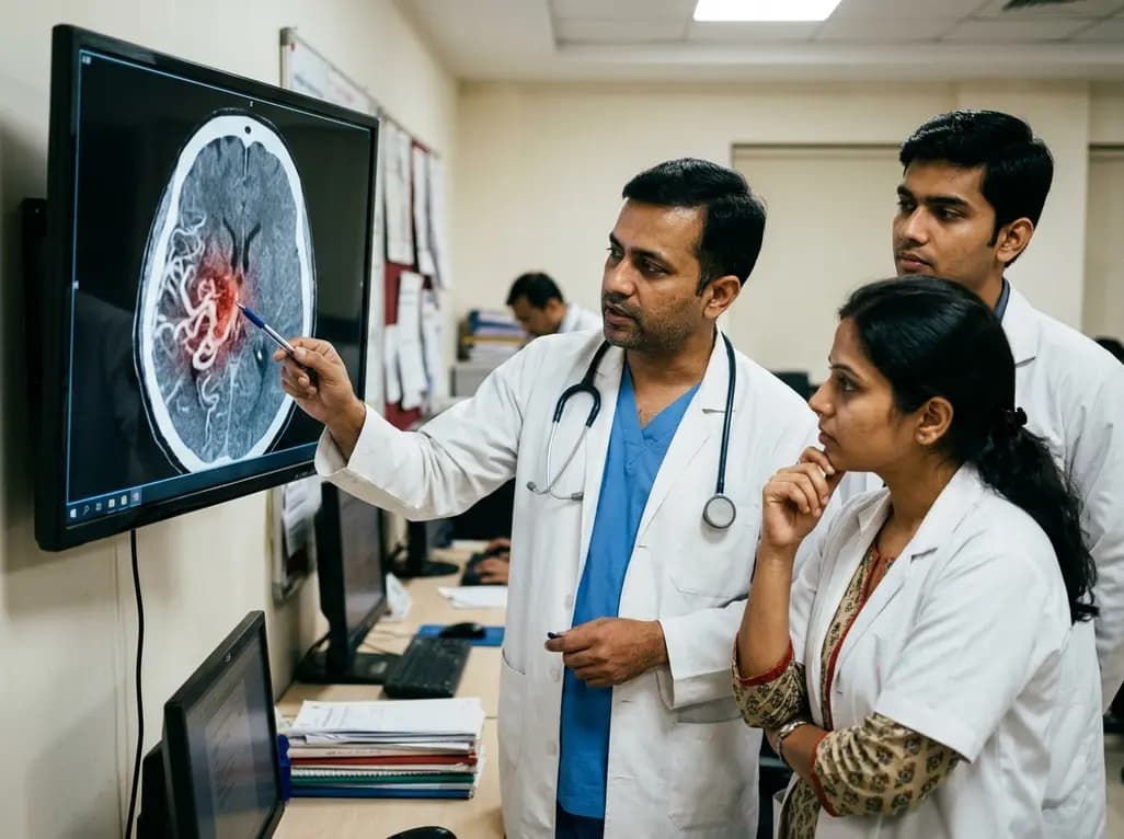
Brain arteriovenous malformations represent complex congenital or acquired vascular lesions characterized by direct shunting between arteries and veins. Because these abnormal connections lack an intervening capillary bed, they subject fragile venous channels to high arterial shear stress. Consequently, patients with brain arteriovenous malformations face a continuous lifelong risk of catastrophic intracranial hemorrhage and severe neurological deficits. Although historically viewed as static congenital anomalies, emerging evidence demonstrates that these vascular lesions undergo active biological remodeling throughout life. Recent translational research identifies persistent localized inflammation as a primary engine driving vascular instability, nidus expansion, and wall degradation. Moreover, a comprehensive review of over 300 experimental and clinical studies confirms that inflammatory cascades govern every critical stage of the disease. Therefore, understanding the molecular mechanisms underlying neurovascular inflammation provides essential clinical insights for improving patient risk stratification, surgical planning, and medical management.
Within the nidus microenvironment, chronic immune stimulation orchestrates significant structural vessel disruption. Specifically, elevated levels of pro-inflammatory cytokines such as interleukin-1beta, interleukin-6, and tumor necrosis factor-alpha perpetuate endothelial cell activation. As a result, activated endothelial cells upregulate key adhesion molecules, which recruit circulating leukocytes into the perivascular space. In addition, this chronic cytokine signaling promotes pathological angiogenesis by disrupting regular endothelial quiescence. Consequently, newly formed vessels exhibit marked wall thinning, discontinuous tight junctions, and severely impaired pericyte coverage. Furthermore, these cytokine cascades activate downstream nuclear transcription factors that inhibit physiological vessel maturation and enhance vascular permeability. Therefore, sustained microvascular inflammation creates fragile channels that are inherently prone to mechanical failure. Clinicians must recognize that persistent cytokine activation maintains an unstable vascular architecture, accelerating lesion growth and increasing spontaneous hemorrhage risk in affected individuals.
Vascular remodeling in cerebrovascular malformations depends on the complex interplay of several cellular signaling pathways. In particular, aberrant activity within the VEGF, TGF-beta/BMP, Notch, NF-kappaB, and KRAS/MAPK-ERK axes directly drives disease progression. For instance, somatic mutations in the KRAS/MAPK pathway induce persistent endothelial proliferation and disrupt organized lumen formation. Meanwhile, elevated VEGF expression stimulates uncontrolled, chaotic capillary sprouting without adequate structural perivascular support. Concurrently, dysregulated TGF-beta signaling impairs extracellular matrix deposition and disrupts smooth muscle cell differentiation across vessel layers. Furthermore, activation of the NF-kappaB pathway amplifies the transcription of matrix metalloproteinases, especially MMP-2 and MMP-9. These proteolytic enzymes rapidly degrade the internal elastic lamina and baseline extracellular matrix scaffolding. Consequently, the structural integrity of the vascular wall deteriorates progressively, rendering the nidus highly vulnerable to arterial pressure surges and catastrophic hemorrhage.
Infiltration of peripheral immune cells plays an active, direct role in precipitating vessel rupture within vascular lesions. In particular, recruited macrophages and neutrophils accumulate in high densities within the perivascular stromal tissue of the nidus. Once recruited, these immune cells release large quantities of reactive oxygen species, pro-inflammatory cytokines, and proteolytic enzymes. Consequently, oxidative stress induces widespread endothelial apoptosis and accelerates smooth muscle degradation along the vessel wall. In addition, activated neutrophils release neutrophil extracellular traps and elastases that directly digest surrounding connective tissues and destabilize mural architecture. Therefore, heightened immune cell infiltration strongly correlates with clinical presentations of micro-hemorrhages and acute intracranial bleeding. Furthermore, systemic inflammatory biomarkers may soon provide non-invasive clinical measures to identify unstable lesions that require urgent surgical evaluation. Ultimately, suppressing leukocyte-mediated proteolytic degradation represents a promising adjunct strategy to prevent life-threatening hemorrhagic events in high-risk patients.
Although chronic inflammation promotes disease pathogenesis, inflammatory pathways also serve as indispensable mediators of successful radiosurgical obliteration. Specifically, stereotactic radiosurgery delivers focused ionizing radiation that induces immediate endothelial cell injury and localized intravascular thrombosis. Consequently, irradiated tissues release localized damage-associated molecular patterns, which stimulate an acute, targeted inflammatory response. In addition, this sterile inflammatory cascade triggers massive recruitment of myofibroblasts and circulating monocytes to the vascular nidus. Over subsequent months, these recruited cells produce abundant collagen and promote progressive concentric intimal hyperplasia. Furthermore, radiation-induced inflammation gradually converts high-flow fistulous shunts into organized, stable fibrotic scar tissue, completely obliterating the lesion. Therefore, a controlled inflammatory reaction is biologically essential for achieving complete nidus occlusion following stereotactic radiosurgery. Modulating this post-radiation immune response may further optimize obliteration rates while minimizing radiation-induced perifocal brain edema in treated patients.
Recognizing inflammation as a major driver of vascular remodeling opens novel avenues for therapeutic innovation in neurovascular practice. Currently, clinical management relies heavily on microsurgical resection, endovascular embolization, and stereotactic radiosurgery. However, incomplete obliteration often leaves residual vessels vulnerable to recurrent hemorrhage and lesion expansion. Therefore, combining targeted anti-inflammatory pharmacotherapy with standard surgical modalities offers substantial clinical promise for high-risk patients. For example, specific small-molecule inhibitors targeting the KRAS/MAPK-ERK pathway or NF-kappaB cascades can stabilize abnormal endothelial walls prior to definitive intervention. Furthermore, selective matrix metalloproteinase inhibitors may decrease the immediate risk of rupture during the latency period following radiosurgery. In addition, targeted anti-cytokine therapies could mitigate harmful radiation-induced brain necrosis while preserving therapeutic vascular fibrosis. Ultimately, translating these molecular insights into everyday clinical practice will significantly refine risk assessment and advance multimodal cerebrovascular management.
Inflammatory cytokines recruit macrophages and neutrophils that release matrix metalloproteinases and reactive oxygen species. Consequently, these enzymes degrade the extracellular matrix, internal elastic lamina, and mural smooth muscle cells. This structural wall degradation drastically increases vascular fragility, making the malformation highly susceptible to spontaneous rupture under arterial pressures.
Ionizing radiation triggers acute endothelial injury and a targeted, sterile inflammatory response. Consequently, this inflammatory cascade recruits immune cells and fibroblasts that stimulate intimal hyperplasia and progressive vascular fibrosis. Ultimately, this controlled inflammatory process safely obliterates abnormal arteriovenous shunts over a period of one to three years.
Anti-inflammatory medications cannot cure brain arteriovenous malformations alone because they do not eliminate high-flow fistulous connections. However, these pharmacological agents can stabilize fragile vascular walls, reduce perivascular edema, and decrease rupture risk. Consequently, they serve as valuable adjuncts alongside definitive surgical, endovascular, or radiosurgical interventions.
Disclaimer: This content is for informational and educational purposes only and should not be considered medical advice. Always consult a qualified healthcare provider for diagnosis and treatment decisions. Refer to the latest local and national guidelines for clinical practice.
References
Mensah EO et al. The molecular basis of arteriovenous malformations (AVMs): Review of inflammation in AVM pathogenesis. Neurosurg Rev. 2026 Aug 24. doi: 10.1007/s10143-026-04457-w. PMID: 42635647.
Kim H et al. Brain arteriovenous malformation biology: from bench to bedside. J Cereb Blood Flow Metab. 2021;41(1):22-38.
Chen W et al. Macrophages and inflammation in brain arteriovenous malformation pathogenesis and therapeutic strategies. Front Immunol. 2022;13:854921.

Read summarized clinical updates, watch expert medical content, and earn CME certifications right from your smartphone.
Open your smartphone camera to scan and install.





A systematic review of 342 studies reveals that chronic inflammation and dysregulated molecular signaling pathways drive brain arteriovenous malformation (bAVM) remodeling, rupture risk, and radiosurgical vascular obliteration.
last month

A study demonstrates an explainable machine learning framework integrating clinical and echocardiographic metrics to predict left atrial appendage thrombus in non-valvular atrial fibrillation, utilizing SHAP analysis for clinical interpretability.
Today

Maternal anaemia contributes to nearly half of pregnancy-related fatalities across India. To prevent complications, Uttar Pradesh has added single-dose parenteral iron to its essential medicines list. This clinical transition aims to restore haemoglobin rapidly and protect high-risk mothers across rural facilities.
Today
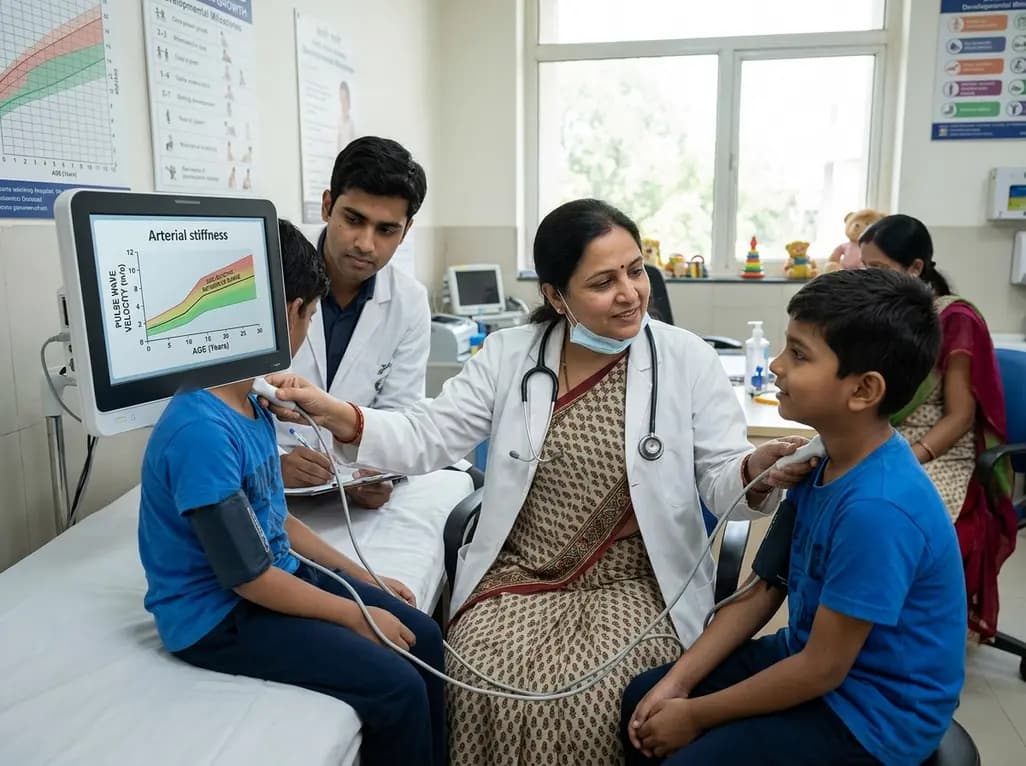
A landmark systematic review and meta-analysis establishes age-specific normative reference values for pediatric pulse wave velocity, revealing a nonlinear trajectory of arterial stiffness from early childhood through adolescence.
Today
A new systematic review evaluates lumbar facet arthroplasty, predominantly the Total Posterior Spine System (TOPS). Evidence shows significant reductions in back and leg pain and preserved segmental mobility, though revision rates and outcome heterogeneity highlight the need for careful patient selection.
Today

A landmark cross-sectional study in Asian adults with type 2 diabetes identifies novel urinary metabolites linked to mild cognitive impairment. Elevated urinary sucrose and altered metabolites highlight non-invasive pathways for early cognitive risk stratification and targeted metabolic management.
Today
