.webp&w=384&q=75)

Loading, please wait...
Loading, please wait...

Evaluating non-accidental trauma in pediatric patients represents one of the most critical responsibilities in clinical medicine. Radiographic skeletal surveys serve as the foundational diagnostic modality for detecting subtle, acute, or healing osseous injuries in young infants presenting with suspected physical abuse. However, radiologists and clinicians frequently encounter equivocal radiographic features that cannot be definitively classified as acute fractures, normal anatomical variations, or benign developmental variants. These uncertain findings, broadly categorized as indeterminate fractures, present substantial diagnostic and clinical management dilemmas. Understanding the prevalence, follow-up imaging yields, and ultimate resolution of these indeterminate features is essential for safeguarding pediatric patients while preventing unnecessary interventions or prolonged administrative holds. Recent clinical investigation illuminates how repeat imaging protocol, subspecialty consultation, and multidisciplinary risk assessment systematically resolve diagnostic ambiguity in suspected abuse evaluations.
Indeterminate fractures on skeletal surveys often involve subtle cortical irregularities, periosteal reaction without an obvious cortical breach, or focal lucencies at metaphyseal junctions. Distinguishing acute traumatic injury from normal physiological periosteal apposition, bone remodeling, or anatomical subluxation requires specialized pediatric imaging expertise. In clinical practice, a significant proportion of initial skeletal surveys yields at least one equivocal finding that necessitates further clarification. These indeterminate findings are most commonly localized to high-risk anatomic sites such as ribs, long bone metaphyses, and classic metaphyseal lesions. The uncertainty surrounding initial diagnostic interpretation frequently triggers serial radiographic evaluations, pediatric orthopedic consultations, and involvement from child protection teams. Standardizing the terminology used in radiology reporting helps clinical teams communicate diagnostic confidence levels accurately to ordering pediatricians and emergency physicians.
To definitively confirm or rule out true osseous trauma, clinical guidelines advocate for targeted follow-up imaging obtained approximately two to three weeks after the index evaluation. Follow-up skeletal surveys leverage the temporal evolution of bone healing, as definitive callus formation or periosteal reaction will mature over time if a true fracture was initially present. Conversely, normal anatomical variants or non-traumatic artifacts remain static on serial imaging, thereby resolving clinical uncertainty. Research indicates that repeat skeletal surveys demonstrate a high diagnostic yield, successfully clarifying a vast majority of initial indeterminate findings. Furthermore, follow-up examinations frequently identify secondary healing fractures that were completely occult on initial presentation due to lack of early radiographically apparent callus. Consequently, protocolized follow-up imaging remains an indispensable element of comprehensive non-accidental trauma protocols.
The identification of indeterminate fractures skeletal surveys heavily influences early multidisciplinary management decisions, including hospital admission length, social work investigation intensity, and child protective service referrals. When diagnostic uncertainty persists, clinical teams must balance the risk of discharging a child back into a potentially hazardous environment against the psychological and social burden imposed on families during extended child abuse investigations. Adopting protocolized pathways that incorporate child abuse pediatrician consultations significantly optimizes clinical management. Multidisciplinary case conferences involving pediatricians, radiologists, orthopedic surgeons, and social workers ensure that indeterminate radiographic findings are interpreted in conjunction with complete clinical histories, physical examination findings, and developmental capabilities. This holistic diagnostic framework minimizes misdiagnosis and optimizes child protection strategy.
While follow-up skeletal surveys provide invaluable diagnostic clarity, repeated exposure to ionizing radiation in infants demands careful consideration. Radiologists and clinical teams must adhere to the ALARA (As Low As Reasonably Achievable) principle while ensuring complete diagnostic efficacy. Evidence suggests that targeted or limited follow-up skeletal surveys, which focus specifically on regions with initial indeterminate findings or high-yield anatomical sites, can frequently achieve diagnostic goals without requiring a full 20-view repeat survey. Omitting low-yield anatomical regions like the skull or spine on follow-up, provided initial projections were completely normal, substantially reduces radiation burden. Standardizing imaging protocols across institutions ensures consistent radiographic quality, reducing the need for repeated views attributable to motion artifacts or improper positioning.
Indeterminate findings on initial skeletal surveys during non-accidental trauma workups present unavoidable diagnostic challenges that require systematic follow-up. Protocolized repeat imaging performed two weeks after initial evaluation remains the gold standard for clarifying diagnostic ambiguity and detecting subacute healing injuries. Integrating expert pediatric radiology review with multidisciplinary clinical consultation guarantees optimal patient safety while avoiding unnecessary medical-legal complications. Continued research into advanced low-dose imaging modalities and automated diagnostic algorithms may further refine our ability to distinguish true abusive skeletal injuries from benign physiological variants in vulnerable pediatric populations.
An indeterminate fracture refers to a radiographic irregularity, such as subtle periosteal reaction, cortical asymmetry, or focal metaphyseal lucency, that cannot be definitively classified as a acute fracture, healing injury, or normal anatomical variant upon initial imaging evaluation.
Follow-up imaging obtained 11 to 14 days later relies on bone healing physiology. True fractures will show progressive callus formation or periosteal thickening over time, whereas normal developmental variants remain unchanged, thus providing diagnostic confirmation.
Indeterminate findings trigger multidisciplinary reviews, child abuse specialist consults, and potential temporary safety plans. Definitive resolution through serial imaging ensures child protection while avoiding unwarranted prolonged hospitalizations or misdiagnoses.
Disclaimer: This content is for informational and educational purposes only and should not be used as a substitute for professional medical advice, diagnosis, or treatment. Refer to the latest local and national guidelines for clinical practice.
References
1. Darwiche SM et al. Outcomes of indeterminate fractures identified on skeletal surveys obtained during non-accidental trauma evaluations. Child Abuse Negl. 2026 Aug 08. doi: undefined. PMID: 42570419.
2. Karmazyn B et al. Yield of follow-up skeletal survey in suspected non-accidental trauma. Pediatr Radiol. 2020;50(3):345-352.
3. Harper NS et al. Radiographic skeletal survey in young children with suspected physical abuse. Pediatrics. 2014;134(6):e1741-e1748.

Read summarized clinical updates, watch expert medical content, and earn CME certifications right from your smartphone.
Open your smartphone camera to scan and install.





This review analyzes the prevalence, clinical significance, and follow-up imaging outcomes of indeterminate fractures identified during skeletal survey evaluations for suspected non-accidental pediatric trauma.
last month
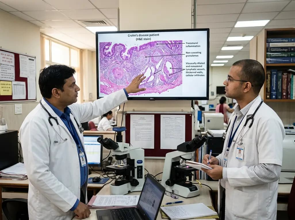
Intestinal lymphatic dysfunction is re-emerging as a fundamental driver of Crohn's disease pathogenesis. Explore how structural remodeling, lymphangitis, and defective clearance shape transmural inflammation and offer novel therapeutic targets.
Today

A school-based intervention using the Health Action Process Approach (HAPA) significantly enhanced adolescents' perceived benefits of consulting healthcare providers and promoted proactive menstrual health behaviors, offering key clinical insights for managing adolescent dysmenorrhea and endometriosis.
Today

A comprehensive analysis of the Translational Maturity Framework for nanomedicine-device combination products, evaluating regulatory complexity, manufacturing hurdles, and clinical readiness across oncology, cardiology, and orthopedics.
Today

A cross-sectional study reveals that sarcopenic obesity independently increases functional impairment and severe disc degeneration in patients with degenerative lumbar spinal stenosis, highlighting the need for body composition assessment.
Today

A landmark dose-response meta-analysis of 8.8 million adults reveals that high ultra-processed food intake significantly elevates risks for cardiovascular disease, cancer, diabetes, depression, and all-cause mortality, highlighting urgent needs for clinical lifestyle interventions.
Today
