

Loading, please wait...
Loading, please wait...

Secondary central nervous system tumors represent a significant cause of morbidity and mortality among oncology patients. Historically, calculating accurate brain metastases incidence has proven difficult because population-based cancer registries routinely prioritize primary malignancies over secondary metastatic deposits. Consequently, national health databases frequently miss subsequent intracranial dissemination. Most registries document only the primary site or generic metastatic spread without identifying the central nervous system as an anatomical site of progression. This systematic documentation gap masks the true epidemiological burden of intracranial secondary disease. Therefore, healthcare providers and policy experts often operate with incomplete epidemiological data when projecting specialized neuro-oncological care demands. To overcome this surveillance challenge, clinical epidemiologists must correlate radiological registries directly with comprehensive histopathological and hospital records. Detailed population-based assessments enable clinicians to understand actual intracranial metastatic disease burdens across defined geographical jurisdictions.
A recent 20-year population study published in the Journal of Neuro-Oncology offers rigorous insights into intracranial tumor dissemination. The investigators analyzed residents of North Savo, Finland, who received a first MRI-confirmed diagnosis of intra-axial secondary tumors between 2004 and 2023. Importantly, the research team identified 516 patients, and 474 individuals possessed histopathologically verified primary or secondary lesions. Lung and breast cancers accounted for 56.8% of all secondary intracranial lesions, confirming their predominant role in neuro-oncology. Across the entire two-decade investigation, the crude incidence rate was 10.22 per 100,000 person-years. Moreover, the age-standardized incidence rate reached 9.20 per 100,000 person-years when matched to the 2013 European Standard population. Crucially, the standardized rate climbed from 5.8 per 100,000 person-years between 2004 and 2008 to 11.8 per 100,000 person-years between 2019 and 2023. Hence, the study demonstrates an alarming doubling in diagnosed secondary brain lesions over twenty years.
Poisson regression analysis adjusted for patient age demonstrated a striking 24% increase in the rate of intracranial metastases per five-year increment. Consequently, demographic aging alone does not explain this marked upward trajectory. Instead, multiple synergistic healthcare advancements drive the escalating brain metastases incidence observed in modern practice. First, modern systemic therapies, including targeted molecular kinase inhibitors and immune checkpoint inhibitors, significantly prolong overall survival for advanced cancer patients. However, the blood-brain barrier often acts as a pharmacological sanctuary site where subclinical micrometastases survive and eventually proliferate. As patients live longer with well-controlled systemic disease, their cumulative lifetime risk of developing symptomatic or radiological central nervous system spread escalates dramatically. Furthermore, widespread adoption of high-field MRI technology allows neuro-radiologists to identify punctate lesions that older computed tomography machines consistently missed. Thus, prolonged systemic control coupled with enhanced diagnostic detection explains this persistent epidemiological increase.
Neuroimaging technology plays a pivotal role in early tumor detection and timely treatment stratification. Thin-slice contrast-enhanced magnetic resonance imaging remains the undisputed gold standard for detecting secondary cerebral pathology. Contrast-enhanced volumetric T1-weighted sequences can reliably reveal intra-axial parenchymal lesions as small as one to two millimeters. In addition, susceptibility-weighted imaging and diffusion-weighted imaging differentiate secondary tumors from radiation necrosis, vascular strokes, or intracranial hemorrhages. Therefore, clinicians should maintain a low threshold for ordering comprehensive neuroimaging in patients who present with subtle headaches or cognitive slowing. Contemporary oncology protocols increasingly advocate for baseline neuroimaging in high-risk cancers such as advanced non-small cell lung cancer and HER2-positive breast cancer. Identifying intracranial disease early prevents acute herniation, preserves baseline neurological performance, and expands therapeutic options beyond palliative interventions.
The documented doubling of secondary intracranial lesions necessitates proactive realignment of clinical workflows across cancer care facilities. In contemporary management, treatment selections depend heavily on performance status, intracranial lesion volume, and actionable tumor biology. Historically, clinicians relied almost exclusively on whole-brain radiation therapy, which caused substantial cognitive deterioration over time. In contrast, modern multidisciplinary teams favor stereotactic radiosurgery for oligometastatic presentations to spare normal brain tissue. Furthermore, neurosurgeons utilize intraoperative neuronavigation and functional mapping to achieve safe maximal resection of large symptomatic lesions causing mass effect. Concurrently, medical oncologists employ third-generation targeted drugs, such as brain-penetrant EGFR inhibitors, ALK inhibitors, and HER2-directed antibody-drug conjugates, which achieve profound intracranial responses. Consequently, close collaboration among medical oncologists, radiation oncologists, neurosurgeons, and radiologists remains vital for optimizing survival outcomes while preserving cognitive function.
Epidemiological patterns established in European cohorts offer vital guidance for clinicians and healthcare administrators worldwide, including high-volume cancer centers across India. In developing and transitional healthcare systems, delayed diagnosis often leads to advanced disease presentation with heavy symptomatic intracranial tumor burdens. Furthermore, access to high-resolution MRI platforms and advanced stereotactic radiosurgery units varies widely across semi-urban and rural regions. Therefore, recognizing the rising frequency of intra-axial secondary lesions highlights the urgent need for structured neuroimaging screening protocols. Clinicians must educate primary care doctors, general practitioners, and patients regarding early signs of intracranial hypertension and focal deficits. In addition, cancer institutions should invest in dedicated multi-modality neuro-oncology tumor boards to standardize care delivery. By adopting systematic surveillance and integrating CNS-penetrant systemic therapies, clinicians can improve clinical survival and maintain patient independence.
The rising incidence resulted primarily from extended patient survival driven by effective systemic oncological therapies. Additionally, widespread implementation of high-resolution contrast-enhanced magnetic resonance imaging protocols significantly improved the detection of small, previously unvisualized intracranial lesions across the aging demographic.
Primary malignancies originating in the lung and breast constitute the majority of secondary intra-axial brain tumors, collectively accounting for over 56% of confirmed cases. Melanoma, renal cell carcinoma, and gastrointestinal malignancies also frequently spread to the brain parenchyma.
Traditional cancer registries generally prioritize initial primary organ diagnoses and do not systematically capture longitudinal disease progression. Consequently, secondary intracranial spread is rarely tracked over time, leading to substantial underestimations of central nervous system metastatic burdens in epidemiological databases.
Disclaimer: This content is for informational and educational purposes only. It is not intended to be a substitute for professional medical advice, diagnosis, or treatment. Always seek the advice of your physician or other qualified health provider with any questions you may have regarding a medical condition. Never disregard professional medical advice or delay in seeking it because of something you have read here. Refer to the latest local and national guidelines for clinical practice.
References

Read summarized clinical updates, watch expert medical content, and earn CME certifications right from your smartphone.
Open your smartphone camera to scan and install.





A 20-year population study reveals a doubling in brain metastases incidence, driven by advanced neuroimaging and prolonged systemic cancer survival. Read key clinical insights.
Today

Review how tailored exercise programs benefit frail older patients with heart failure. Explore pathophysiological mechanisms, individualized training prescriptions, and strategies to overcome adherence barriers in clinical practice.
Today
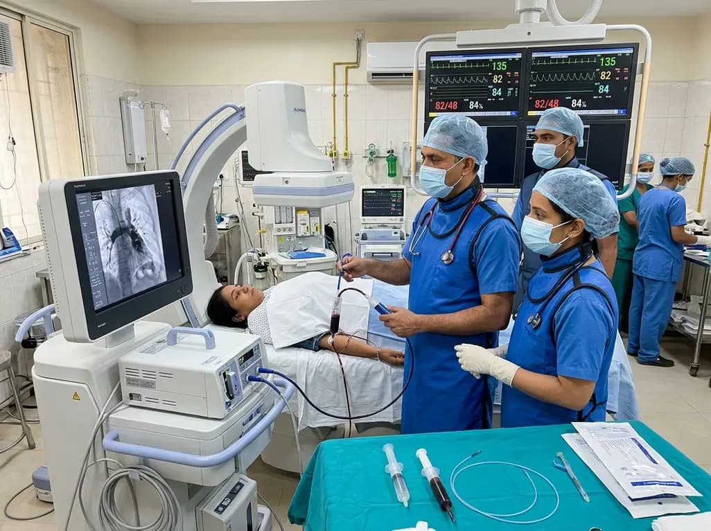
In high-risk pulmonary embolism complicated by severe anemia and active uterine bleeding, systemic thrombolysis is contraindicated. Large-bore aspiration thrombectomy provides rapid hemodynamic rescue without fibrinolysis, supported by inferior vena cava filter placement and multidisciplinary critical care.
Today

A cross-sectional study in Zanzibar reveals that 46.5% of adults with type 2 diabetes experience multidomain autonomic dysfunction on the COMPASS-31 tool, primarily orthostatic intolerance, while 45.9% exhibit cardiovascular autonomic neuropathy, highlighting the urgent need for structured clinical screening.
Today

This comparative study evaluates posterior labral tears with and without glenohumeral instability, highlighting distinct demographic patterns, MRI underestimation in stable shoulders, and successful arthroscopic repair outcomes.
Today

A recent case-control study reveals that urinary clusterin provides superior diagnostic performance for early diabetic kidney disease detection in pediatric type 1 diabetes mellitus, identifying tubular damage before conventional microalbuminuria occurs.
Today
