.webp&w=384&q=75)

Loading, please wait...
Loading, please wait...
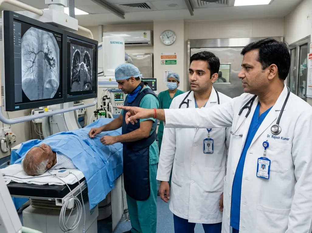
Endovascular transcatheter occlusion serves as the definitive standard of care for treating pulmonary vascular anomalies. Recently, the integration of image fusion guidance has emerged as a transformative technique to enhance interventional safety and procedural precision during the treatment of pulmonary arteriovenous malformations. Pulmonary arteriovenous malformations represent direct, low-resistance communications between pulmonary arteries and veins that bypass the intervening capillary network. Consequently, these lesions produce a right-to-left circulatory shunt that predisposes patients to severe complications, including hypoxemia, paradoxical cerebral embolism, stroke, and life-threatening hemoptysis. While transcatheter coil or plug embolization effectively obliterates the abnormal vascular nidus, traditional fluoroscopy requires substantial volumes of iodinated contrast media and prolonged radiation exposure.
Interventional radiologists historically rely on repeated digital subtraction angiography acquisitions to navigate tortuous pulmonary branches and pinpoint the exact angioarchitecture of vascular malformations. However, this conventional approach exposes patients to cumulative ionizing radiation and potential contrast-induced renal injury. Image fusion guidance integrates pre-procedural three-dimensional computed tomography angiography datasets directly onto real-time fluoroscopic screens. Therefore, operators obtain a dynamic spatial roadmap that accurately tracks anatomical structures during catheter manipulation. Furthermore, this multimodal overlay aligns seamlessly with patient movement, compensating for respiratory shifts and cardiac oscillations. As a result, clinicians navigate complex pulmonary arterial trees with greater anatomical confidence. Additionally, the technology allows operators to visualize optimal working projection angles before stepping on the fluoroscopy pedal. Consequently, catheter engagement into targeted feeding vessels occurs rapidly, minimizing the need for preliminary diagnostic test injections and repetitive roadmap series throughout the procedure.
A recent observational comparative analysis evaluated thirty-eight consecutive endovascular embolization procedures performed for pulmonary arteriovenous malformations across a tertiary care university hospital. The investigators compared twenty-two procedures conducted using image fusion guidance against sixteen historical control interventions performed with standard fluoroscopic guidance. The results demonstrated striking reductions across primary safety endpoints. Specifically, the median contrast media dose decreased from 0.62 grams of iodine per kilogram in the control group to just 0.18 grams of iodine per kilogram in the fusion cohort, achieving high statistical significance. Moreover, radiation exposure dropped substantially under fusion navigation. The median dose-area product fell from 86.6 Gray-centimeters squared in standard procedures to 22.4 Gray-centimeters squared in fusion-assisted cases. In addition, the median fluoroscopy time shortened from 55.7 minutes down to 31.2 minutes, confirming significant procedural efficiency without compromising technical success.
Several technological and ergonomic factors explain the marked reduction in contrast media volume and radiation dose during fused endovascular procedures. First, 3D anatomical overlays delineate the exact location, caliber, and orientation of the feeding arteries, the aneurysmal sac, and draining veins. Because the interventional team can view these structures simultaneously in augmented reality, they avoid performing multi-angle test angiograms. Second, operators utilize the pre-procedural volume rendering to preselect precise C-arm gantry angles. This alignment prevents vessel foreshortening and superimposition before catheter advancement begins. Furthermore, microcatheter tracking becomes markedly more intuitive, allowing interventionalists to negotiate acute arterial bifurcations swiftly. Therefore, the duration of continuous fluoroscopic screening drops dramatically. By reducing iodinated contrast requirements, this strategy provides crucial renal protection for high-risk patients, especially those presenting with baseline renal impairment or underlying hereditary hemorrhagic telangiectasia who require multiple staged embolizations throughout their lifetime.
Minimizing cumulative radiation and nephrotoxic contrast agents is vital in managing pulmonary vascular diseases. A majority of pulmonary arteriovenous malformations occur in association with hereditary hemorrhagic telangiectasia, an autosomal dominant disorder characterized by widespread vascular dysplasia. Because these individuals frequently manifest multiple lung malformations, they often undergo repeated screening studies and sequential transcatheter embolization sessions beginning in childhood or early adulthood. Consequently, the lifetime cumulative radiation dose presents a genuine concern regarding radiation-induced malignancy risks. By adopting advanced image fusion guidance, clinical teams can significantly lower procedural radiation burdens for young patients. Moreover, pediatric patients exhibit heightened sensitivity to ionizing radiation, making dose optimization an ethical and clinical imperative. Adopting image overlay platforms aligns completely with radiation safety principles, preserving long-term organ health while maintaining optimal occlusion rates and minimal complication profiles.
Although the clinical advantages of image fusion guidance are undeniable, widespread clinical adoption requires addressing several practical challenges. First, successful 3D image fusion demands high-quality pre-procedural volumetric computed tomography data and rigorous software co-registration. Inaccurate image alignment due to varying respiratory phases or patient positioning can lead to navigational errors. Therefore, operators must maintain rigorous verification protocols using minimal contrast confirmation prior to releasing permanent metallic coils or vascular plugs. Additionally, hybrid interventional suites equipped with advanced fusion processing tools require substantial capital investment and specialized staff training. Nevertheless, emerging software updates now feature automated 2D-3D matching algorithms and real-time respiratory motion compensation. As prospective randomized multicenter trials validate these findings, image fusion guidance will likely establish itself as the mandatory standard of care in advanced pulmonary vascular interventions worldwide.
Image fusion guidance synchronizes high-resolution pre-procedural computed tomography angiography scans with live fluoroscopic imaging in real time. The software accurately aligns three-dimensional vascular models over the patient anatomy on the interventional monitor. This setup provides an augmented spatial roadmap, allowing operators to track catheters, steer microcatheters into targeted feeding arteries, and deploy embolization coils efficiently without requiring frequent contrast injections or continuous high-dose fluoroscopy.
Reducing iodinated contrast volume protects patients from contrast-induced acute kidney injury, particularly individuals with pre-existing renal compromise or diabetes mellitus. Furthermore, patients with hereditary vascular diseases often require staged or repeated embolization procedures throughout their lives. Minimizing contrast exposure during each intervention preserves baseline renal function, reduces systemic toxicities, lowers hydration requirements, and supports faster post-procedural recovery without compromising clinical outcomes.
No, clinical evidence demonstrates that image fusion guidance maintains outstanding procedural success rates and excellent embolization accuracy. The three-dimensional overlay actually enhances vessel visualization and clarifies complex vascular bifurcations. Consequently, operators position vascular plugs and microcoils with greater precision, achieving complete lesion occlusion while simultaneously shortening total fluoroscopy time, decreasing contrast consumption, and preserving surrounding healthy lung parenchyma.
Disclaimer: This content is for informational and educational purposes only and is not intended as medical advice. Always consult a qualified healthcare professional regarding any medical condition or treatment. Refer to the latest local and national guidelines for clinical practice.
References

Read summarized clinical updates, watch expert medical content, and earn CME certifications right from your smartphone.
Open your smartphone camera to scan and install.





A comparative study reveals that image fusion guidance during endovascular embolization of pulmonary arteriovenous malformations significantly decreases contrast media volume, dose-area product, and fluoroscopy duration without compromising therapeutic success.
4 weeks back
Discover how to identify superimposed entrapment neuropathy in diabetic patients. This review details clinical evaluation, surgical decompression evidence, and a structured algorithm for primary care referrals to prevent ulcers and preserve limb function.
Today

A retrospective cohort study evaluated 1,000 low-risk singleton births to assess maternal and neonatal outcomes during office-hours versus off-hours. Off-hours deliveries were significantly associated with higher odds of intrapartum cesarean sections and episiotomies, highlighting critical obstetric staffing considerations.
Today
New clinical evidence demonstrates a shift away from strict exercise restrictions in genetic heart disease. While patients with channelopathies engage in more vigorous activity than those with cardiomyopathies, shared decision-making and individualized risk assessments ensure safe, beneficial exercise participation.
Today

Researchers have engineered an artificial sensory-pain integrated receptor using a multi-threshold organic synaptic transistor. The system distinguishes benign touch from noxious stimuli, enabling safer, more responsive prosthetic limbs with built-in memory and simplified hardware architecture.
Today

Medical robotics is transitioning beyond operative suites into diagnostics, cardiology, and endocrinology. Explore how innovations in tele-robotics, AI integration, and nanomedicine are overcoming geographical barriers and transforming precision healthcare delivery across India's evolving disease landscape.
Today
