.webp&w=384&q=75)

Loading, please wait...
Loading, please wait...

Glioblastoma represents one of the most aggressive and biologically complex primary central nervous system malignancies in adults. Clinicians constantly face therapeutic resistance driven primarily by profound intratumoral hypoxia and microenvironmental chaos. To address this diagnostic challenge, hypoxia-targeted BOLD MRI has emerged as a promising non-contrast imaging modality designed to characterize functional tumor physiology. Beyond assessing standard signal amplitudes, evaluating signal heterogeneity offers deeper insights into microvascular disruption and invasive cellular niches. Consequently, this advanced functional imaging framework provides neuro-oncologists and neurosurgeons with a refined non-invasive window into high-grade glioma biology.
Glioblastoma microenvironments suffer from severe oxygen deprivation because rapidly proliferating neoplastic cells quickly outpace their disorganized vascular supply. Dysfunctional neoangiogenesis yields aberrant capillaries with defective endothelial junctions, which compromises regional blood delivery. Consequently, profound hypoxia triggers hypoxia-inducible factors that upregulate pro-angiogenic factors like vascular endothelial growth factor. This cellular cascade drives extensive tissue invasion and induces marked resistance to both standard radiotherapy and alkylating chemotherapy. Furthermore, hypoxic niches foster glioma stem-like cell populations that readily survive cytotoxic interventions and trigger inevitable tumor recurrence.
Conventional structural neuroimaging often fails to capture the true metabolic and physiological variation across distinct tumor compartments. Contrast-enhanced scans outline macroscopic blood-brain barrier disruption, yet they reliably miss subtle functional gradients within the tumor core and the infiltrating edge. Therefore, advanced physiological imaging must step in to map these hostile microenvironments accurately. By delineating regional hypoxic stresses, clinicians can anticipate radioresistance patterns and tailor localized therapeutic escalation. Ultimately, resolving intratumoral oxygenation differences remains essential for designing more effective patient-specific treatment strategies.
Hypoxia-targeted BOLD MRI leverages blood oxygen level-dependent contrast to assess microvascular responsiveness without exogenous contrast agents. During this dynamic protocol, the patient inhales a tightly controlled, transient hypoxic gas mixture to perturb baseline tissue oxygenation. Paramagnetic deoxyhemoglobin within the microcirculation alters local magnetic susceptibility, which directly modulates transverse relaxation rates. Therefore, functional signal fluctuations directly mirror regional oxygen delivery capacity and intrinsic capillary network compliance. Healthy cerebral tissue typically maintains balanced vasoreactivity, whereas malformed glioma microvessels display aberrant and delayed signal responses.
In high-grade gliomas, vascular abnormalities produce marked spatial and temporal signal variations during isocapnic hypoxic challenges. Highly cellular tumor regions with abnormal microvessels respond differently compared to peritumoral edema or healthy parenchyma. Consequently, recording voxel-wise signal trajectories enables quantitative evaluation of dynamic oxygenation physiology across the entire intracranial space. Clinicians can thus map regional vascular vulnerability and impaired oxygen exchange without administering gadolinium-based contrast agents. Thus, this respiratory challenge framework delivers rich physiological information that substantially complements standard anatomic magnetic resonance imaging sequences.
Accurately decoding glioma complexity requires sophisticated post-processing platforms capable of segmenting distinct functional microenvironments. Researchers utilize automated frameworks such as Oncohabitats to divide tumors into reproducible hemodynamic habitats. By integrating conventional anatomical magnetic resonance scans with dynamic susceptibility contrast perfusion imaging, these algorithms objectively delineate subregions. Specifically, this segmentation separates the highly angiogenic tumor core from the low angiogenic tumor rim and adjacent peritumoral edema. Additionally, investigators generate mirrored contralateral control tissue to provide internal baseline references for physiological comparisons.
Within each predefined volume of interest, algorithms compute voxel-wise hypoxia-targeted BOLD MRI signal changes across the hypoxic exposure. Evaluating both the magnitude of signal response and its internal distribution uncovers subtle microenvironmental phenotypes. For instance, researchers utilize the interquartile range and kernel density estimation-derived entropy to quantify spatial heterogeneity robustly. These statistical dispersion metrics highlight subtle physiological instability that simple mean or median metrics overlook. Consequently, automated habitat segmentation provides an objective spatial blueprint for comprehensive multiregional glioma characterization.
Recent clinical pilot data demonstrate that distinct glioblastoma habitats exhibit markedly different physiological responses under controlled hypoxic conditions. Specifically, the highly angiogenic tumor habitat shows the greatest magnitude of BOLD signal changes compared to low angiogenic tumor areas, edema, and normal tissue. This heightened signal deflection directly reflects dense yet dysmorphic vascular beds that respond erratically to respiratory challenges. Furthermore, statistical analysis confirms that these signal elevations achieve strict significance across all compared intracranial volumes.
Beyond mere response magnitude, heterogeneity metrics uncover critical microenvironmental complexity within aggressive glioma regions. Both the interquartile range and entropy values rise significantly within highly angiogenic tumor zones compared to edema and contralateral control tissue. Higher entropy values indicate elevated randomness and irregular oxygenation distribution within the actively vascularized neoplastic core. Conversely, low angiogenic tumor habitats exhibit intermediate dispersion values, reflecting transitional physiological phenotypes. Therefore, incorporating statistical heterogeneity alongside response magnitude provides a highly sensitive, multidimensional assessment of glioblastoma vascular pathology.
The clinical integration of hypoxia-targeted BOLD MRI signal metrics carries profound implications for individualized neuro-oncology management. Radiation oncologists frequently struggle with localized radioresistance in poorly oxygenated tumor sectors, leading to central treatment failures. By identifying high-entropy, severely hypoxic subregions pre-therapeutically, clinicians can design targeted dose-painting radiotherapy boost protocols. Consequently, radiation teams could selectively escalate radiation doses to resistant angiogenic clusters while sparing adjacent normal brain parenchyma.
Neurosurgical planning also benefits substantially from non-invasive physiological habitat characterization. Mapping vascular heterogeneity helps surgeons identify aggressive tumor margins that extend beyond visible contrast-enhancing borders on conventional imaging. Additionally, neurosurgeons can select biologically informative biopsy targets, ensuring representative tissue sampling from highly proliferative, hypoxic niches. Longitudinal monitoring with this non-contrast method further allows clinicians to detect early treatment resistance or malignant transformation before morphological tumor progression manifests. Thus, functional hypoxia mapping bridges the critical gap between molecular biology and everyday clinical decision-making.
Although these pilot findings offer compelling proof of concept, larger multicenter prospective clinical trials remain necessary for broad clinical adoption. Future studies must correlate hypoxia-targeted BOLD MRI metrics directly with histopathological hypoxia biomarkers, such as hypoxia-inducible factor expression. Furthermore, combining respiratory BOLD challenges with advanced PET tracers like fluoromisonidazole will help cross-validate non-invasive oxygenation maps. Machine learning algorithms may also combine habitat heterogeneity features with genomic profiles to predict patient survival outcomes accurately.
Moreover, streamlining respiratory challenge delivery systems will enhance patient compliance and simplify clinical scanning protocols in routine hospital settings. Standardizing automated post-processing pipelines ensures uniform habitat segmentation across different MRI scanner manufacturers and field strengths. As non-contrast functional imaging continues to evolve, physiological heterogeneity mapping will become an indispensable tool in precision neuro-oncology. Ultimately, translating these advanced imaging biomarkers into standard clinical practice promises to improve risk stratification, guide adaptive therapies, and enhance overall survival for patients with glioblastoma.
Hypoxia-targeted BOLD MRI measures dynamic tissue oxygenation changes during a controlled hypoxic gas challenge without exogenous contrast agents. Unlike conventional MRI that only highlights structural disruption, this non-invasive functional technique maps microvascular responsiveness and spatial oxygenation heterogeneity across diverse tumor habitats in real time.
Intratumoral hypoxia severely limits the efficacy of ionizing radiation and alkylating chemotherapy by stabilizing hypoxia-inducible factors, promoting aggressive invasion, and supporting radioresistant cancer stem cells. Consequently, poorly oxygenated tumor niches frequently escape cytotoxic therapy, driving early local tumor recurrence and reduced overall patient survival.
Signal magnitude reflects overall vascular reactivity, but heterogeneity metrics like entropy and interquartile range capture microenvironmental chaos and disorganized angiogenesis. Quantifying this internal variation identifies aggressive, radioresistant subregions that standard average metrics obscure, thereby providing deeper prognostic insights for targeted treatment planning.
Disclaimer: This content is for informational and educational purposes only. It is not intended to be a substitute for professional medical advice, diagnosis, or treatment. Always seek the advice of your physician or other qualified health provider with any questions you may have regarding a medical condition. Refer to the latest local and national guidelines for clinical practice.
References

Read summarized clinical updates, watch expert medical content, and earn CME certifications right from your smartphone.
Open your smartphone camera to scan and install.





A prospective pilot study reveals that hypoxia-targeted BOLD MRI signal heterogeneity and magnitude accurately characterize glioblastoma subregions, offering a non-contrast imaging biomarker for tumor microenvironment mapping and radiotherapy planning.
last month

Advances in pediatric cardiology have allowed most patients with congenital heart defects to reach adulthood. However, acquired cardiovascular risk factors like hypertension and diabetes now significantly drive late mortality in adult congenital heart disease, requiring proactive cardiometabolic intervention.
Today

A ten-year cohort study of ABU case logs reveals that URPS fellowship training significantly drives higher annual case volumes, advanced surgical complexity, and increased female representation in pelvic floor reconstruction, underscoring the vital need for structured subspecialty training.
Today
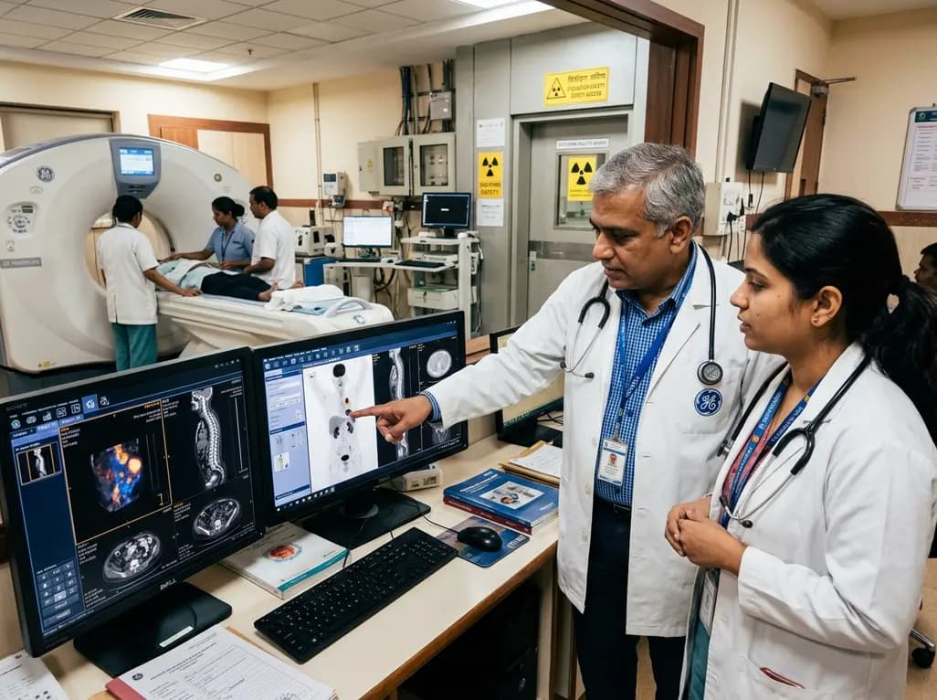
GE HealthCare has agreed to acquire Sofie Biosciences in a 945 million dollar all-cash transaction. The deal secures global rights to the investigational pan-cancer tracer FAPI-74 and expands cyclotron manufacturing infrastructure to accelerate precision theranostics and clinical molecular imaging workflows.
Today

Dual-nanofiber interpenetrating network scaffolds combining silica and PLLA/gelatin nanofibers with sodium alginate achieve complementary mechanical stability, immunomodulatory M2 macrophage polarization, and enhanced osteogenesis for advanced bone repair.
Today

New research reveals that amylin receptor signalling in the laterodorsal tegmental nucleus controls food intake through inhibitory projections to the ventral tegmental area. This study demonstrates how midbrain reward circuits regulate feeding, presenting crucial targets for next-generation obesity pharmacotherapy.
Today
