

Loading, please wait...
Loading, please wait...

The emergence of an acutely ruptured M1 fusiform aneurysm presents one of the most formidable emergencies in modern cerebrovascular surgery. Unlike common saccular lesions, these complex vascular pathologies lack an identifiable neck and incorporate the circumferential parent vessel wall into the diseased segment. Consequently, standard microsurgical clip reconstruction often remains impossible without sacrificing downstream flow. Moreover, acute subarachnoid hemorrhage introduces severe brain swelling and elevated intracranial pressure, which severely restricts safe surgical exposure. Traditional endovascular flow diversion also presents serious limitations in this acute setting, primarily because immediate dual antiplatelet therapy significantly increases hemorrhagic complications. In addition, flow diverters rarely provide immediate occlusion, leaving the vulnerable rupture site exposed to high rebleeding risks. Therefore, vascular specialists must deploy sophisticated multimodal strategies to achieve definitive dome exclusion. A one-stage hybrid intervention that pairs bypass revascularization with endovascular parent artery occlusion offers an elegant solution. By utilizing a hybrid neurosurgical suite, interventionalists and microsurgeons can restore distal perfusion and isolate the hemorrhagic lesion simultaneously. This coordinated approach prevents recurrent catastrophic rupture while protecting vulnerable hemispheric territories from critical ischemic damage during emergency neurovascular rescue.
Treating lesions along the proximal middle cerebral artery requires meticulous anatomical respect for the lenticulostriate perforating vessels. Specifically, these critical end-arteries supply the posterior limb of the internal capsule, basal ganglia, and adjacent motor pathways. Any unintended occlusion of these tiny vessels invariably produces dense, permanent hemiplegia and catastrophic functional deficits. Furthermore, giant fusiform aneurysms often stretch or incorporate these tiny perforating branches directly within the aneurysmal sac wall. Microsurgeons face immense technical hurdles when attempting to dissect and preserve these fragile structures under tight brain conditions. Similarly, attempting endovascular coiling without protective strategies risks catastrophic backward coil migration into the parent trunk and perforator origins. Consequently, clinicians must obtain high-resolution rotational digital subtraction angiography to map the exact relationship between the aneurysm and all visualised lenticulostriate arteries. In addition, intraoperative neuromonitoring provides real-time functional surveillance of the corticospinal pathways during temporary parent artery manipulation. By clearly delineating perforator anatomy before initiating vessel occlusion, surgical teams ensure that parent vessel sacrifice occurs strictly distal or selectively around perforator origins. Therefore, thorough anatomical evaluation underpins every tactical decision throughout this complex hybrid cerebrovascular intervention.
Parent vessel sacrifice demands robust and reliable revascularization to prevent massive cerebral infarction across the middle cerebral artery territory. Although a single-barrel superficial temporal artery bypass provides modest flow, it frequently fails to satisfy the extensive metabolic demands of the entire hemisphere. Therefore, surgical teams utilize a double-barrel superficial temporal artery to middle cerebral artery bypass to deliver superior hemodynamic flow replacement. Specifically, surgeons harvest both the frontal and parietal branches of the superficial temporal artery through a meticulous pterional or preauricular dissection. Because adult males frequently possess larger caliber donor vessels, these branches can provide substantial revascularization volume. Subsequently, the microsurgeon anastomoses the two donor branches separately to distinct recipient cortical branches, such as the anterior and posterior divisions of the M2 segment. In addition, intraoperative indocyanine green videoangiography and microvascular Doppler ultrasonography confirm patent anastomoses with vigorous downstream flow. This dual revascularization effectively restores independent blood supply to both temporal and frontal opercular territories prior to parent vessel obliteration. Consequently, the brain safely tolerates deliberate proximal arterial occlusion without encountering hypoperfusion, ischemia, or watershed infarction.
Following successful microsurgical revascularization, the hybrid team transitions immediately to endovascular isolation of the diseased vascular segment. Because surgical clip placement around giant fusiform anatomy carries severe risk of tearing friable atherosclerotic vessels, endovascular occlusion provides a safer alternative. The neurointerventionalist establishes transfemoral or transradial access and navigates microcatheters directly into the aneurysmal lumen. Specifically, teams frequently employ a dual-microcatheter technique to achieve stable, controlled coil packing within complex and irregular aneurysmal sacs. One microcatheter stabilizes the primary frame, while the second microcatheter navigates into the daughter sac to eliminate the fragile rupture point. Subsequently, the operator advances dense detachable coils progressively throughout the aneurysmal segment, culminating in deliberate parent artery occlusion. Moreover, this controlled endovascular deployment minimizes mechanical traction on the recently ruptured vessel wall. Immediate post-embolization angiographic runs confirm complete obliteration of the aneurysm while demonstrating retrograde filling of distal branches via the active bypass. Thus, endovascular embolization achieves definitive hemostasis and permanently protects the patient against lethal rebleeding events.
The integration of microsurgical and endovascular techniques within a single hybrid operating suite represents a major paradigm shift in vascular neurosurgery. Historically, patients requiring combined bypass and endovascular sacrifice underwent staged procedures across different hospital rooms or separate operative days. However, staged interventions leave patients vulnerable to catastrophic aneurysm rerupture during the fragile waiting interval between revascularization and vessel occlusion. Furthermore, moving critically ill patients with external ventricular drains between standard operating rooms and radiology angiography suites creates severe physiological instability. In contrast, the one-stage hybrid operating suite eliminates inter-facility transit risks completely. Microsurgeons can perform bypass anastomoses under the operating microscope, and the angiography system immediately verifies graft patency and baseline hemispheric hemodynamics. Additionally, if the bypass exhibits sluggish flow, the team can immediately diagnose and correct technical issues before initiating parent vessel embolization. Once the team confirms adequate cortical perfusion, endovascular parent artery occlusion proceeds without delay under high-resolution fluoroscopy. Therefore, the hybrid suite maximizes patient safety, reduces cumulative anesthetic exposure, and streamlines critical emergency decision-making.
After completing the hybrid intervention, the patient requires dedicated neurocritical care monitoring in a specialized intensive care unit. Critical care teams maintain normotensive to slightly hypertensive blood pressure targets to promote bypass graft patency without stressing non-occluded vessels. Furthermore, clinicians utilize serial transcranial Doppler sonography and continuous arterial line monitoring to evaluate graft velocities and cerebral autoregulation. Because the procedure avoids immediate high-potency dual antiplatelet therapy, clinicians can safely place or maintain intracranial pressure monitors and external ventricular drains. In addition, aggressive fluid management prevents vasospasm-induced ischemia, which frequently complicates ruptured subarachnoid hemorrhage cases during the second week post-ictus. If neurological deterioration occurs, clinicians perform emergency computed tomography angiography or perfusion studies to confirm bypass patency and rule out delayed vasospasm. Ultimately, this comprehensive perioperative management ensures that the surgical revascularization matures smoothly while brain tissue recovers from the primary hemorrhagic insult. Through synchronized multidisciplinary protocols, high-risk patients achieve favorable functional outcomes and avoid long-term neurological debility.
A double-barrel bypass provides substantially greater flow volume by utilizing both frontal and parietal superficial temporal artery branches. Because parent artery occlusion permanently sacrifices antegrade M1 circulation, a single branch rarely supplies sufficient flow. Connecting two donor branches to separate M2 divisions adequately revascularizes the entire hemispheric territory, preventing cerebral ischemia.
Surgeons utilize fine-cut three-dimensional digital subtraction angiography and intraoperative electrophysiological monitoring to map all visualised perforators. Furthermore, coil occlusion is restricted strictly to the diseased segment distal to or clear of critical perforator origins. Preserving antegrade blood flow into proximal perforator trunks prevents deep basal ganglia infarction and permanent hemiparesis.
A one-stage hybrid procedure combines open revascularization and endovascular occlusion in a single operative session. This approach eliminates the waiting period between procedures, minimizing catastrophic aneurysm rebleeding risks. Furthermore, it avoids moving critically unstable subarachnoid hemorrhage patients between suites while enabling immediate intraoperative angiographic confirmation of bypass flow and aneurysm thrombosis.
Disclaimer: This content is for informational and educational purposes only and does not constitute medical advice, diagnosis, or treatment regimens. Healthcare professionals must evaluate clinical scenarios independently. Treatment decisions should align with individual patient assessments, institutional capabilities, and applicable professional standards. Refer to the latest local and national guidelines for clinical practice.
References

Read summarized clinical updates, watch expert medical content, and earn CME certifications right from your smartphone.
Open your smartphone camera to scan and install.





A one-stage hybrid strategy combining double-barrel STA-MCA bypass with endovascular coiling offers a safe and definitive solution for managing ruptured giant M1 fusiform aneurysms lacking a clippable neck.
Today

Novel glucose-responsive mPEG-PSA nanocarriers deliver insulin aspart on demand, achieving dual postprandial and basal glycemic control without hypoglycemia in diabetic models. This biomaterial breakthrough offers promising clinical implications for physiologic diabetes management.
Today

A novel folate-conjugated tecto-dendrimer nanoplatform successfully delivers siRNA to achieve targeted HK2 silencing, disrupting ovarian cancer glycolysis and suppressing lactate output.
Today
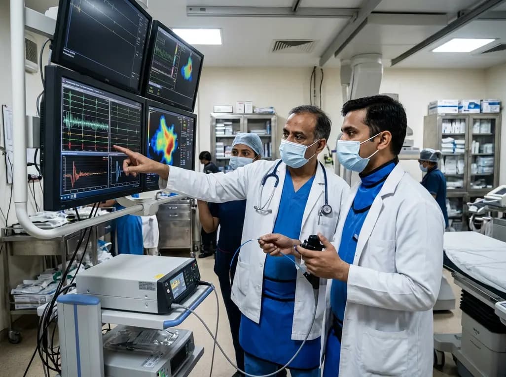
A comparative non-contact study reveals that high-power radiofrequency delivery with the QDOT MICRO catheter induces an artifactual contact force rise. Electrophysiologists must understand this power-dependent thermal and electrical phenomenon to avoid inadvertent catheter withdrawal and ineffective ablation.
Today

A novel proof-of-concept study presents a six-degree-of-freedom articulated manikin neck equipped with linear potentiometers. The system provides real-time kinematic motion feedback during cervical immobilisation, delivering sub-degree tracking accuracy to improve pre-hospital trauma training and patient safety.
Today

A retrospective comparative cohort study evaluated genitourinary safety in HFpEF between dapagliflozin and empagliflozin over six months. Rates of symptomatic UTI and asymptomatic bacteriuria showed no statistically significant differences, supporting comparable safety profiles across both SGLT2 inhibitors.
Today
