.webp&w=384&q=75)

Loading, please wait...
Loading, please wait...
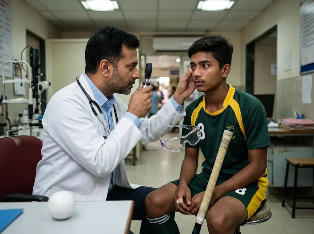
Sports-related eye injuries represent a major cause of preventable vision impairment across various age groups worldwide. Among high-impact athletic activities, ice and field hockey present significant risks of serious periocular and intraocular damage. A recent multi-decade cross-sectional investigation published in JAMA Ophthalmology highlights essential patterns in hockey ocular trauma within emergency departments. The study utilized national surveillance data covering twenty years to quantify the incidence, demographic distribution, and primary mechanisms driving these severe ophthalmic injuries. Clinicians frequently encounter players presenting with blunt impact, high-velocity projectile strikes, or sharp contact during athletic participation. Recognizing these clinical presentations enables physicians to quickly classify ocular structural risk and initiate timely therapeutic measures. Furthermore, understanding epidemiological trends helps public health authorities and sports organizations refine protective gear mandates. Because young athletes and male participants experience a disproportionate share of ocular physical harm, targeted educational interventions remain essential. Prompt medical evaluation prevents catastrophic sight-threatening complications, including persistent hyphema, traumatic cataract, or severe retinal damage. Therefore, healthcare providers must understand these underlying injury patterns to optimize immediate patient care and promote comprehensive prevention guidelines.
Analyzing demographic variables provides key clinical insights into which populations suffer high rates of ophthalmic structural injury. The study demonstrated that male athletes account for nearly four-fifths of all reported cases, reflecting elevated participation rates and aggressive physical play styles. Additionally, pediatric and young adult players represent a major portion of hospital emergency presentations. Younger athletes often display underdeveloped peripheral awareness and slower reflexive responses to flying equipment. Furthermore, non-professional and recreational settings generate a substantial volume of severe facial and eye injuries compared to professional leagues. In professional environments, strict equipment requirements and trained medical staff help limit acute structural harm. Conversely, amateur leagues often lack mandatory full-face visors or rigid physical enforcement, which significantly increases individual exposure. Understanding these demographic nuances allows emergency specialists and sports medicine clinicians to screen high-risk individuals effectively. Moreover, targeted safety campaigns should specifically focus on youth leagues, school teams, and amateur adult organizations to reduce preventable hospital visits. By prioritizing structured educational programs and mandatory protective equipment, healthcare leaders can safeguard vulnerable young players from irreversible vision impairment.
The mechanisms responsible for ocular harm in hockey stem primarily from high-energy collisions involving sticks, pucks, or physical contact. High-speed puck impacts typically deliver high-force blunt trauma directly to the orbit, frequently causing facial fractures and severe contusions. Conversely, accidental stick contact often inflicts sharp lacerations, corneal abrasions, or open-globe disruptions. Clinicians evaluating these emergency presentations encounter diverse diagnostic conditions, ranging from superficial adnexal trauma to vision-threatening posterior segment damage. Traumatic hyphema remains a frequent anterior segment complication, requiring close monitoring to prevent secondary glaucoma or corneal blood staining. In addition, severe mechanical impacts frequently precipitate vitreous hemorrhage, commotio retinae, traumatic iritis, or traumatic cataract formation. Posterior pole involvement, including full-thickness retinal tears or detachments, demands urgent vitreoretinal evaluation to preserve visual function. Furthermore, periocular soft tissue lacerations and orbital blowout fractures often accompany internal eyeball damage. Consequently, comprehensive slit-lamp examination, indirect ophthalmoscopy, and urgent radiological imaging are crucial diagnostic steps. Systematic clinical assessment ensures that physicians identify occult intraocular pathology quickly, minimizing long-term structural and functional deficits.
Prompt triage and rigorous severity assessment dictate the long-term visual prognosis following acute ocular physical trauma. Emergency room physicians and ophthalmologists must rapidly differentiate between minor adnexal injuries and sight-threatening open-globe trauma. Initial stabilization requires immediate protection of the global structure using a rigid, non-pressure eye shield. Clinicians should explicitly avoid topical ointments, cycloplegic drops, or ocular manipulation if open-globe rupture is suspected. Furthermore, systemic antiemetic and analgesic agents help prevent sudden spikes in intraocular pressure caused by valsalva maneuvers or vomiting. Comprehensive imaging, such as non-contrast computed tomography of the orbits, confirms orbital floor fractures or intraocular foreign bodies. For severe closed-globe injuries with traumatic hyphema, strict bed rest, elevation of the head, and topical corticosteroid therapy are essential initial strategies. Serial intraocular pressure monitoring prevents optic nerve damage secondary to elevated tension. When vitreoretinal or corneal lacerations occur, immediate surgical consultation ensures timely primary repair. Close coordination between emergency physicians, pediatricians, and vitreoretinal specialists optimizes patient outcomes. Standardized acute care algorithms significantly reduce long-term vision impairment, allowing athletes a safer path toward functional recovery.
Preventing sports-related ophthalmic physical harm relies heavily on mandatory, high-quality protective gear and robust policy enforcement. Historical evidence demonstrates that mandating full-face shields or polycarbonate visors significantly decreases severe eye injuries across amateur and professional leagues. Polycarbonate shields offer exceptional impact resistance, effectively absorbing high-velocity energy from sticks and pucks. However, partial visors leave the lower facial structure and inferior orbital region vulnerable to deflected equipment. Therefore, sports medicine experts strongly advocate for full-face cage protection, especially among pediatric and adolescent participants. Beyond individual equipment, educational programs must teach young players proper stick-handling techniques and aggressive play regulation. Referees and league administrators play a crucial role by strictly penalizing high-sticking and reckless physical contact. Healthcare providers should actively counsel athletic patients and parents during routine physical examinations regarding proper protective equipment standards. Moreover, national athletic associations must continuously update safety regulations to align with emerging epidemiological findings. By fostering a strong safety culture and enforcing universal eyewear compliance, sports organizations and medical professionals can virtually eliminate catastrophic, preventable visual loss on the ice.
The most common ocular injuries in hockey include corneal abrasions, periocular lacerations, traumatic hyphema, and eyelid contusions. High-energy impacts from pucks or sticks can also cause orbital blowout fractures, traumatic iritis, vitreous hemorrhage, or retinal tears. While superficial soft tissue trauma responds well to conservative care, high-impact blunt force often causes severe intraocular structural damage that demands urgent ophthalmic evaluation to preserve long-term visual acuity.
Pediatric players face higher risk due to developing motor skills, reduced peripheral visual field processing, and slower reflexive responses during fast-paced play. Additionally, young athletes frequently participate in recreational or informal leagues where protective gear standards and officiating rules are less strictly enforced. Because youth eyes are anatomically smaller and still growing, blunt physical impacts often cause disproportionately severe intraocular disruption, emphasizing the vital necessity of mandatory full-face protective visors.
Immediate management requires stopping play and inspecting the eye without applying pressure to the globe. If severe trauma or open-globe injury is suspected, place a protective rigid eye shield over the eye immediately. Avoid applying topical medications, ointments, or chemical rinses. Keep the patient calm, elevate their head, and prohibit eating or drinking in case urgent surgical intervention is required. Transport the patient immediately to an emergency department with ophthalmology coverage.
Disclaimer: This content is for informational and educational purposes only and should not be used as a substitute for professional medical advice, diagnosis, or treatment. Always seek the advice of a physician or other qualified health provider with any questions you may have regarding a medical condition. Refer to the latest local and national guidelines for clinical practice.
References

Read summarized clinical updates, watch expert medical content, and earn CME certifications right from your smartphone.
Open your smartphone camera to scan and install.





A 20-year NEISS study published in JAMA Ophthalmology characterizes national trends in hockey-related ocular trauma, highlighting high risks among male and pediatric participants, primary mechanisms including puck and stick impacts, and the imperative role of protective eyewear mandates in injury prevention.
3 months ago

Novel diimide and hydrazone scaffolds, particularly compound #C4, synergistically reactivate latent HIV-1 when combined with JQ1 or PKC activators. Demonstrating donor-matched proviral transcription ex vivo and non-canonical signaling, these findings advance shock and kill strategies toward reservoir eradication.
Today

A comparative cohort study evaluates vNOTES vs TLH hysterectomy for benign conditions, demonstrating shorter operative duration, faster return to daily activities, and equivalent female sexual function scores without increased dyspareunia.
Today

Review how tailored exercise programs benefit frail older patients with heart failure. Explore pathophysiological mechanisms, individualized training prescriptions, and strategies to overcome adherence barriers in clinical practice.
Yesterday

This comparative study evaluates posterior labral tears with and without glenohumeral instability, highlighting distinct demographic patterns, MRI underestimation in stable shoulders, and successful arthroscopic repair outcomes.
Yesterday

A clinical study demonstrates that early intervention with tocilizumab, either as monotherapy or bridged with corticosteroids, achieves significant proptosis reduction and resolves active inflammation in treatment-naive thyroid eye disease.
Today
