

Loading, please wait...
Loading, please wait...

The evaluation of HMGA1 in gliomas provides essential insights into chromatin dysregulation in central nervous system malignancies. High-Mobility Group A1 (HMGA1) functions as a non-histone, chromatin-associated protein that dynamically coordinates nuclear architecture and gene transcription. Under physiological conditions, HMGA1 regulates vital cellular processes, including DNA replication, genetic recombination, cellular differentiation, and genomic repair mechanisms. Embryonic tissues express abundant HMGA1 to facilitate rapid fetal organogenesis. In contrast, differentiated adult human tissues largely silence this architectural factor. However, diverse human cancers aberrantly reactivate HMGA1, driving malignant transformation, aggressive cellular proliferation, local tissue invasion, and neo-angiogenesis. In neuro-oncology, malignant gliomas represent exceptionally aggressive primary brain tumors with poor historical survival outcomes. Despite multimodal interventions involving maximal safe resection, ionizing radiotherapy, and cytotoxic temozolomide chemotherapy, tumor recurrence remains nearly inevitable. Clinicians urgently require validated molecular biomarkers to refine prognostic stratification and uncover novel therapeutic vulnerabilities. Therefore, exploring HMGA1 in gliomas clarifies the critical transcriptional networks governing astrocytic malignancy. Furthermore, recent molecular investigations demonstrate that HMGA1 promotes glioma stem cell self-renewal and therapeutic resistance. Consequently, analyzing HMGA1 provides meaningful biological perspectives for risk assessment and individualized patient management.
Most existing neuro-oncological genomic studies evaluate Caucasian or East Asian populations, leaving South Asian demographics critically underrepresented. To bridge this significant evidence gap, investigators conducted a focused observational cohort study in Pakistan. The surgical team collected 75 distinct glioma tissue specimens from patients undergoing craniotomy at Aga Khan University Hospital. Simultaneously, the researchers harvested 10 non-neoplastic normal adjacent tissue samples to establish baseline physiological controls. The laboratory team immediately preserved all resected specimens to safeguard total RNA integrity. Following extraction, the scientists performed real-time quantitative reverse transcription polymerase chain reaction to measure relative HMGA1 mRNA abundance accurately. The pathologists classified and graded all tumors according to standard World Health Organization criteria. Additionally, the clinical team systematically monitored overall patient survival durations across the entire cohort. For statistical computations, the investigators utilized advanced bioinformatic packages within the R software environment. Furthermore, the researchers applied the Kaplan-Meier estimator to generate survival curves and evaluate prognostic differences between expression subgroups. This methodological framework ensured stringent analytical quality control from surgical tissue harvest to downstream computational modeling. Therefore, this investigation provides dependable molecular pathology data from an understudied regional population.
Quantitative analysis revealed pronounced disparities in HMGA1 transcript levels between neoplastic tissues and normal brain parenchyma. Specifically, glioma specimens exhibited marked upregulation of HMGA1 compared to normal adjacent controls, achieving high statistical significance (p < 0.001). Moreover, HMGA1 transcript levels demonstrated a clear, stepwise elevation that correlated with increasing histological malignancy (p = 0.015). Grade IV glioblastoma multiforme cases displayed the highest transcriptional expression, whereas low-grade astrocytomas displayed modest baseline levels. Notably, the authors documented a statistically significant positive correlation between elevated HMGA1 expression and Ki-67 proliferation indices. The Ki-67 protein represents a standard clinical marker reflecting active mitotic division within neuroepithelial tumors. Therefore, this biological correlation indicates that HMGA1 actively fuels neoplastic cell cycle progression and astrocytic proliferation. In addition, previous molecular studies confirm that HMGA1 upregulates vascular endothelial growth factor and matrix metalloproteinases. Consequently, tumors exhibiting high HMGA1 levels frequently demonstrate aggressive microvascular proliferation and diffuse parenchymal infiltration. These laboratory findings illustrate that HMGA1 gene elevation closely mirrors histological aggression and aggressive tumor biology. Thus, quantifying HMGA1 offers neuro-pathologists an objective parameter to corroborate tumor proliferation and aggressive malignant behavior.
Beyond histological and proliferative correlations, HMGA1 expression significantly influenced overall patient survival in the cohort. Survival analysis using the Kaplan-Meier plotter demonstrated markedly shortened survival durations in patients with high HMGA1 transcripts (p = 0.0014). Furthermore, multivariable Cox proportional hazards modeling revealed that elevated HMGA1 expression conferred a 3.5-fold higher risk of mortality. The calculated 95% confidence interval ranged from 1.5 to 7.9, confirming substantial statistical robustness (p = 0.003). To evaluate the diagnostic and prognostic accuracy of this biomarker, researchers performed receiver operating characteristic analysis. Notably, the receiver operating characteristic curve generated an area under the curve of 0.752. This metric reflects dependable prognostic discrimination for classifying clinical outcomes. Consequently, HMGA1 acts as an independent molecular predictor of poor survival in glioma patients. Individuals with low transcriptional expression achieved prolonged survival times following standard surgical debulking and chemoradiation. In contrast, patients exhibiting marked HMGA1 upregulation experienced accelerated disease progression and early clinical mortality. Therefore, incorporating HMGA1 into post-resection molecular evaluations could assist clinicians in identifying high-risk individuals who require intensified clinical surveillance. Ultimately, these survival data underline the clinical value of HMGA1 as a potent risk-stratification biomarker.
To validate the findings beyond the single-center Pakistani cohort, the authors performed computational cross-validation utilizing the Chinese Glioma Genome Atlas. Interestingly, external validation across this extensive multi-omics database confirmed the initial findings with striking consistency. Elevated HMGA1 transcripts correlated with advancing tumor grade and worse patient survival across distinct geographic populations. Therefore, HMGA1 appears to drive aggressive biological behavior independent of regional or ethnic variations. These insights carry direct clinical relevance for neuro-oncologists and neurosurgeons practicing across South Asia, including India. In resource-conscious healthcare settings, quantitative PCR provides a cost-effective, reproducible tool for molecular diagnostics. While next-generation sequencing requires expensive infrastructure, qPCR assays for HMGA1 offer rapid prognostic guidance in regional neurosurgical centers. Furthermore, established molecular markers like IDH mutation status and MGMT promoter methylation do not fully explain patient survival heterogeneity. Measuring HMGA1 expression provides complementary prognostic precision alongside established World Health Organization classification standards. Additionally, preclinical researchers are actively evaluating pharmacological agents that disrupt HMGA1 chromatin interactions to overcome chemoresistance. However, clinical teams must conduct prospective multi-center clinical trials to establish standardized clinical threshold values. Ultimately, HMGA1 represents an actionable biomarker that could enhance precision neuro-oncology.
Elevated HMGA1 expression strongly correlates with advanced tumor grades, increased proliferative activity, and significantly poorer overall survival in glioma patients. Specifically, high expression confers a 3.5-fold higher risk of mortality. Consequently, HMGA1 functions as a potent independent prognostic biomarker that effectively identifies patients facing rapid tumor progression.
HMGA1 expression demonstrates a robust positive correlation with Ki-67 labeling indices across glioma specimens. Because Ki-67 directly reflects ongoing cellular proliferation and mitotic index, elevated HMGA1 signifies aggressive tumor replication. Furthermore, high HMGA1 levels correlate with matrix metalloproteinase expression, driving enhanced microvascular invasion and extensive local infiltration.
Currently, HMGA1 testing remains primarily within translational research and validation environments rather than standard clinical protocols. Although real-time qPCR and immunohistochemistry reliably detect HMGA1, clinicians require standardized cut-off values and validation through prospective multi-institutional trials before integrating this biomarker into formal clinical treatment guidelines.
Disclaimer: This content is for informational and educational purposes only... Refer to the latest local and national guidelines for clinical practice.
References

Read summarized clinical updates, watch expert medical content, and earn CME certifications right from your smartphone.
Open your smartphone camera to scan and install.





A Pakistani cohort study reveals that HMGA1 gene expression is significantly upregulated in gliomas and correlates with advanced tumor grade, Ki-67 proliferation, and poor survival. Patients with high HMGA1 exhibit a 3.5-fold higher mortality risk, highlighting its potential as an independent prognostic biomarker.
Today

Review how tailored exercise programs benefit frail older patients with heart failure. Explore pathophysiological mechanisms, individualized training prescriptions, and strategies to overcome adherence barriers in clinical practice.
Today
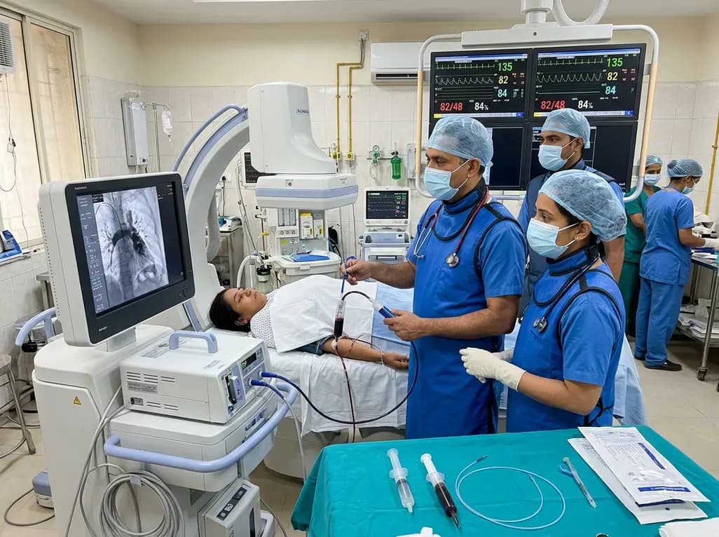
In high-risk pulmonary embolism complicated by severe anemia and active uterine bleeding, systemic thrombolysis is contraindicated. Large-bore aspiration thrombectomy provides rapid hemodynamic rescue without fibrinolysis, supported by inferior vena cava filter placement and multidisciplinary critical care.
Today

A cross-sectional study in Zanzibar reveals that 46.5% of adults with type 2 diabetes experience multidomain autonomic dysfunction on the COMPASS-31 tool, primarily orthostatic intolerance, while 45.9% exhibit cardiovascular autonomic neuropathy, highlighting the urgent need for structured clinical screening.
Today

This comparative study evaluates posterior labral tears with and without glenohumeral instability, highlighting distinct demographic patterns, MRI underestimation in stable shoulders, and successful arthroscopic repair outcomes.
Today

A recent case-control study reveals that urinary clusterin provides superior diagnostic performance for early diabetic kidney disease detection in pediatric type 1 diabetes mellitus, identifying tubular damage before conventional microalbuminuria occurs.
Today
