.webp&w=384&q=75)

Loading, please wait...
Loading, please wait...
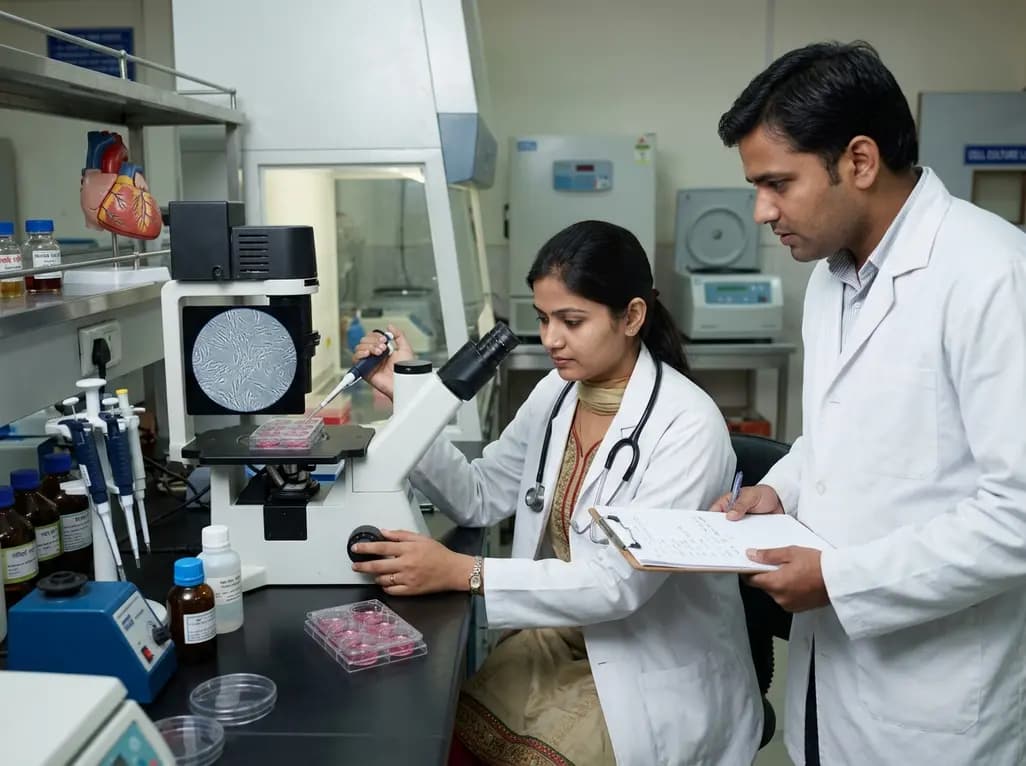
Researchers at the Sanjay Gandhi Postgraduate Institute of Medical Sciences (SGPGIMS) in Lucknow have achieved a remarkable milestone by culturing human heart valve interstitial cells in a laboratory environment. Lead investigators Dr. Shantanu Pande and Dr. Alok Kumar harvested discarded valvular tissues from consenting surgical patients to culture these specialized cells. Heart valve interstitial cells maintain the structural integrity, elasticity, and repair mechanism of native human cardiac valves. Consequently, culturing these cells in vitro provides scientists with an unprecedented opportunity to study pathological processes directly on living human tissue. The multidisciplinary effort involved experts from cardiovascular surgery, molecular medicine, clinical immunology, and pathology. Supported by the Department of Biotechnology, Government of India, this groundbreaking work opens new pathways for cardiovascular research. Furthermore, this initiative bridges basic scientific research with clinical applications in tissue engineering. By establishing a robust cell culture model, the research team can now investigate cellular responses to hemodynamic stress and metabolic changes. Additionally, this in vitro platform minimizes reliance on animal models for preliminary cardiac studies. Thus, the successful isolation of these primary cells marks a major technical advancement in Indian biomedical research.
Rheumatic heart disease remains a severe global health concern, particularly across developing countries like India. Globally, India bears forty to fifty percent of the total disease burden and up to forty percent of related mortality. Furthermore, epidemiological studies demonstrate that disease prevalence among Indian schoolchildren ranges between 0.5 and 5 per thousand children. Consequently, chronic inflammatory damage leading to valvular stenosis and regurgitation frequently requires costly surgical interventions. Repeated streptococcal pharyngitis triggers an autoimmune response that progressively damages the mitral and aortic valve structures. However, traditional surgical valve replacement options present significant long-term clinical challenges for young patients. Mechanical valves mandate lifelong oral anticoagulation, whereas biological prostheses suffer from structural degeneration over time. Therefore, understanding the microscopic mechanisms of valvular scarring is essential for developing disease-modifying medical therapies. Cultured heart valve interstitial cells allow investigators to observe how inflammatory cascades alter cellular behavior in real time. Subsequently, researchers can evaluate specific molecular interactions that lead to collagen deposition and leaflet stiffening. Ultimately, this foundational work aims to reduce the crushing burden of advanced valvular disease across vulnerable patient populations.
During the investigation, the SGPGIMS scientific team identified two primary signaling pathways that drive valvular inflammation and progressive tissue fibrosis. Specifically, the Transforming Growth Factor-beta pathway plays a pivotal role through its intracellular downstream messenger protein known as SMAD3. Under physiological conditions, this signaling cascade regulates tissue healing and matrix turnover after minor tissue injuries. However, persistent activation of the pathway triggers aberrant myofibroblast differentiation and excessive deposition of extracellular matrix components. Consequently, the affected valvular cusps suffer from progressive thickening, contracture, and loss of mechanical pliability. Researchers observed that sustained TGF-beta activation directly accelerates cellular scarring in human interstitial cell cultures. Moreover, blocking specific receptors along this axis could potentially halt the relentless progression of fibrotic valvular remodeling. Therefore, targeting SMAD3 signaling represents a promising therapeutic target for pharmacological intervention in early-stage rheumatic heart disease. Additionally, studying this pathway in cultured human tissue provides clearer insights than previous animal models offered. As a result, clinicians may eventually utilize targeted molecular therapies to preserve endogenous valve architecture before irreversible structural damage occurs.
In addition to the TGF-beta pathway, the research team highlighted the key contribution of Extracellular Signal-Regulated Kinases 1 and 2. These intracellular enzymatic messengers actively transmit extracellular signals from cell surface receptors directly into the nucleus. Consequently, the ERK1/2 network regulates fundamental cellular processes, including cell proliferation, survival, growth, and structural matrix organization. However, chronic inflammatory stimuli in rheumatic heart disease turn this physiological repair system into a destructive fibrotic process. In cultured heart valve interstitial cells, hyperactivation of ERK1/2 enzymes correlates directly with cellular hypertrophy and matrix stiffening. Furthermore, this signaling cascade interacts dynamically with inflammatory cytokines to reinforce the fibrotic feedback loop within valvular tissue. Therefore, inhibiting excessive ERK1/2 phosphorylation could significantly attenuate tissue rigidity and preserve leaflet flexibility over extended periods. Researchers are currently evaluating small-molecule inhibitors to determine whether targeting this enzymatic pathway can arrest valvular degeneration. Moreover, understanding cross-talk between ERK1/2 and other signaling cascades will refine future drug development strategies. Thus, unraveling intracellular kinase pathways provides a clearer roadmap for targeted pharmacological therapies in valvular heart disease.
The successful cultivation of human cardiac cells holds vast potential for the emerging field of cardiovascular tissue engineering. Currently, patient outcomes following valve replacement remain limited by mechanical failure, thrombosis, or bioprosthetic degradation. However, tissue-engineered human heart valves constructed using autologous interstitial cells could dramatically overcome these clinical limitations. Ideally, engineered living valves could grow, repair, and adapt alongside the patient, eliminating the need for repeated reoperations. Furthermore, this bioengineering paradigm is especially crucial for pediatric and adolescent patients suffering from severe rheumatic lesions. Cultured cells can be seeded onto biocompatible three-dimensional scaffolds to construct functional, living valvular substitutes in laboratory bioreactors. Additionally, these living constructs exhibit superior immunocompatibility compared to foreign artificial materials or xenografts. Consequently, patients receiving tissue-engineered implants would no longer require lifelong systemic anticoagulation therapy. As research progresses, clinicians anticipate that laboratory-grown valvular tissues will transform reconstructive cardiovascular surgery. Therefore, the foundational achievements at SGPGIMS represent a vital step toward practical clinical applications in regenerative medicine.
Beyond regenerative surgical applications, cultured interstitial cell lines serve as an efficient biological platform for high-throughput pharmacological testing. Currently, drug discovery for valvular heart disease is hindered by the lack of human-relevant preclinical screening models. However, testing candidate therapeutic compounds directly on cultured human cells provides precise physiological data regarding drug efficacy and toxicity. Moreover, researchers can analyze unique cellular secretions and gene expression profiles to discover novel diagnostic biomarkers. Consequently, identifying early circulating biomarkers could allow clinicians to diagnose rheumatic valve involvement long before structural stenosis becomes clinically manifest. Furthermore, early detection enables timely initiation of disease-modifying medical therapies that slow or arrest fibrotic progression. In addition, this research framework fosters interdisciplinary collaboration among cardiologists, molecular biologists, and pharmaceutical researchers across India. Institutional leaders at SGPGIMS have affirmed robust ongoing support to expand these molecular heart valve research programs. Therefore, continued academic and clinical investment in this laboratory model will accelerate translation from basic science to bedside care. Ultimately, these innovations promise to reduce surgical morbidity and improve quality of life for cardiovascular patients worldwide.
Q1: What are heart valve interstitial cells and why are they important?
Heart valve interstitial cells are the predominant cell type residing within cardiac valve tissue. They actively maintain structural integrity, regulate extracellular matrix homeostasis, and repair minor cellular injuries. In disease conditions like rheumatic heart disease, these cells become hyperactive and transform into myofibroblasts. Consequently, understanding their behavior helps scientists develop targeted treatments to prevent valvular scarring and avoid complex replacement surgeries.
Q2: How do TGF-beta and ERK1/2 pathways contribute to rheumatic valve damage?
The TGF-beta pathway, operating via SMAD3, and the ERK1/2 kinase network regulate cellular growth and matrix repair. However, persistent inflammation in rheumatic heart disease overactivates these pathways. Consequently, interstitial cells produce excessive collagen and inflammatory proteins, leading to severe valvular fibrosis and stiffening. Target-specific inhibition of these pathways may help preserve leaflet flexibility and prevent progressive structural degeneration.
Q3: How will lab-grown valve cells impact future heart valve replacement therapies?
Lab-grown human interstitial cells enable researchers to engineer living, tissue-based heart valves on biocompatible scaffolds. Unlike mechanical or animal-derived prostheses, living engineered valves can potentially repair themselves and grow with the patient. Furthermore, they eliminate the need for long-term blood thinners and reduce reoperation risks. Additionally, these cultured cells provide a human-relevant platform for testing new anti-fibrotic drugs.
Disclaimer: This content is for informational and educational purposes only. It does not constitute medical advice or replace professional judgment. Refer to the latest local and national guidelines for clinical practice.
References

Read summarized clinical updates, watch expert medical content, and earn CME certifications right from your smartphone.
Open your smartphone camera to scan and install.





Researchers at SGPGIMS Lucknow have isolated and cultured human heart valve interstitial cells from donor tissues. This milestone advances understanding of rheumatic heart disease pathways like TGF-beta/SMAD3 and ERK1/2, laying the foundation for tissue-engineered heart valves and drug testing.
last month

A quality-improvement study reveals how hospital pharmacy leadership improves medication safety at home. By eliminating concentrated electrolytes, standardizing kits, and auditing drug labeling, the model successfully aligned home acute care with international patient safety standards.
Today
A recent case-control study identified key protein changes in peritoneal fluid-derived small extracellular vesicles, highlighting candidates like S100-A10 and ALDOA as potential biomarkers to overcome diagnostic delays in endometriosis.
Today
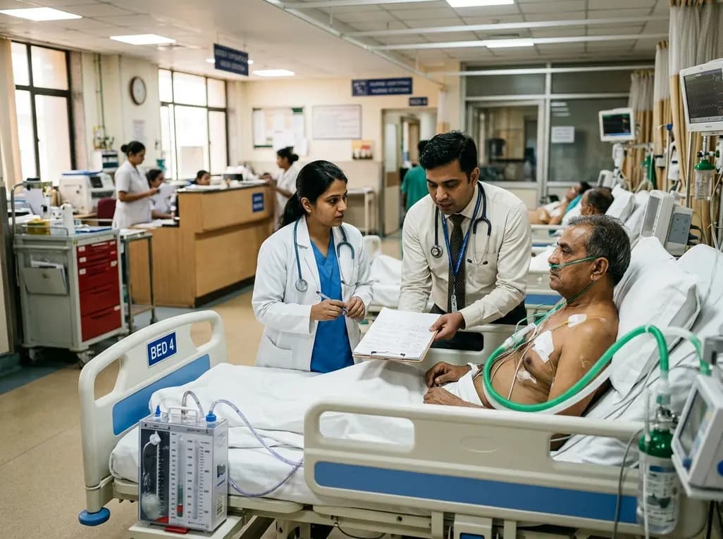
Managing refractory hypoxemia after cardiothoracic surgery becomes perilous when prolonged air leak limits positive-pressure ventilation. This case-based review details how high-flow nasal cannula, paired with awake rehabilitation, enabled successful extubation and avoided invasive re-intubation.
Today
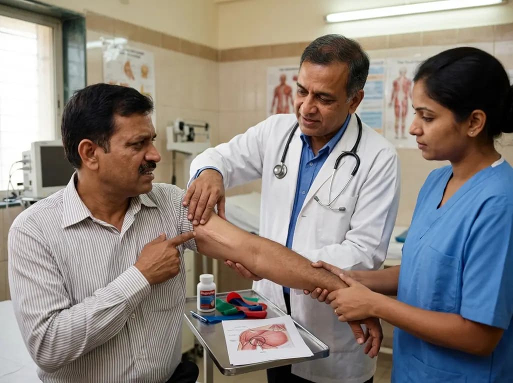
Idiopathic axillary web syndrome (IAWS) is a rare cause of shoulder pain and mobility restriction characterized by palpable axillary cording without prior surgery or trauma. Early diagnosis, nonsteroidal anti-inflammatory therapy, and physical rehabilitation lead to complete resolution of symptoms.
Today

SWEDEPAD-1 trial insights show paclitaxel-coated devices do not improve long-term limb salvage in patients with chronic limb-threatening ischemia and tissue loss. While one-year reinterventions decreased, the devices were linked to a higher risk of major amputation at three months.
Today
