

Loading, please wait...
Loading, please wait...

Good's syndrome represents an intriguing and complex clinical crossroads where thoracic oncology converges with adult immunodeficiency. Robert Good first documented this disorder more than seven decades ago, linking epithelial thymic tumors with severe immune dysfunction. Although clinicians historically associated this condition with an abysmal prognosis, recent diagnostic breakthroughs have transformed clinical outcomes. Today, vigilant physicians recognize that early identification protects patients from life-threatening infections and dramatically enhances long-term survival.
Recent scientific discoveries have shed critical light on the elusive pathogenesis of Good's syndrome. For decades, researchers struggled to clarify why an anterior mediastinal tumor triggers profound immune dysregulation. However, investigators have recently discovered high titers of neutralizing autoantibodies targeting endogenous cytokines in affected individuals. Specifically, patients exhibit autoantibodies directed against interleukin-12, interleukin-23, interleukin-6, and both type I and type II interferons. These neutralizing antibodies severely disrupt critical signaling cascades, thereby impairing cytokine pathways necessary for host defense against opportunistic pathogens. Furthermore, defective thymic architecture impairs the normal process of negative selection. Consequently, autoreactive T-cell clones escape into the peripheral circulation while normal B-cell maturation ceases within the bone marrow. Bone marrow aspirates frequently demonstrate a complete absence of pre-B cells and mature B lymphocytes. In addition, the persistent cytokine neutralization compromises cell-mediated immunity against intracellular pathogens and opportunistic fungi. Therefore, the condition represents a multifaceted combined immunodeficiency rather than an isolated humoral defect. As researchers uncover additional cytokine pathways, these molecular insights provide novel targets for therapeutic monitoring.
Clinicians classify thymoma-associated immunodeficiency into classical and nonclassical clinical subtypes based on distinct laboratory findings. In the classical syndrome, patients present with marked hypogammaglobulinemia accompanied by a severe reduction or total absence of peripheral blood B cells. These patients exhibit precipitously low serum levels of IgG, IgA, and IgM, mirroring severe agammaglobulinemia. Conversely, nonclassical presentations involve prominent cellular immunity defects with preserved or moderately reduced immunoglobulin levels. In these atypical cases, patients exhibit profound CD4-positive T-cell lymphopenia and inverted CD4-to-CD8 ratios. Moreover, natural killer cell dysfunction and impaired delayed-type hypersensitivity responses often complicate nonclassical disease. Because phenotypic expression varies significantly across individuals, specialists must maintain a high index of suspicion during routine evaluations. Furthermore, some patients initially display normal immunoglobulin profiles but subsequently develop progressive antibody depletion over several years. Therefore, clinicians cannot rule out the diagnosis based on a single laboratory evaluation. Instead, medical teams must perform longitudinal serological and flow cytometric assessments in all patients diagnosed with thymoma.
The clinical picture of thymoma-associated immunodeficiency encompasses recurrent infections, widespread autoimmunity, and secondary neoplasms. Recurrent sinopulmonary infections represent the most frequent initial clinical manifestation. Encapsulated bacterial pathogens such as Streptococcus pneumoniae and Haemophilus influenzae frequently cause recurrent pneumonia, sinusitis, and progressive bronchiectasis. Additionally, impaired cell-mediated immunity renders patients highly susceptible to opportunistic viral, fungal, and mycobacterial pathogens. Patients often experience severe cytomegalovirus reactivation, Pneumocystis jirovecii pneumonia, mucocutaneous candidiasis, and atypical mycobacterial disease. Beyond infectious vulnerability, severe autoimmune conditions arise in nearly half of all affected patients. Clinicians frequently encounter myasthenia gravis, pure red cell aplasia, lichen planus, systemic lupus erythematosus, and autoimmune enteropathy. Persistent chronic diarrhea, secondary to either mucosal autoimmunity or opportunistic infections like Giardia lamblia, causes debilitating malabsorption and weight loss. Furthermore, patients face a heightened lifetime risk of secondary solid organ and hematological malignancies. Consequently, physicians must treat any persistent or unusual symptom aggressively to prevent irreversible end-organ damage.
Establishing an accurate diagnosis requires a comprehensive immunological workup integrated with high-resolution thoracic imaging. When an adult presents with recurrent sinopulmonary infections or unexplained opportunistic disease, clinicians should immediately examine quantitative serum immunoglobulin levels. Severe pan-hypogammaglobulinemia provides the earliest biochemical clue to the underlying syndrome. Subsequently, flow cytometry plays an indispensable role by quantifying peripheral lymphocyte subsets. Flow cytometric analysis typically reveals virtually absent CD19-positive and CD20-positive B cells alongside abnormal CD4-to-CD8 T-cell ratios. In addition, contrast-enhanced computed tomography of the chest helps identify asymptomatic or subtle anterior mediastinal lesions. Histopathological evaluation of the resected mass usually confirms type A, AB, or B thymoma according to World Health Organization criteria. Moreover, specialized centers increasingly evaluate anti-cytokine autoantibody panels to support complex diagnostic evaluations. Clinicians must also distinguish this syndrome from common variable immunodeficiency. Unlike common variable immunodeficiency, this disorder features absent circulating B lymphocytes, thymic neoplasia, and adult onset. Therefore, rigorous baseline testing secures an accurate diagnosis and expedites life-saving interventions.
Effective clinical management requires a coordinated, multimodal therapeutic approach centered on surgery and lifelong replacement therapy. First, thoracic surgeons must perform a complete thymectomy to prevent local tumor invasion and paraneoplastic complications. However, surgical resection of the thymoma does not restore immunological competence or normalize serum immunoglobulin concentrations. In fact, immune dysfunction and severe hypogammaglobulinemia persist indefinitely even after curative surgical excision. Consequently, lifelong immunoglobulin replacement therapy serves as the undisputed cornerstone of medical management. Clinicians administer intravenous or subcutaneous immunoglobulin infusions to maintain physiological trough IgG levels. This regular replacement therapy dramatically reduces bacterial pneumonia episodes, prevents bronchiectasis progression, and limits hospitalizations. Furthermore, clinicians must prescribe targeted antimicrobial prophylaxis against Pneumocystis jirovecii when severe CD4 lymphopenia exists. Decades ago, 15-year survival rates hovered near zero percent; however, modern prompt recognition and immunoglobulin replacement have elevated 20-year survival beyond 60 percent. Long-term surveillance with periodic computed tomography and pulmonary function testing remains imperative for sustaining these impressive survival gains.
No, surgical thymectomy does not reverse or cure the underlying immunodeficiency. Although surgical excision removes the anterior mediastinal tumor and prevents local thoracic invasion, humoral and cellular immune defects persist indefinitely. The peripheral B-cell depletion and severe hypogammaglobulinemia remain unchanged postoperatively. Therefore, all affected patients require lifelong immunoglobulin replacement therapy alongside continuous infectious surveillance, regardless of whether the surgical team achieves complete tumor resection.
While both disorders manifest as adult-onset hypogammaglobulinemia and recurrent infections, their immunological profiles differ distinctly. Patients with Good's syndrome consistently exhibit a total absence or profound reduction of circulating mature B cells, whereas patients with CVID usually maintain normal B-cell counts. Furthermore, Good's syndrome specifically associates with thymoma, frequent autoimmune red cell aplasia, and neutralizing anti-cytokine autoantibodies, which do not typically occur in common variable immunodeficiency.
Recent research reveals that neutralizing autoantibodies directed against endogenous cytokines significantly exacerbate immune dysfunction in Good's syndrome. Investigators frequently detect autoantibodies targeting interleukin-6, interleukin-12, interleukin-23, and interferons in these patients. By neutralizing these vital signaling molecules, autoantibodies impair intracellular pathogen defense, blunt inflammatory cascades, and compromise mucosal immunity. Consequently, testing for these autoantibodies provides valuable mechanistic insights and may soon guide individualized targeted management strategies for refractory cases.
Disclaimer: This content is for informational and educational purposes only. It is not intended to substitute professional medical advice, diagnosis, or treatment. Refer to the latest local and national guidelines for clinical practice.
References

Read summarized clinical updates, watch expert medical content, and earn CME certifications right from your smartphone.
Open your smartphone camera to scan and install.





Good's syndrome links thymoma with severe humoral and cellular immunodeficiency. While autoantibodies against cytokines like IL-12 and interferons drive pathology, early diagnosis and prompt immunoglobulin replacement therapy have elevated 20-year survival rates above 60%.
Today
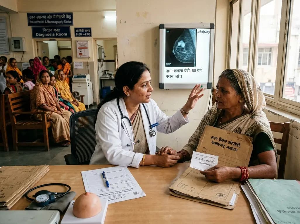
A KGMU study reveals over half of women with breast abnormalities fail to undergo diagnostic confirmation, leading to advanced-stage detection. Discover the socio-cultural barriers, clinical risks of incomplete resection, and community-level solutions needed to bridge the critical screening-to-diagnosis gap in India.
Today

A meta-analysis of 18 studies reveals that bariatric surgery does not significantly alter circulating homocysteine levels, regardless of follow-up duration. Clinicians must maintain vigilance over post-surgical micronutrient supplementation to prevent hyperhomocysteinemia.
Today

Researchers have engineered a self-powered Zn/C micro-galvanic electrospun membrane that couples sustained zinc release with endogenous microcurrents. This bioelectric scaffold stimulates M2 macrophage polarization and accelerates bone deposition to achieve robust cranial defect regeneration.
Today

A nationwide UK Cystic Fibrosis Registry study demonstrates that higher updated mean HbA1c levels correlate with a graded, significant increase in all-cause mortality among individuals with cystic fibrosis-related diabetes, reinforcing the critical need for proactive glycemic monitoring and intervention.
Today
