.webp&w=384&q=75)

Loading, please wait...
Loading, please wait...

Gluteal lymphedema is a complex and often debilitating condition that frequently develops in patients suffering from severe secondary lower extremity lymphedema. This condition is particularly prevalent among cancer survivors who have undergone extensive pelvic surgeries or radiation therapy. Understanding the precise anatomical pathways of lymphatic drainage in the hip region is essential for effective diagnosis and the development of targeted therapeutic interventions. Recent research into gluteal lymphatic watershed mapping has provided significant clarity regarding how lymph moves between the back and the thigh. Specifically, the study of collecting lymphatic vessels using indocyanine green (ICG) lymphography allows clinicians to visualize these pathways in real-time. By delineating the watershed lines, medical professionals can better predict the progression of edema and tailor treatments to the individual anatomical variations of each patient. This mapping is not merely an academic exercise; it serves as a critical roadmap for both surgical planning and manual lymphatic drainage techniques in clinical practice.
Indocyanine green (ICG) lymphography has revolutionized the field of lymphology by providing a safe, minimally invasive, and highly sensitive method for visualizing superficial lymphatic channels. In the context of gluteal lymphatic watershed mapping, this technology involves the intradermal injection of ICG at specific anatomical landmarks. Researchers typically focus on points such as the tip of the coccygeal bone, the gluteal fold, and various crosspoints connecting the medial hip to the lateral thigh. Once injected, the fluorescent dye is taken up by the collecting lymphatic vessels, which can then be observed using a specialized near-infrared camera. This process reveals the directionality of lymph flow and identifies any areas of dermal backflow, which are indicative of lymphatic dysfunction. Consequently, ICG lymphography provides a dynamic view of the lymphatic system that traditional imaging modalities, such as lymphoscintigraphy, often fail to capture with the same level of detail. By observing these linear patterns, clinicians can identify the unique watersheds that divide different drainage territories in the gluteal region.
The results of recent anatomical studies have highlighted two primary pathways for lymphatic drainage in the gluteal region: the posterolateral and the posteromedial pathways. The posterolateral pathway typically carries lymph toward the lateral aspects of the hip and upper thigh, eventually draining into the regional inguinal lymph nodes. Conversely, the posteromedial pathway tends to follow a more internal route toward the medial groin. Interestingly, the distribution of these pathways can vary significantly among individuals, which underscores the importance of personalized gluteal lymphatic watershed mapping. In many cases, the posteromedial pattern is more dominant, particularly from points closer to the midline, such as the coccygeal region. Furthermore, an anteromedial pattern has also been observed in a subset of patients, showing that lymph can sometimes cross toward the front of the body. These findings suggest that the lymphatic system in the hip is far more interconnected than previously thought. Recognizing these distinct pathways allows for a more nuanced understanding of how lymphedema might spread from the lower leg up into the gluteal area and vice versa.
For oncologists and surgeons specializing in pelvic and gynecological cancers, the knowledge of lymphatic watersheds is paramount. Secondary lower extremity lymphedema is a common complication following lymph node dissection or radiation therapy for cancers such as cervical, prostate, or melanoma. When the standard pathways are disrupted, the body may attempt to reroute lymph through collateral channels, often crossing established watershed lines. Therefore, performing gluteal lymphatic watershed mapping in cancer survivors helps in identifying these compensatory mechanisms. If a patient develops gluteal swelling, it often indicates that the proximal lymphatic system is severely compromised. By using ICG mapping, surgeons can identify viable collecting vessels that may be suitable for lymphaticovenular anastomosis (LVA). Moreover, understanding which specific territories are affected allows for more accurate staging of the lymphedema. This proactive approach can significantly improve the quality of life for cancer survivors by addressing swelling before it leads to irreversible tissue changes or infections like cellulitis.
Secondary lower extremity lymphedema (LEL) often manifests as swelling in the feet and legs, but in advanced stages, it frequently involves the gluteal region. The presence of dermal backflow in the hip area on an ICG scan is a clear indicator of high-pressure lymphatic failure. Furthermore, the identification of the watershed line between the back and the thigh helps therapists understand where the "bottleneck" of fluid accumulation is occurring. Specifically, the gluteal region acts as a transitional zone where lymph must find its way toward functional nodes in either the groin or the trunk. If the inguinal nodes are removed or damaged, the gluteal lymphatic watershed mapping may show that lymph is being forced across the sagittal or horizontal watersheds. Consequently, the clinical management of LEL must account for these proximal changes. Treatments that focus only on the lower leg without addressing the gluteal pathways may result in incomplete reduction of edema. Addressing the gluteal region through targeted therapies ensures that the entire limb and its associated territories are adequately drained.
The practical application of gluteal lymphatic watershed mapping is most evident in the administration of manual lymphatic drainage (MLD) and the planning of reconstructive surgeries. MLD relies on the therapist's ability to move lymph from congested areas to functional drainage territories. By knowing the precise location of the gluteal watersheds, a therapist can avoid pushing fluid into dead-end pathways and instead direct it toward active collateral routes identified via ICG lymphography. Additionally, for patients undergoing surgical interventions like LVA, mapping provides a preoperative blueprint. Surgeons can locate the most functional collecting vessels in the gluteal region and bypass the obstructed segments. This precision reduces surgical time and increases the likelihood of a successful outcome. As our understanding of the hip's lymphatic anatomy continues to evolve, these mapping techniques will likely become standard protocol in specialized lymphedema clinics. Ultimately, the integration of advanced imaging with traditional physical therapy offers the best chance for long-term management of this chronic and complex condition.
ICG lymphography is instrumental because it provides real-time visualization of the superficial lymphatic system. By injecting a fluorescent dye, clinicians can observe the movement of lymph and identify areas of dermal backflow or obstruction in the gluteal region. This allows for a more accurate diagnosis than physical examination alone, as it reveals the functional state of the collecting vessels and identifies specific watershed lines that guide treatment.
Cancer survivors often face lymphatic disruptions due to surgery or radiation. Gluteal lymphatic watershed mapping helps identify how the body is compensating for damaged lymph nodes. It allows medical professionals to see if lymph is being rerouted through alternative pathways. This information is critical for preventing the progression of secondary lymphedema and for planning surgical interventions that can restore proper lymphatic drainage and improve overall patient outcomes.
Research has identified two main pathways: the posterolateral and the posteromedial pathways. The posterolateral pathway generally directs lymph toward the lateral thigh and inguinal region, while the posteromedial pathway moves toward the medial groin. Understanding these routes is vital because lymphedema often follows these specific anatomical lines. Mapping these pathways ensures that therapeutic measures like manual drainage are directed toward functional areas rather than towards zones of high resistance.
Disclaimer: This content is for informational and educational purposes only and does not constitute medical advice, diagnosis, or treatment. Always seek the advice of your physician or other qualified health provider with any questions you may have regarding a medical condition. Refer to the latest local and national guidelines for clinical practice.
References
Sakai H et al. Watershed of Collecting Lymphatic Vessels of The Hip; Between Back and Thigh. Lymphat Res Biol. 2026 Jul 09. doi: 10.1177/15578585261461969. PMID: 42423049.
Suami H, Scaglioni MF. Anatomy of the Lymphatic System and the Lymphosome Concept. Handchir Mikrochir Plast Chir. 2018;50(6):380-387.
Yamamoto T et al. Indocyanine green (ICG) lymphography for evaluation of lymphedema. J Plast Reconstr Aesthet Surg. 2011;64(11):1479-1486.

Read summarized clinical updates, watch expert medical content, and earn CME certifications right from your smartphone.
Open your smartphone camera to scan and install.





A detailed review of gluteal lymphatic watershed mapping, focusing on identifying the specific pathways of collecting lymphatic vessels in the hip. This research is vital for managing secondary lower extremity lymphedema and improving surgical and rehabilitative outcomes for cancer survivors.
3 months ago
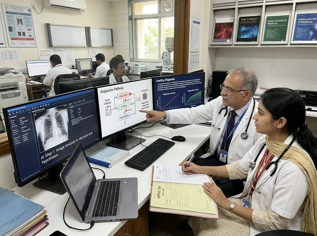
A breakthrough peer-reviewed study demonstrates that a sequential AI diagnosis system can mirror real-world clinical reasoning. By ordering stepwise tests and updating hypotheses dynamically, this multi-agent orchestrator achieved superior diagnostic accuracy while reducing investigative costs.
Today

A prenatal case highlights severe glutaric acidemia type II presenting as bilateral hyperechoic cystic kidneys at 18 weeks. Whole-exome sequencing revealed a novel homozygous ETFDH frameshift variant, demonstrating the power of rapid genetic testing in fetal renal anomalies.
Today

Review how tailored exercise programs benefit frail older patients with heart failure. Explore pathophysiological mechanisms, individualized training prescriptions, and strategies to overcome adherence barriers in clinical practice.
Yesterday

This comparative study evaluates posterior labral tears with and without glenohumeral instability, highlighting distinct demographic patterns, MRI underestimation in stable shoulders, and successful arthroscopic repair outcomes.
Yesterday

A clinical study demonstrates that early intervention with tocilizumab, either as monotherapy or bridged with corticosteroids, achieves significant proptosis reduction and resolves active inflammation in treatment-naive thyroid eye disease.
Yesterday
