

Loading, please wait...
Loading, please wait...

Glossopharyngeal neuralgia presents as severe, lancinating pain distributed along the sensory branches of the glossopharyngeal and vagus nerves. Although medical therapy provides initial symptomatic control, drug-resistant cases often require surgical intervention. Therefore, clinicians increasingly favor glossopharyngeal neuralgia MVD to address underlying vascular compression at the root entry zone of cranial nerves IX and X. Furthermore, a recent systematic review and meta-analysis offers robust evidence regarding offending vascular patterns and long-term surgical efficacy. Consequently, these pooled findings provide critical anatomical guidance for neurosurgeons operating in the cerebellomedullary cistern.
Glossopharyngeal neuralgia represents a rare yet severely disabling cranial rhizopathy. Patients experience intense paroxysmal electric-shock sensations in the posterior pharynx, base of the tongue, tonsillar pillar, and external auditory canal. In addition, routine physiological actions like swallowing, coughing, or speaking frequently trigger these excruciating pain episodes. In severe presentations, the glossopharyngeal-vagal reflex arc provokes profound bradycardia, hypotension, or syncope. Pharmacotherapy serves as the primary conservative approach. Anticonvulsants such as carbamazepine, oxcarbazepine, and gabapentin commonly stabilize neural membranes. However, many patients develop intolerable pharmacological toxicity or intractable pain breakthroughs over time. When conservative medical management fails, surgical intervention becomes mandatory. Surgeons may evaluate destructive options such as percutaneous rhizotomy or open rootlet section. Nevertheless, microvascular decompression remains the definitive curative modality because it preserves neurological function while correcting structural compression. Accurate preoperative evaluation is paramount for optimal outcomes. High-resolution magnetic resonance imaging demonstrates the course of neurovascular bundles within the posterior fossa. In particular, specialized steady-state sequences reveal detailed cistern architecture. However, radiographic findings do not always expose every conflicting vessel. Therefore, comprehensive surgical knowledge of anatomical patterns remains essential for successful decompressive procedures.
The landmark meta-analysis pooled nineteen observational studies comprising 1,000 surgical patients to determine offending vascular anatomy. Fifteen studies specifically detailed vessel-level pathology across 674 patients undergoing decompression. Interestingly, the investigation highlights marked differences in how various vascular structures impinge upon the glossopharyngeal-vagal complex. The posterior inferior cerebellar artery emerged as the overwhelming primary culprit. Specifically, the posterior inferior cerebellar artery was implicated, either alone or alongside other vessels, in 87% of patients. This estimate exhibited minimal heterogeneity across published series, demonstrating remarkable anatomical consistency. The vertebral artery served as the second most common arterial offender, affecting 12% of surgical cases. In addition, the anterior inferior cerebellar artery caused compression in 7% of treated individuals. Venous compression represented a much smaller surgical subgroup. Specifically, isolated or contributory venous conflicts occurred in only 3% of patients. Venous conflicts often require delicate mobilization or selective coagulative sacrifice to relieve mechanical tension. Moreover, the meta-analysis documented that 14% of patients presented with multiple offending vessels simultaneously. Consequently, relying on a single vascular culprit can lead to incomplete decompression. Neurosurgeons must thoroughly inspect the entire root entry zone to identify secondary conflicting branches.
Understanding the biomechanical behavior of the posterior inferior cerebellar artery is critical during skull base dissection. This cerebellar artery often forms an elongated upward loop that enters the cerebellomedullary cistern. Consequently, the loop impinges directly against the rootlets of cranial nerve IX and the superior rootlets of cranial nerve X. Furthermore, severe arterial elongation can trap the delicate cranial nerves against the skull base or brainstem surface. In 14% of cases, patients harbor compound conflicts involving more than one vessel. For instance, an elongated vertebral artery frequently displaces the origin of the posterior inferior cerebellar artery. This dynamic pushes both arterial vessels directly into the glossopharyngeal root exit zone. In such scenarios, decompressing only one artery fails to relieve the mechanical distortion completely. Therefore, the operating surgeon must untether both vascular structures from the brainstem. Moreover, arachnoid adhesions frequently encase the neurovascular conflict, creating substantial mechanical rigidity. Surgeons must dissect these thick arachnoid bands sharply to ensure complete mobilization. However, aggressive retraction of the cerebellum or brainstem can compromise fragile perforating branches that supply the medulla oblongata. Thus, maintaining continuous visualization of brainstem perforators prevents ischemic complications during vessel transposition. Using specialized sling techniques or inert shredded Teflon felt provides durable separation while preserving vital microvascular blood flow.
Microvascular decompression yields outstanding therapeutic results for medically refractory glossopharyngeal neuralgia. The systematic review revealed that immediate postoperative pain relief occurred in 94% of patients. This remarkable relief demonstrates the profound curative capability of direct anatomical decompression. Patients typically experience complete cessation of their excruciating paroxysms immediately after waking from general anesthesia. Furthermore, long-term therapeutic durability remains highly favorable across evaluated surgical cohorts. Across a weighted mean follow-up period of 66 months, approximately 90% of patients maintained satisfactory pain freedom. This long-lasting efficacy highlights that resolving the mechanical neurovascular conflict addresses the true root cause of the disease. In contrast, medical therapies merely suppress symptoms and frequently lose efficacy over prolonged periods. Nevertheless, late symptom recurrence can occur in a small minority of surgical cases. The pooled recurrence rate stood at only 4% across long-term follow-up intervals. Recurrence typically stems from incomplete initial decompression, graft migration, or progressive vascular elongation. In addition, postoperative arachnoid scar formation can generate renewed tethering of the nerve roots. If significant symptoms recur, neurosurgeons must re-evaluate patients thoroughly with advanced neuroimaging. Repeat exploration or adjuvant stereotactic radiosurgery can provide durable salvage relief for recurrent presentations.
Despite manipulating critical posterior fossa neurovascular structures, microvascular decompression exhibits an acceptable modern safety profile. In the meta-analysis of 1,000 surgical procedures, overall mortality was 0.5%, representing five reported deaths. Importantly, four of these five fatalities occurred in earlier surgical eras characterized by older microsurgical instrumentation. Modern neurosurgical techniques, refined illumination, and advanced neuroanesthesia have significantly reduced life-threatening risks. However, cranial nerve morbidity remains a recognized clinical consideration that requires vigilant surgical technique. Because cranial nerves IX and X mediate swallowing, phonation, and airway protection, rootlet manipulation can cause temporary deficits. Patients occasionally experience transient dysphagia, hoarseness, or diminished gag reflex postoperatively. Consequently, clinicians must evaluate swallowing function carefully before resuming oral intake to prevent pulmonary aspiration. Fortunately, permanent lower cranial nerve deficits remain uncommon in experienced centers. Cerebellar retraction injury and cerebrospinal fluid leaks also represent potential perioperative complications. To minimize cerebellar trauma, surgeons employ dynamic retraction rather than continuous fixed retractors. In addition, meticulous water-tight dural closure with autologous grafts or synthetic sealants prevents postoperative pseudomeningocele formation. Hemodynamic instability can also arise intraoperatively due to glossopharyngeal and vagal stimulation. Therefore, continuous intraoperative neurophysiological monitoring and tight coordination with neuroanesthesiologists ensure patient safety throughout the operative intervention.
The synthesized findings offer critical practical insights for practicing neurosurgeons and multidisciplinary clinical teams. First, surgeons must recognize that vascular conflict is rarely restricted to isolated or obvious single compression points. Because 14% of patients have multiple compressing vessels, surgeons should avoid terminating exploration upon finding the first conflict. Instead, complete microvascular inspection along the entire rootlet trajectory from the brainstem to the jugular foramen is necessary. Second, the relationship between the glossopharyngeal nerve and vagal rootlets requires meticulous handling. Because the upper vagal rootlets contribute significantly to pharyngeal sensation and motor reflex arcs, compressing vessels frequently distort both nerves. Therefore, gentle mobilization of vascular loops away from the glossopharyngeal-vagal complex is critical. Transposition should maintain vascular patency without creating acute angulation or stretching of perforating medullary branches. Finally, long-term postoperative surveillance remains an integral component of comprehensive patient management. Although 90% of patients maintain durable pain relief over five years, late recurrences require timely intervention. Establishing multidisciplinary collaboration between neurologists, otolaryngologists, and neurosurgeons optimizes diagnostic precision and perioperative safety. Consequently, patients with drug-refractory neuralgic pain can achieve sustained symptom relief and improved quality of life through standardized microsurgical decompression.
The posterior inferior cerebellar artery is the primary offending vessel, identified in 87% of patients undergoing microvascular decompression. Other vascular culprits include the vertebral artery in 12%, the anterior inferior cerebellar artery in 7%, and venous compression in 3%. Multiple compressing vessels occur in 14% of surgical cases.
Microvascular decompression demonstrates high surgical efficacy, with 94% of patients experiencing immediate pain relief postoperatively. Long-term follow-up across a weighted mean of 66 months confirms sustained pain relief in 90% of individuals. The pooled recurrence rate remains low at 4%, supporting microvascular decompression as an effective definitive treatment.
Neurosurgeons must inspect the entire glossopharyngeal-vagal root entry zone because approximately one in seven patients has multiple offending vessels. Relying solely on the first identified vascular loop risks leaving secondary compressive vessels untreated. Complete anatomical exploration ensures comprehensive neurovascular decompression and minimizes the likelihood of persistent pain or early recurrence.
Disclaimer: This content is for informational and educational purposes only... Refer to the latest local and national guidelines for clinical practice.
References

Read summarized clinical updates, watch expert medical content, and earn CME certifications right from your smartphone.
Open your smartphone camera to scan and install.





A systematic review and meta-analysis of 1,000 patients reveals that the posterior inferior cerebellar artery causes 87% of neurovascular conflicts in glossopharyngeal neuralgia. Microvascular decompression achieved 94% immediate pain relief and 90% long-term freedom with low complication rates.
Today

The 2025 Australian meningococcal surveillance report shows a 13% decline in invasive meningococcal disease notifications to 118 cases. Genogroup B accounted for 82.2% of typed infections, predominantly impacting infants and young adults, highlighting critical vaccination and rapid diagnostic imperatives.
Today
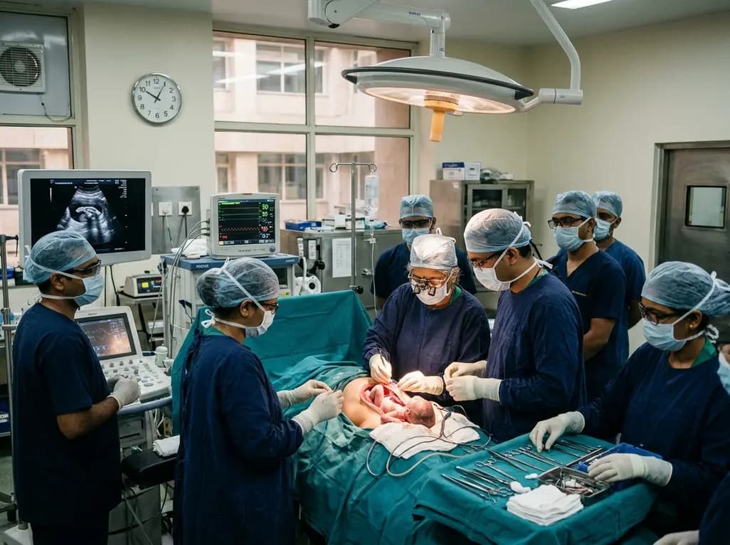
Doctors at AIIMS New Delhi have achieved a historic milestone by performing India's first open prenatal myelomeningocele repair on a 25-week unborn fetus. This landmark multidisciplinary procedure provides vital neuroprotective benefits, lowers hydrocephalus risks, and transforms maternal-fetal medicine nationwide.
Today

The ACE-PAS registry evaluated 21,612 patients fitted with the ASSURE wearable cardioverter defibrillator. The study demonstrated a 100% ventricular arrhythmia conversion rate, an inappropriate shock incidence of 0.0065 per patient-month, and high daily compliance of 23.1 hours, confirming real-world safety.
Today

A clinical overview of resveratrol supplementation in mitigating exercise-induced muscle damage. This narrative review explores physiological mechanisms, reductions in soreness markers like creatine kinase, dosage strategies, and bioavailability enhancements for sports medicine and nutrition practitioners.
Today

Childhood insulinoma is a rare cause of recurrent hyperinsulinemic hypoglycemia that often eludes early diagnosis. Advanced GLP-1 receptor-targeted PET/CT imaging using 68Ga-exendin-4 complements anatomic MRI to localize occult pancreatic lesions, facilitating focused, curative surgical enucleation.
Today
