.webp&w=384&q=75)

Loading, please wait...
Loading, please wait...
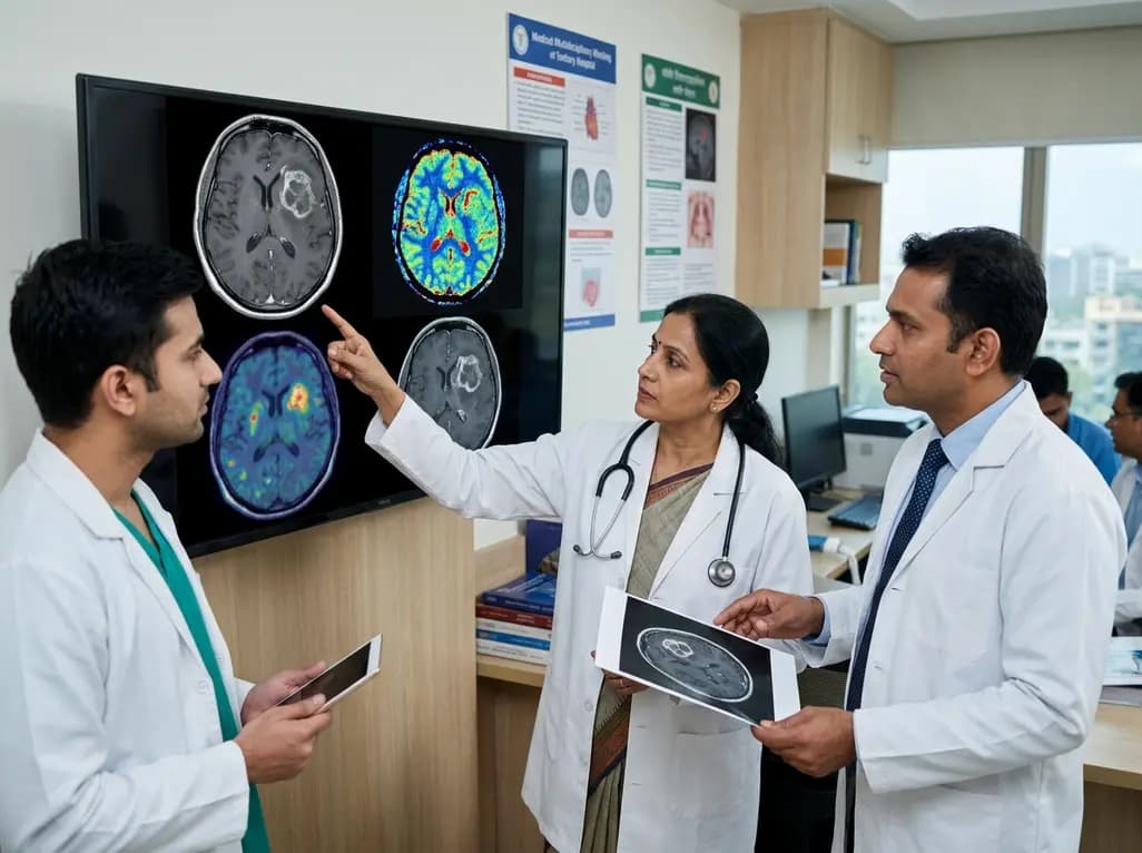
Evaluating disease status in patients with glioblastoma after chemoradiation remains one of the most challenging tasks in neuro-oncology. Differentiating glioblastoma true progression from benign treatment-induced changes—such as pseudoprogression and radiation necrosis—is crucial for guiding therapy. Conventional magnetic resonance imaging (MRI) frequently falls short, failing to deliver clear diagnostic certainty in up to 40% of post-treatment evaluations. Because false interpretation can lead to premature cessation of effective therapy or unnecessary exposure to toxic treatments, clinicians require a structured, evidence-graded approach. This comprehensive article reviews an updated diagnostic framework designed to refine pretest probability, optimize advanced imaging modalities, and foster multidisciplinary decision-making.
Conventional contrast-enhanced MRI relies heavily on blood-brain barrier disruption to identify tumor recurrence. However, radiation and concomitant temozolomide chemotherapy frequently disrupt the microvasculature, generating transient contrast enhancement that closely mimics tumor growth. Pseudoprogression typically manifests within the first three to six months following chemoradiation completion. Conversely, radiation necrosis usually presents as a late treatment effect, developing months to years after high-dose radiotherapy.
In approximately 30% to 40% of ambiguous post-treatment cases, standard T1-weighted contrast-enhanced and T2-FLAIR sequences cannot reliably distinguish viable neoplastic tissue from treatment-induced inflammation. Mistaking pseudoprogression for genuine disease advancement might cause clinicians to discontinue beneficial adjuvant temozolomide prematurely. On the other hand, misinterpreting progressive tumor growth as pseudoprogression delays necessary second-line interventions, including surgical resection, re-irradiation, or alternative systemic agents.
Given these diagnostic hurdles, relying on a single imaging modality or an isolated specialty opinion often yields sub-optimal patient care. Successful evaluation requires integrating longitudinal clinical trajectories, molecular tumor profiles, and advanced functional neuroimaging. Neuro-oncology teams must actively synthesize data across neurosurgery, neuroradiology, radiation oncology, and medical oncology to establish an accurate diagnosis and tailor treatment strategies effectively.
To address diagnostic ambiguity, experts have proposed a tiered, step-wise imaging framework to evaluate suspected glioblastoma true progression. The initial evaluation begins with Tier 1 standard RANO 2.0 criteria using contrast-enhanced MRI. If conventional imaging yields equivocal findings, clinicians escalate to Tier 2 advanced neuroimaging, which incorporates perfusion-weighted MRI, diffusion-weighted imaging, and MR spectroscopy. Perfusion MRI measures relative cerebral blood volume (rCBV), where elevated hyperperfusion indicates hypervascular tumor tissue, whereas hypoperfusion supports treatment effects.
When Tier 2 findings remain inconclusive, Tier 3 amino acid positron emission tomography (PET) offers metabolic clarification. Amino acid radiotracers, such as fluoroethyl-L-tyrosine (FET) or F-fluciclovine, exploit upregulated amino acid transporters in active glioma cells, offering superior target-to-background contrast compared to FDG-PET. Finally, if non-invasive imaging across all tiers fails to yield definitive answers, Tier 4 involves surgical tissue sampling or closely monitored empirical therapy.
This sequential structure helps clinicians systematically gather evidence while avoiding unnecessary invasive procedures. However, researchers emphasize that this tiered framework serves as a prospective evaluation structure rather than an absolute clinical decision tool. Clinicians must weigh each diagnostic tier alongside clinical context to ensure optimal care.
A central pillar of the new framework involves modifying pretest probability based on key patient-specific clinical parameters. MGMT (O-6-methylguanine-DNA methyltransferase) promoter methylation status represents a critical genetic modifier. Tumors with MGMT promoter methylation demonstrate enhanced sensitivity to temozolomide, which significantly increases the likelihood of pseudoprogression compared to unmethylated tumors.
The time interval since completing chemoradiation also dramatically influences diagnostic suspicion. Contrast enhancement appearing within twelve weeks of completing radiotherapy strongly favors pseudoprogression. In contrast, new enhancing lesions arising six months or more after treatment carry a far higher pretest probability of true recurrence.
Additional clinical modifiers include neurologic trajectory, concurrent medication exposure, and lesion location. Patients who remain clinically stable or show symptomatic improvement despite radiological worsening are more likely experiencing treatment effects. Furthermore, prior exposure to antiangiogenic agents like bevacizumab or immune checkpoint inhibitors alters imaging characteristics, generating rebound enhancement or immune-related inflammatory changes. Previous re-irradiation history and deep structural lesion locations further complicate interpretation, requiring careful contextual integration before assigning diagnostic certainty.
Diagnostic accuracy studies demonstrate that combining advanced MRI techniques with amino acid PET yields impressive diagnostic performance. Literature syntheses report areas under the receiver operating characteristic curve (AUC) ranging from 0.90 to 0.95 for combined advanced MRI and amino acid PET. In contrast, conventional MRI alone achieves AUC values between 0.65 and 0.72. These statistics highlight the superior discriminatory capability of metabolic and physiological imaging.
However, clinical experts urge cautious interpretation of these diagnostic performance metrics. Many published studies evaluate broad patient cohorts containing straightforward, easily classified cases alongside complex ones. Consequently, reported AUC values likely overstate real-world diagnostic accuracy when applied exclusively to the ambiguous subset of patients who actually require advanced imaging.
Access to amino acid PET radiotracers also remains a practical hurdle, particularly within specific geographic regions and community practice settings. While European guidelines routinely incorporate FET-PET, access in the United States has historically faced regulatory and logistical limitations. Recent prospective, multicenter diagnostic-accuracy data for alternative agents, such as F-fluciclovine, are expanding clinical access. Identifying true progression conditional on equivocal perfusion MRI remains a critical knowledge gap that ongoing multicenter prospective trials aim to resolve.
Managing post-chemoradiation glioblastoma effectively requires robust collaboration within a multidisciplinary tumor board. Neuroradiologists, neuro-oncologists, radiation oncologists, and neurosurgeons must collectively evaluate clinical trends, molecular markers, and advanced neuroimaging. Because no single diagnostic technique offers absolute sensitivity and specificity, consensus discussion reduces diagnostic errors and prevents premature treatment termination.
Despite significant technological progress, several unresolved diagnostic gaps persist in neuro-oncology literature. The most prominent unresolved question centers on how amino acid PET performs specifically after a perfusion MRI returns equivocal findings. While advanced PET imaging demonstrates high overall sensitivity, its incremental diagnostic value in the exact subset of perfusion-ambiguous lesions requires further prospective validation.
Furthermore, integrating radiomics and artificial intelligence machine-learning algorithms into standard clinical workflows offers promising avenues for quantitative image analysis. Ongoing prospective trials aim to standardise radiomic feature extraction and validate automated predictive models against histopathological benchmarks. Until these prospective trials conclude, multidisciplinary teams should continue using evidence-graded, tiered frameworks to synthesize clinical modifiers, perfusion metrics, and metabolic imaging for every patient.
MGMT promoter methylation silences DNA repair enzymes, increasing tumor sensitivity to temozolomide chemoradiation. This heightened therapeutic responsiveness induces local tissue inflammation and vascular permeability, resulting in a significantly higher frequency of pseudoprogression compared to unmethylated glioblastomas.
Conventional contrast-enhanced MRI relies on blood-brain barrier disruption rather than true cellular proliferation. Because radiation and chemotherapy disrupt vascular integrity, treatment-induced necrosis and inflammation produce contrast enhancement indistinguishable from active tumor progression in up to 40% of cases.
Amino acid PET radiotracers target active amino acid transporters that are highly overexpressed in glioma cells. Because normal brain parenchyma exhibits low baseline amino acid uptake, amino acid PET delivers superior tumor-to-background contrast compared to FDG-PET, which suffers from high normal cortical glucose metabolism.
Disclaimer: This content is for informational and educational purposes only. It does not constitute formal medical advice, diagnosis, or treatment recommendations. Clinical decisions in neuro-oncology require individualized assessment by qualified healthcare professionals based on multidisciplinary team evaluation. Refer to the latest local and national guidelines for clinical practice.
References

Read summarized clinical updates, watch expert medical content, and earn CME certifications right from your smartphone.
Open your smartphone camera to scan and install.





Distinguishing true progression from treatment effects in glioblastoma after chemoradiation remains a complex clinical challenge. This article reviews a practical, evidence-graded imaging framework that integrates clinical modifiers, advanced MRI, and amino acid PET to optimize multidisciplinary decision-making.
last month

A quality-improvement study reveals how hospital pharmacy leadership improves medication safety at home. By eliminating concentrated electrolytes, standardizing kits, and auditing drug labeling, the model successfully aligned home acute care with international patient safety standards.
Today
A recent case-control study identified key protein changes in peritoneal fluid-derived small extracellular vesicles, highlighting candidates like S100-A10 and ALDOA as potential biomarkers to overcome diagnostic delays in endometriosis.
Today

Managing refractory hypoxemia after cardiothoracic surgery becomes perilous when prolonged air leak limits positive-pressure ventilation. This case-based review details how high-flow nasal cannula, paired with awake rehabilitation, enabled successful extubation and avoided invasive re-intubation.
Today

Idiopathic axillary web syndrome (IAWS) is a rare cause of shoulder pain and mobility restriction characterized by palpable axillary cording without prior surgery or trauma. Early diagnosis, nonsteroidal anti-inflammatory therapy, and physical rehabilitation lead to complete resolution of symptoms.
Today

SWEDEPAD-1 trial insights show paclitaxel-coated devices do not improve long-term limb salvage in patients with chronic limb-threatening ischemia and tissue loss. While one-year reinterventions decreased, the devices were linked to a higher risk of major amputation at three months.
Today
