

Loading, please wait...
Loading, please wait...

Glioblastoma remains the most aggressive primary malignant brain tumor encountered in neuro-oncology. In particular, isocitrate dehydrogenase wild-type glioblastoma carries a notoriously poor prognosis despite aggressive maximal surgical resection, radiotherapy, and temozolomide chemotherapy. Intratumoral heterogeneity and treatment resistance continue to pose formidable barriers to effective clinical management. To overcome these therapeutic challenges, researchers have increasingly investigated the tumor microenvironment to understand how malignant cells interact with host neural and immune cells. A breakthrough multi-institutional investigation has now decoded the glioblastoma spatial landscape using GBmap, an expansive single-cell and spatially resolved transcriptomic atlas. Consequently, this benchmark resource provides unprecedented biological insights into glioblastoma architecture, cellular plasticity, and patient survival outcomes.
Historically, genomic profiling categorized glioblastomas into bulk transcriptional subtypes. However, bulk tissue sequencing inevitably obscures profound single-cell diversity and spatial organization. The revolutionary GBmap platform resolves this limitation by combining deep computational harmonization with high-resolution spatial transcriptomics. Specifically, the authors harmonized twenty-six distinct transcriptomic cohorts into a unified data reference. This framework allows researchers to perform label transfer, reference mapping, and cellular annotation with exceptional fidelity. Moreover, investigators can project newly sequenced clinical cohorts onto this reference map seamlessly. Therefore, researchers can rapidly characterize unstudied patient samples without repeating massive integration workflows. Furthermore, the atlas preserves critical biological variance while eliminating technical batch effects across independent laboratories. By systematically organizing over one million single cells, the study establishes an enduring foundation for translational neuro-oncology. Clinicians and scientists can now interrogate how distinct cellular phenotypes organize across intact tumor tissue. Thus, GBmap transforms our conceptual understanding of malignant brain tumor architecture. Importantly, this resource bridges the persistent gap between computational biology and clinical practice. Neuropathologists can evaluate molecular diversity alongside histopathological features to understand therapy resistance.
To build this comprehensive reference, the study integrated single-cell RNA sequencing data from over 1.1 million individual cells derived from 240 glioblastoma patients. Consequently, the sheer scale of GBmap empowered the authors to discover rare cell states that standard studies consistently overlook. For instance, the analysis successfully uncovered rare populations of tumor-associated neutrophils infiltrating the microenvironment. In addition, the team identified distinct states of homeostatic microglia alongside pro-inflammatory and immunosuppressive myeloid cells. Traditionally, researchers struggled to distinguish native brain microglia from circulating bone marrow-derived macrophages. However, GBmap provides explicit transcriptional definitions that clearly segregate these diverse myeloid compartments. Furthermore, neoplastic cells displayed diverse transcriptional states matching neural progenitor-like, oligodendrocyte progenitor-like, astrocyte-like, and mesenchymal-like programs. These malignant states coexist dynamically within the same patient specimen. Because plastic glioma cells switch dynamically between programs under therapeutic pressure, therapeutic targeting remains notoriously difficult. Therefore, capturing this granular continuum of cellular states proves essential for designing effective multi-targeted therapies. Ultimately, this comprehensive harmonization illuminates previously invisible immunological and neoplastic subsets inside lethal human brain tumors.
Beyond single-cell cataloging, spatial transcriptomics provided crucial positional context for understanding cellular interactions in glioblastoma. By resolving tissue sections spatially, the investigators uncovered seven recurrent and distinct tumor niches. Each niche possesses a unique microenvironment governed by specific cell types, extracellular matrix components, and signaling pathways. Importantly, hypoxia-dependent niches emerged as the most critical determinants of disease aggression. These hypoxic compartments harbor necrotic foci and hyperplastic microvasculature, mimicking classic pseudopalisading necrosis seen on histology. Moreover, mesenchymal-like tumor cells heavily concentrate within these hypoxic environments. In contrast, neural progenitor-like cells predominantly inhabit well-perfused, non-hypoxic cortical zones. The study revealed that hypoxia directly reprograms malignant cells toward stem-like and invasive phenotypes. Furthermore, these oxygen-deprived regions recruit immunosuppressive myeloid populations that deactivate infiltrating cytotoxic T cells. As a result, hypoxia-driven niches generate potent localized barriers against host immune surveillance and standard chemoradiation. Consequently, patients exhibiting high frequencies of hypoxia-dominated niches suffer significantly worse overall survival. Understanding these niche-specific dynamics therefore exposes novel opportunities to combine hypoxia-activated prodrugs or angiogenesis inhibitors with modern immunotherapy regimens.
To translate spatial observations into measurable clinical metrics, the investigators engineered an innovative computational metric known as the Tumor Structure Score. This score mathematically quantifies the spatial organization, vascular patterning, and niche compartmentalization of glioblastoma tissue. Notably, higher scores reflect highly structured tumor architecture featuring distinct microvascular proliferation and prominent hypoxic boundaries. In clinical correlations, tumors demonstrating high organization exhibited significantly worse survival outcomes compared to less organized counterparts. Although counterintuitive, this finding indicates that structured tumor architecture represents an evolutionary adaptation that supports aggressive malignant growth. Specifically, structured niches protect stem-like glioma cells from therapeutic assault while facilitating efficient metabolic adaptation. Meanwhile, disorganized tumors display lower levels of coordinated niche protection and altered treatment resistance patterns. Thus, the Tumor Structure Score provides prognostic value that surpasses standard histopathological grading alone. Clinicians can utilize this metric to identify patients who harbor aggressive hypoxia-driven architecture upon initial resection. In the near future, integrating this scoring paradigm into routine neuropathological evaluation could refine risk stratification. Accordingly, oncologists could tailor postoperative monitoring frequencies and clinical trial eligibility based on individual tumor architecture.
The discoveries generated by GBmap introduce exciting translational avenues for overcoming therapeutic resistance in malignant glioma. Currently, standard care for isocitrate dehydrogenase wild-type glioblastoma relies on surgery, radiation, and temozolomide. However, recurrent tumors invariably emerge because residual infiltrating cells survive in protective spatial sanctuaries. By delineating cell-cell communication networks within distinct niches, researchers can pinpoint specific ligand-receptor interactions that promote tumor cell survival. For example, interrupting signaling pathways between immunosuppressive neutrophils, macrophages, and hypoxic glioma cells could re-sensitize resistant tumors to immunotherapy. In addition, spatial mapping identifies targetable metabolic vulnerabilities unique to hypoxic microenvironments, including carbonic anhydrase and hypoxia-inducible factor signaling pathways. Furthermore, neurosurgeons can correlate advanced intraoperative imaging with spatial niche patterns to optimize surgical margins. Medical oncologists can also leverage transfer learning tools from GBmap to accelerate prospective clinical trial biomarker discovery. Ultimately, resolving the cellular composition and structural organization of glioblastoma moves neuro-oncology closer to personalized therapeutic strategies. Rather than treating glioblastoma as a uniform entity, clinicians can target the specific spatial microenvironments that sustain treatment resistance.
GBmap serves as a harmonized single-cell and spatial reference atlas integrating data from 240 patients and over 1.1 million cells. Furthermore, it enables reference mapping and transfer learning. Consequently, researchers can accurately annotate cellular subsets, investigate intercellular crosstalk, and analyze patient-specific tumor microenvironments without repeating complex bioinformatic processing.
The Tumor Structure Score quantifies the spatial organization, hypoxic boundaries, and microvascular architecture of glioblastoma tissue. Notably, higher scores indicate highly organized tumor compartments. Because organized hypoxic niches foster therapy-resistant glioma stem cells and immune evasion, patients with elevated tumor structure scores experience significantly worse overall survival than those with disorganized tumors.
Hypoxia-dependent niches represent critical drivers of glioblastoma aggression and treatment failure. Specifically, severe oxygen deprivation induces mesenchymal transition, stimulates abnormal microvascular proliferation, and recruits immunosuppressive myeloid cells. Consequently, these protective niches shield malignant glioma cells from chemoradiation and host immune attacks, driving persistent local recurrence and poor patient prognosis.
Disclaimer: This content is for informational and educational purposes only... Refer to the latest local and national guidelines for clinical practice.
References

Read summarized clinical updates, watch expert medical content, and earn CME certifications right from your smartphone.
Open your smartphone camera to scan and install.





GBmap integrates single-cell RNA sequencing from over 1.1 million cells across 240 glioblastoma patients. By pairing single-cell resolution with spatial transcriptomics, researchers mapped seven cellular niches and established the Tumor Structure Score to quantify tissue architecture and predict patient survival.
Today

Novel glucose-responsive mPEG-PSA nanocarriers deliver insulin aspart on demand, achieving dual postprandial and basal glycemic control without hypoglycemia in diabetic models. This biomaterial breakthrough offers promising clinical implications for physiologic diabetes management.
Today
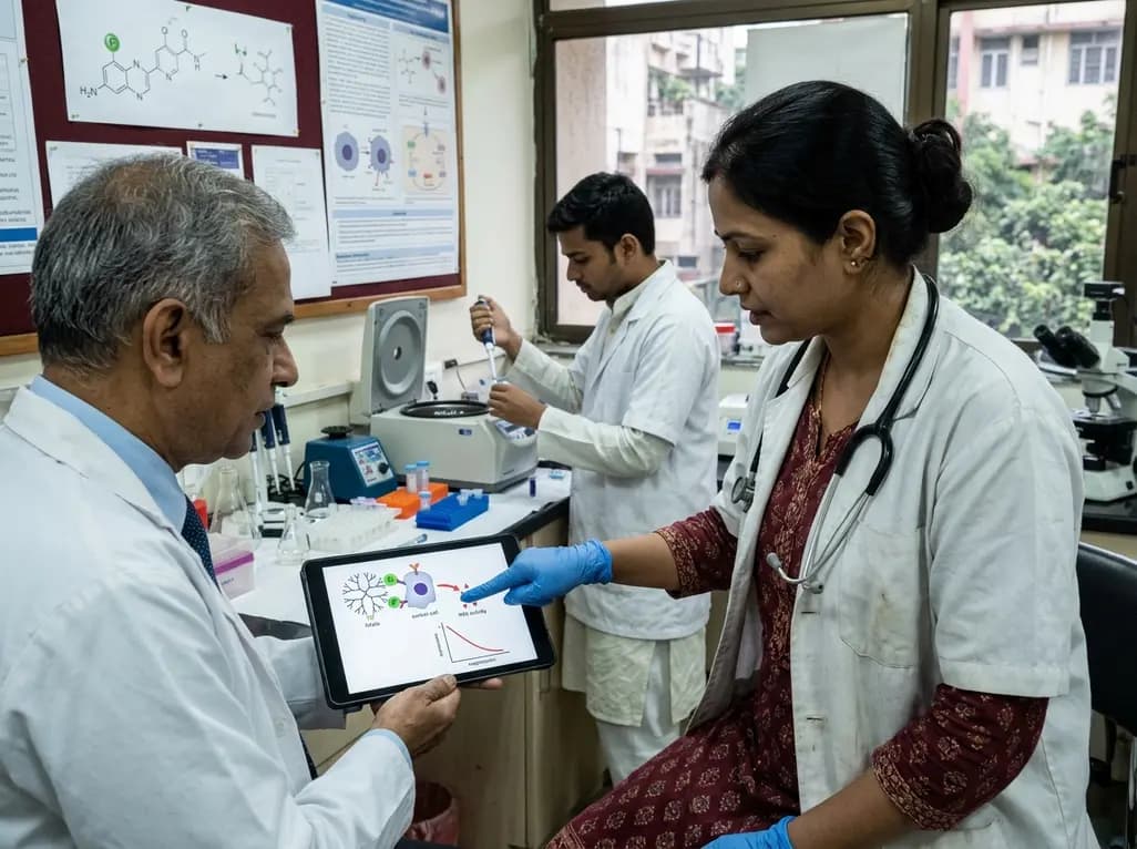
A novel folate-conjugated tecto-dendrimer nanoplatform successfully delivers siRNA to achieve targeted HK2 silencing, disrupting ovarian cancer glycolysis and suppressing lactate output.
Today

A comparative non-contact study reveals that high-power radiofrequency delivery with the QDOT MICRO catheter induces an artifactual contact force rise. Electrophysiologists must understand this power-dependent thermal and electrical phenomenon to avoid inadvertent catheter withdrawal and ineffective ablation.
Today

A novel proof-of-concept study presents a six-degree-of-freedom articulated manikin neck equipped with linear potentiometers. The system provides real-time kinematic motion feedback during cervical immobilisation, delivering sub-degree tracking accuracy to improve pre-hospital trauma training and patient safety.
Today

A retrospective comparative cohort study evaluated genitourinary safety in HFpEF between dapagliflozin and empagliflozin over six months. Rates of symptomatic UTI and asymptomatic bacteriuria showed no statistically significant differences, supporting comparable safety profiles across both SGLT2 inhibitors.
Today
