

Loading, please wait...
Loading, please wait...

Modern neurovascular specialists recognize that giant intracranial aneurysm treatment represents one of the most formidable frontiers in clinical neurosurgery. By definition, a giant intracranial aneurysm measures 25 millimeters or greater at its maximal diameter. Consequently, these massive vascular lesions impose catastrophic clinical risks, including fatal subarachnoid hemorrhage and severe local mass effect. Furthermore, the natural history of untreated giant aneurysms remains notoriously poor. Untreated patients routinely face progressive neurological decline or death within several years of diagnosis. However, neurosurgeons face extraordinary technical hurdles during both open surgical clipping and endovascular intervention. The sheer volume of the aneurysmal sac distorts surrounding cerebral architecture. In addition, extensive intraluminal thrombus, calcification, and fragile arterial walls frequently prevent straightforward neck clipping. Standalone endovascular coiling also rarely achieves permanent obliteration because large luminal volumes permit recurrent coil compaction. Therefore, neurovascular teams must carefully avoid rigid, uniform management protocols. Instead, multidisciplinary specialists must tailor every intervention to individual vascular anatomy, collateral circulation, and baseline neurological status. Ultimately, patient-specific treatment planning offers the most reliable strategy to prevent devastating hemorrhage and preserve neurological function.
Recent retrospective evidence spanning seven years highlights the complex clinical presentation of giant intracranial lesions. In this single-center cohort, the mean maximal aneurysm diameter reached 29.4 millimeters, excluding rare serpentine lesions. The majority of lesions originated along the internal carotid artery, while the middle cerebral and anterior cerebral arteries comprised the remainder. Morphologically, saccular configurations predominated, whereas serpentine structures appeared in a smaller subset of patients. In addition, seventy percent of patients presented with severe acute or progressive symptoms. Specifically, acute subarachnoid hemorrhage occurred in forty percent of individuals, with over sixty percent of these experiencing concurrent intracerebral hemorrhage. This high incidence of parenchymal hematoma illustrates the immense mechanical stress exerted during aneurysmal rupture. Furthermore, compression of adjacent cranial nerves caused cranial neuropathies in numerous internal carotid artery cases. Several patients also presented with direct carotid-cavernous fistulas. Notably, twenty-five percent of patients harbored multiple intracranial aneurysms alongside the primary giant lesion. Consequently, neurovascular teams must perform comprehensive six-vessel angiography and advanced magnetic resonance imaging during initial workups to map complex vascular networks and collateral pathways accurately.
Determining the appropriate intervention requires balanced evaluation between open microsurgical techniques and endovascular embolization. In the 7-year cohort, forty percent of patients initially underwent direct microsurgical management. Open surgery offers immediate vascular exclusion and relieves debilitating mass effect on nearby brain parenchyma. However, wide neck anatomy, calcified arterial walls, and severe vessel tortuosity frequently preclude simple surgical clipping. In contrast, endovascular specialists utilize advanced flow-diverting stents and coil embolization systems to reconstruct diseased segments. These innovative catheter-based devices redirect blood flow and promote progressive aneurysmal thrombosis while preserving adjacent perforating branches. Nevertheless, endovascular flow reconstruction introduces persistent risks of delayed rupture and transient perilesional edema during thrombus formation. Therefore, neurosurgeons and interventional neuroradiologists increasingly collaborate to design individualized hybrid strategies. For instance, teams frequently evacuate acute intracranial hematomas surgically before performing endovascular coil embolization. In addition, clinicians carefully assess patient age, systemic comorbidities, and baseline clinical grade when selecting therapeutic modalities. Overall, unbiased multidisciplinary collaboration between microsurgeons and interventionalists optimizes procedural safety and procedural efficacy across diverse patient cohorts.
When direct reconstruction remains anatomically unfeasible, deconstructive strategies provide a reliable therapeutic solution. In the retrospective study, clinicians selected parent artery occlusion, performed with or without bypass surgery, as the final approach in forty percent of patients. Parent artery sacrifice eliminates hemodynamic stress within the aneurysm sac, effectively preventing catastrophic rupture. However, permanent occlusion of a major cerebral trunk introduces significant risk of devastating cerebral infarction. Consequently, rigorous hemodynamic testing remains essential prior to vessel sacrifice. Specialists utilize balloon occlusion tests and quantitative perfusion imaging to evaluate native collateral circulation. When collateral capacity proves insufficient, neurosurgeons construct extracranial-to-intracranial bypasses, including superficial temporal artery-to-middle cerebral artery revascularization or high-flow saphenous vein grafts. Furthermore, modern microvascular techniques maintain excellent long-term bypass graft patency. Thus, surgical revascularization restores vital distal cerebral perfusion before the operator occludes the diseased parent artery. In addition, parent artery occlusion combined with targeted vascular bypass promotes progressive aneurysm shrinkage, which relieves cranial nerve compression. Therefore, combining microvascular bypass with therapeutic vessel sacrifice represents an indispensable neurosurgical strategy for complex vascular lesions.
Managing complex giant aneurysms frequently necessitates staged, sequential interventions rather than a single definitive procedure. In this retrospective review, thirty percent of patients required two or three treatment steps to achieve complete vascular isolation. For example, neurovascular teams frequently secure an acute rupture site during initial emergency surgery and complete formal flow diversion weeks later. Similarly, interventionalists often perform preliminary coil reduction followed by staged parent artery sacrifice. Notably, this tailored, staged management paradigm yielded favorable clinical outcomes in seventy percent of patients at one-year follow-up, defined by a modified Rankin Scale score of two or lower. These robust results demonstrate that planned procedural staging safely controls surgical risk without compromising functional recovery. Furthermore, long-term angiographic follow-up confirmed stable aneurysm exclusion across the treated population. Nevertheless, ten to fifteen percent of patients experienced procedural complications, including thromboembolism and transient neurological deficits. Therefore, rigorous post-intervention surveillance in specialized neurointensive care units remains vital. Dedicated neurocritical teams optimize blood pressure targets and monitor cerebral hemodynamics to mitigate ischemic risks. Ultimately, systematic follow-up ensures durable vascular obliteration and functional independence.
Clinicians define a giant intracranial aneurysm as any cerebral aneurysm measuring 25 millimeters or greater at its maximal diameter. These massive lesions frequently involve internal carotid or middle cerebral arteries. Furthermore, they carry severe risks of catastrophic rupture, cranial neuropathy, and parenchymal hemorrhage, demanding individualized neurovascular evaluation and advanced intervention.
Surgeons recommend parent artery occlusion with bypass revascularization when severe calcification, wide necks, or fusiform geometries preclude primary clipping or flow diversion. Furthermore, neurosurgeons perform preoperative balloon occlusion testing to confirm adequate collateral reserve. Constructing an extracranial-to-intracranial bypass prevents cerebral ischemia while achieving permanent, complete exclusion of the aneurysmal sac from systemic circulation.
Giant aneurysms often require staged interventions because acute presentations with hemorrhage require immediate life-saving stabilization before definitive reconstruction. In addition, neurovascular teams may combine initial coil reduction with subsequent flow diversion or surgical bypass. Staging reduces periprocedural complication rates, preserves critical perforating arteries, and allows brain tissue to adapt gradually to major hemodynamic shifts.
Disclaimer: This content is for informational and educational purposes only... Refer to the latest local and national guidelines for clinical practice.
References

Read summarized clinical updates, watch expert medical content, and earn CME certifications right from your smartphone.
Open your smartphone camera to scan and install.





A 7-year retrospective study examines treatment strategies and clinical outcomes for giant intracranial aneurysms. Findings highlight parent artery occlusion with bypass and tailored, staged surgical and endovascular interventions, achieving favorable 1-year outcomes in 70% of treated patients.
Today
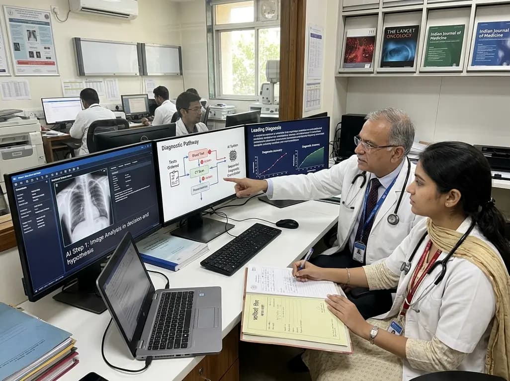
A breakthrough peer-reviewed study demonstrates that a sequential AI diagnosis system can mirror real-world clinical reasoning. By ordering stepwise tests and updating hypotheses dynamically, this multi-agent orchestrator achieved superior diagnostic accuracy while reducing investigative costs.
Today

A prenatal case highlights severe glutaric acidemia type II presenting as bilateral hyperechoic cystic kidneys at 18 weeks. Whole-exome sequencing revealed a novel homozygous ETFDH frameshift variant, demonstrating the power of rapid genetic testing in fetal renal anomalies.
Today

Review how tailored exercise programs benefit frail older patients with heart failure. Explore pathophysiological mechanisms, individualized training prescriptions, and strategies to overcome adherence barriers in clinical practice.
Yesterday

This comparative study evaluates posterior labral tears with and without glenohumeral instability, highlighting distinct demographic patterns, MRI underestimation in stable shoulders, and successful arthroscopic repair outcomes.
Yesterday

A clinical study demonstrates that early intervention with tocilizumab, either as monotherapy or bridged with corticosteroids, achieves significant proptosis reduction and resolves active inflammation in treatment-naive thyroid eye disease.
Yesterday
