.webp&w=384&q=75)

Loading, please wait...
Loading, please wait...

For decades, classic neuroanatomy treated hemispheric dominance as an immutable biological trait. However, modern neuroimaging demonstrates that functional connectivity language lateralization represents a highly dynamic network property. In most individuals, language functions predominantly reside within the left perisylvian network. Nevertheless, intrinsic connectivity analyses reveal that interhemispheric communication constantly modulates this lateralization. Functional magnetic resonance imaging captures these synchronous hemodynamic fluctuations between distributed cortical territories. Consequently, hemispheric specialization reflects coordinated interaction across complex cerebral networks rather than isolated regional modules. Neurobiologists now recognize that language architecture balances stable structural backbones with fluid functional interactions. Specifically, resting-state connectivity reveals that right-hemisphere homologues actively participate in linguistic computation. In addition, these bilateral circuits preserve communicative capacity while maintaining processing efficiency. Therefore, language lateralization functions as an adaptive, multi-tiered process rather than a static state. Moreover, modern analytical toolboxes quantify dynamic variations in connectivity strength across distinct temporal windows. Thus, language dominance manifests as an evolving equilibrium between left-hemisphere segregation and bilateral integration.
The establishment of hemispheric dominance follows a protracted developmental trajectory from infancy through adolescence. Initially, young children demonstrate bilateral functional connectivity across frontal and temporal language territories. However, synaptic pruning and progressive myelination gradually strengthen left-hemisphere pathways. Consequently, intrinsic intrahemispheric connections consolidate, driving progressive functional lateralization. Furthermore, early sensory experiences and rich linguistic immersion accelerate this network maturation. For instance, bilingual acquisition modulates dynamic connectivity patterns by demanding enhanced cognitive control. In bilingual individuals, the brain recruits bilateral prefrontal circuits to negotiate language switching. Therefore, developmental experience continually refines the spatial and temporal architecture of language processing. In contrast, atypical developmental pathways, such as autism spectrum disorders, often exhibit diminished leftward connectivity. Notably, disruptions in structural white matter tracts, including the arcuate fasciculus, directly influence this maturational trajectory. Moreover, neuroplasticity ensures that early brain maturation accommodates linguistic demands through dynamic network calibration. As a result, language lateralization reflects both genetic programming and environmental adaptation over decades.
Beyond long-term developmental changes, functional connectivity undergoes rapid, transient fluctuations across seconds and minutes. While global dominance remains relatively stable, local network recruitment adapts swiftly to immediate communicative demands. For example, demanding semantic ambiguity tasks prompt immediate recruitment of right-hemisphere homologues. Consequently, transient interhemispheric coupling increases to assist semantic selection and resolution. Moreover, dynamic resting-state fMRI studies capture spontaneous shifts in laterality without external stimuli. Researchers designate these microstate changes as spontaneous laterality fluctuations. Notably, higher frequency of moderate laterality fluctuations correlates positively with superior verbal performance and cognitive flexibility. In contrast, excessive or uncoordinated laterality reversals correlate with impaired executive control. Thus, healthy cognition depends upon a fine-tuned balance between stability and flexibility. Neuromodulatory systems, particularly ascending noradrenergic and dopaminergic projections, likely govern these rapid shifts. In addition, local metabolic demands continuously fine-tune transient regional synchrony. Therefore, short-term language lateralization acts as a flexible physiological repertoire. Furthermore, electrophysiological investigations verify that these transient state transitions occur across sub-second timescales. As a result, language networks adapt instantaneously to incoming syntactic and semantic complexities.
Pathological insults induce profound alterations in functional connectivity language lateralization across intermediate and prolonged timescales. Following an ischemic stroke in the left middle cerebral artery territory, acute aphasia emerges immediately. However, the injured brain rapidly initiates compensatory network reorganization. Initially, functional neuroimaging often demonstrates disinhibition and hyperactivation of contralateral right-hemisphere homologues. Consequently, transcallosal inhibitory pathways undergo significant remodeling after focal tissue infarction. Over subsequent months, successful speech recovery often requires re-establishing connectivity within preserved left-hemisphere perisylvian nodes. Similarly, chronic focal epilepsy dramatically alters intrinsic language network architecture. In patients with left temporal lobe epilepsy, recurring epileptiform discharges disrupt normal functional connectivity. Therefore, language dominance frequently shifts toward bilateral or atypical right-hemisphere representation. Moreover, non-invasive therapeutic neuromodulation can actively guide these compensatory network shifts. For example, repetitive transcranial magnetic stimulation selectively modulates excitability across interhemispheric language targets. Thus, clinicians witness substantial plastic reorganization following both destructive lesions and therapeutic interventions. In addition, speech therapy reinforces adaptive intrahemispheric circuits while suppressing maladaptive contralateral interference. As a result, longitudinal connectivity mapping directly informs individualized neurorehabilitation strategies.
Understanding dynamic language lateralization has paramount clinical relevance for modern neurosurgery and neurological practice. In India and worldwide, neurosurgeons routinely perform awake craniotomies for dominant hemisphere gliomas. Historically, clinicians relied upon the invasive intracarotid sodium amobarbital procedure to identify language dominance. Today, functional magnetic resonance imaging provides a safe and non-invasive alternative for presurgical evaluation. However, static task-based fMRI may misclassify dominance in uncooperative patients, young children, or individuals with severe aphasia. Resting-state functional connectivity overcomes these patient limitations by measuring intrinsic hemodynamic synchrony without requiring explicit tasks. Furthermore, dynamic connectivity analysis reveals whether language representation remains strictly unilateral or dynamically distributed. Consequently, surgical teams can optimize resection margins while safeguarding critical cortical and subcortical language tracts. In addition, identifying bilateral network participation helps predict postoperative linguistic deficits after anterior temporal lobectomy. Neurologists also utilize connectivity profiles to monitor disease progression in primary progressive aphasia. Therefore, integrating dynamic functional connectivity into clinical workflows enhances neurosurgical precision and patient safety. Moreover, advanced neuroimaging centers increasingly adopt resting-state protocols to streamline preoperative evaluations. As a result, personalized connectivity profiles preserve cognitive quality of life following complex brain resections.
Resting-state functional connectivity evaluates language dominance by measuring spontaneous blood-oxygen-level-dependent signal correlations across bilateral cerebral hemispheres without requiring active tasks. Clinicians place seeds in classic expressive and receptive regions, such as Broca and Wernicke areas. Consequently, algorithms compute laterality indices by contrasting intrahemispheric and interhemispheric connection strengths. This non-invasive approach provides critical dominance evaluations for pediatric patients, uncooperative individuals, or patients suffering from severe expressive aphasia prior to elective neurosurgical interventions.
Yes, significant network reorganization often occurs following acute ischemic damage to left-hemisphere language cortices. Initially, decreased transcallosal inhibition allows contralateral right-hemisphere homologues to exhibit marked hyperactivity. However, long-term functional recovery depends substantially upon the lesion extent and specific rehabilitative therapies. While mild strokes achieve optimal recovery by re-engaging surviving left perisylvian nodes, massive left-sided infarctions rely permanently upon right-hemisphere compensatory networks. Therefore, functional connectivity mapping tracks this dynamic neuroplastic reorganization throughout rehabilitation.
Chronic focal epilepsy alters normal network development through repetitive aberrant electrical discharges across temporal pathways. When recurrent seizures originate in the language-dominant hemisphere during early childhood, abnormal neural activity interferes with typical synaptic maturation. Consequently, functional connectivity shifts linguistic processing toward unaffected contralateral regions or establishes atypical bilateral representation. Identifying this atypical lateralization through preoperative functional neuroimaging prevents devastating postoperative verbal memory deficits and speech impairments during resective surgery.
Disclaimer: This content is for informational and educational purposes only... Refer to the latest local and national guidelines for clinical practice.
References

Read summarized clinical updates, watch expert medical content, and earn CME certifications right from your smartphone.
Open your smartphone camera to scan and install.





Recent neuroimaging reveals that language lateralization is not a fixed anatomical trait but a dynamic network process. This article explores multiscale functional connectivity dynamics across developmental, contextual, and pathological timescales, highlighting applications in epilepsy and neurosurgery.
Today
Evaluating caffeine intake in lower urinary tract symptoms (LUTS) requires more than cutting coffee. This 5-step clinical framework details symptom classification, intake quantification across hidden sources, CYP1A2 pharmacokinetic modifiers, and individualized weaning schedules for improved urological outcomes.
Today

Tamil Nadu public health authorities are ramping up immunization efforts to vaccinate 700,000 adolescent girls before stocks expire in January 2028. To tackle digital bottlenecks and age cut-offs, the state is introducing manual consent forms and coordinating with schools to ensure robust cervical cancer prevention.
Today
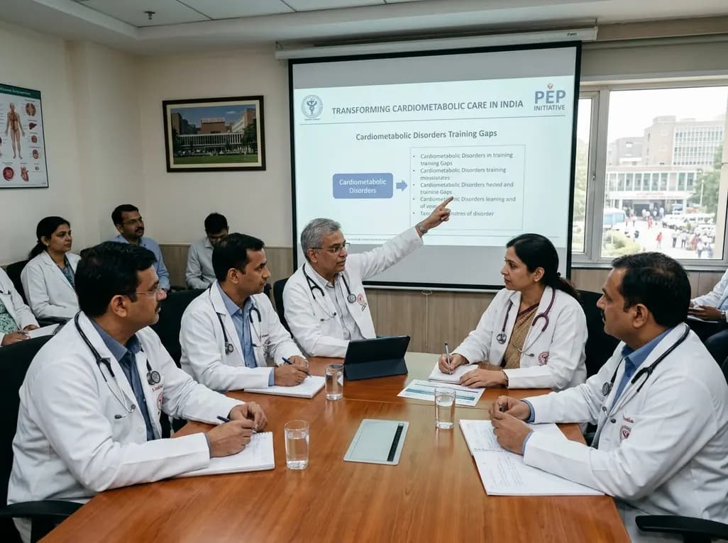
The All India Institute of Medical Sciences has convened national health experts, policymakers, and state nodal officers to transform medical training for cardiometabolic disorders. This collaborative effort under the PEP initiative addresses training gaps to elevate chronic disease care across Indian healthcare.
Today

A systematic review and meta-analysis of 21 RCTs reveals that platelet-rich plasma provides small but statistically superior pain relief and functional improvements compared to hyaluronic acid at 12 months in knee osteoarthritis, influenced by platelet concentration and leukocyte enrichment.
Today

A multicenter real-world prospective study in Taiwan demonstrates that liraglutide effectively reduces body weight and BMI standard deviation score in adolescents with obesity. The therapy shows a favorable safety profile with mild gastrointestinal adverse events and zero treatment discontinuations due to intolerance.
Today
