

Loading, please wait...
Loading, please wait...
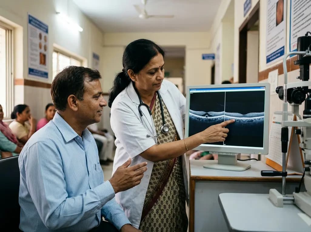
Premacular hemorrhage represents an acute vitreoretinal emergency that abruptly diminishes central vision. In many patients, blood collects in the potential space beneath the internal limiting membrane (ILM). While clinicians frequently observe complete clearance, structural complications occasionally develop in the underlying retina. A rare yet critical anatomical sequela is foveal neurosensory dehiscence beneath a preserved ILM. Previously reported cases of such dehiscence typically demonstrated persistent separation or progression toward macular holes. Consequently, vitreoretinal surgeons often consider prompt pars plana vitrectomy or laser hyaloidotomy. However, emerging clinical evidence demonstrates that certain non-tractional defects can resolve without invasive treatment. When the detached internal limiting membrane spontaneously reapposes against the retinal layers, intrinsic neuroglial healing proceeds undisturbed. Therefore, understanding the precise natural history of foveal neurosensory dehiscence helps specialists avoid premature surgery. Conservative management offers significant visual recovery while eliminating operative complications. As advanced spectral-domain optical coherence tomography (OCT) imaging evolves, clinicians can now track microstructural healing with remarkable precision.
Sudden painless loss of central vision typically defines acute premacular bleeding. For example, a 51-year-old hypertensive male presented with a sudden central scotoma and counting-fingers visual acuity. Notably, clinical interview identified no preceding Valsalva event, physical exertion, or ocular trauma. Fundus examination revealed a dense premacular hemorrhage centered directly over the fovea. Furthermore, spectral-domain OCT demonstrated a classic hyperreflective dome-shaped elevation with dense posterior acoustic shadowing. The central subfield thickness measured 571 micrometers, compared to 223 micrometers in the unaffected fellow eye. Fluorescein angiography confirmed complete hypofluorescence due to optical blockage from the dense blood pool. Moreover, angiography revealed no active macroaneurysm, retinal vascular occlusion, or choroidal neovascular membrane in the visualized arcade vessels. Because blood obscured the underlying foveal circulation, clinicians could not definitively exclude an occult ruptured macroaneurysm. Nevertheless, the vitreoretinal team chose careful observation rather than immediate pneumatic displacement or vitrectomy. Serial follow-up evaluations demonstrated gradual erythrocyte breakdown and progressive clearance of the entrapped blood.
The accumulation of blood beneath the internal limiting membrane creates substantial hydrostatic tension. In addition, dense hemorrhage induces mechanical cleavage between the inner retinal layers and the fragile foveal architecture. This forceful hemo-dissection disrupts the external limiting membrane and the delicate ellipsoid zone. Historically, researchers believed that prolonged blood entrapment always produced permanent neurosensory necrosis. Indeed, free hemoglobin and iron generate cytotoxic reactive oxygen species that damage photoreceptors. However, mechanical factors appear equally decisive in determining ultimate anatomical outcomes. If the distended internal limiting membrane remains detached and fibrotic, persistent tangential traction pulls the foveal margins outward. In stark contrast, when the membrane relaxes and reapposes against the retinal surface, tangential traction dissipates. In this patient, the blood fully cleared by week eight. OCT imaging subsequently revealed that the internal limiting membrane had returned to its native anatomical position. Although an extensive neurosensory gap remained, the reapposed membrane provided a structural roof. Thus, eliminating mechanical vector forces created a permissive microenvironment for cellular repair. As a result, neuroretinal tissue could bridge the foveal floor effectively.
Accurate structural classification remains vital for determining appropriate vitreoretinal management strategies. According to the International Vitreomacular Traction Study Group, a full-thickness macular hole requires complete interruption through all neurosensory retinal layers. That definition entails a foveal defect extending from the internal limiting membrane down to the retinal pigment epithelium. In contrast, the lesion observed in this patient exhibited neurosensory separation beneath an intact, continuous internal limiting membrane. Therefore, the defect did not satisfy consensus criteria for a full-thickness macular hole. Recognizing this crucial distinction fundamentally alters the expected clinical prognosis. Specifically, a true macular hole exposes the pigment epithelium directly to the vitreous cavity, inducing subretinal fluid accumulation. Conversely, an intact internal limiting membrane isolates the photoreceptor layer from vitreous fluid currents and enzymatic factors. Furthermore, preserved Müller cell footplates anchored to the basement membrane maintain regenerative biological potential. When ophthalmologists detect an intact membrane during OCT review, they should recognize the high likelihood of spontaneous healing. Hence, clinicians must avoid hasty classification of these defects as conventional operative holes.
The cellular timeline of foveal repair demonstrates the remarkable plastic capacity of the human retina. At eight weeks post-presentation, the patient exhibited eccentric fixation with a visual acuity of 20/60. Detailed OCT scans at that stage confirmed photoreceptor disruption and an extensive neurosensory gap. However, by 22 weeks, the defect had closed completely without any medical, laser, or surgical therapy. Repeat OCT imaging demonstrated restored foveal contour with a central hyperreflective vertical column. Pathologically, this distinct hyperreflective column reflects robust Müller cell proliferation and glial bridging across the foveola. In addition, the external limiting membrane and ellipsoid zone reconstituted their linear continuity beneath the sealed foveal floor. Consequently, the patient achieved an exceptional best-corrected visual acuity of 20/20. This functional restoration proves that neurosensory foveal gaps can re-establish synaptic connections once mechanical stress abates. Moreover, glial bridging provides adequate structural stability for photoreceptor realignment. Thus, complete anatomical restoration remains achievable even after profound, full-depth foveal disruption. Clinicians can confidently communicate these positive prognostic milestones to patients presenting with similar non-tractional configurations.
This remarkable clinical course provides actionable guidance for managing premacular hemorrhage and secondary foveal defects. Traditionally, vitreoretinal specialists favored early surgical intervention to prevent irreversible photoreceptor atrophy. For instance, many surgeons advocate early vitrectomy with membrane peeling for persistent sub-ILM blood. Similarly, Nd:YAG membranotomy remains popular for rapid blood evacuation into the vitreous body. Nevertheless, invasive interventions introduce risks of iatrogenic hole formation, retinal detachment, endophthalmitis, and cataract progression. This case demonstrates that conservative observation represents a safe, viable first-line alternative when the ILM remains intact. Specifically, clinicians should monitor the mechanical behavior of the internal limiting membrane via frequent OCT scans. If the membrane reapposes and relieves tangential vector stress, the fovea retains remarkable self-healing capacity. Conversely, persistent membrane detachment, contracture, or secondary epiretinal proliferation warrants surgical exploration. Therefore, retinal specialists should individualize management based on precise membrane mechanics rather than immediate visual acuity. In conclusion, careful observation preserves retinal architecture and achieves excellent visual outcomes in carefully selected patients.
A full-thickness macular hole involves complete anatomical disruption of all retinal layers, including the internal limiting membrane, exposing the retinal pigment epithelium to the vitreous cavity. In contrast, foveal neurosensory dehiscence features a separation of the neurosensory retinal layers while the internal limiting membrane remains completely intact and continuous overhead. This critical structural preservation prevents vitreous fluid entry, relieves fluid turbulence, and preserves the essential basement membrane scaffold needed for spontaneous glial repair.
Sub-internal limiting membrane hemorrhages exert acute hydrostatic pressure directly onto the delicate foveola, causing mechanical hemo-dissection between neuroretinal cells. Furthermore, trapped blood isolates the foveal photoreceptors. Erythrocyte breakdown subsequently releases free iron and oxidative compounds that induce focal cellular toxicity. When blood dissects beneath the tight membrane, intense outward tension separates neuroretinal tissue. Consequently, neurosensory dehiscence develops, even though the robust basement membrane withstands rupture.
Observation is strongly favored when optical coherence tomography demonstrates an intact, reapposed internal limiting membrane without persistent tangential traction or full-thickness hole formation. In addition, spontaneous resorption of hemorrhage frequently occurs within several months, especially in younger or non-diabetic patients. If regular imaging confirms progressive visual improvement and stable retinal architecture, clinicians can safely avoid surgery. Vitrectomy remains reserved for persistent dense clots, secondary traction, or progressive full-thickness holes.
Disclaimer: This content is for informational and educational purposes only... Refer to the latest local and national guidelines for clinical practice.
References

Read summarized clinical updates, watch expert medical content, and earn CME certifications right from your smartphone.
Open your smartphone camera to scan and install.





Discover how a foveal neurosensory dehiscence beneath an intact internal limiting membrane spontaneously closed after premacular hemorrhage, achieving complete 20/20 visual recovery without surgery.
Today

A cross-sectional study of 543 patients evaluated the clinical and economic impact of hemophilia inhibitors, revealing higher annual bleeding rates, increased treatment complexity, and severe orthopedic disability, highlighting the need for routine inhibitor screening and modern prophylactic strategies.
Today
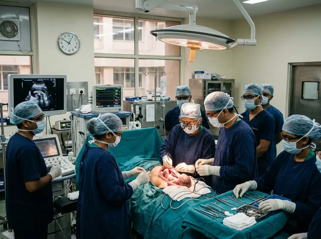
Doctors at AIIMS New Delhi have achieved a historic milestone by performing India's first open prenatal myelomeningocele repair on a 25-week unborn fetus. This landmark multidisciplinary procedure provides vital neuroprotective benefits, lowers hydrocephalus risks, and transforms maternal-fetal medicine nationwide.
Today

A randomized crossover trial reveals that daily fresh watermelon consumption significantly reduces baseline TMAO levels and favorably modulates lipid profiles, lowering triglycerides and LDL cholesterol in adults with overweight and obesity.
Today
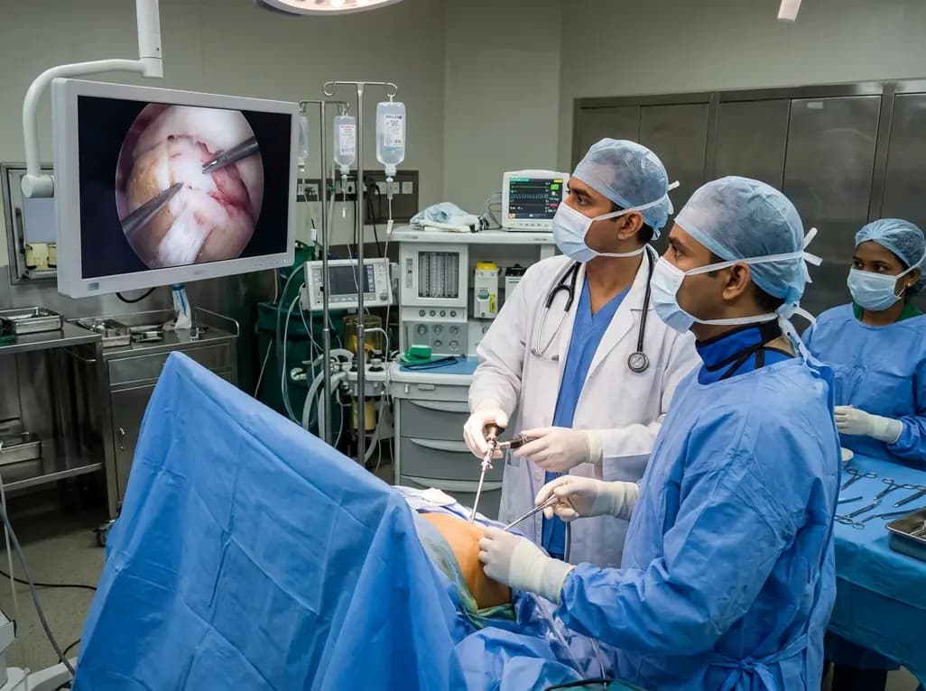
Discover how all-arthroscopic repair using a resorbable bioinductive bovine collagen implant offers a reproducible biologic solution for partial-thickness upper border subscapularis tears, enhancing healing while preserving native tendon architecture.
Today

A large-scale retrospective cohort study demonstrates that GLP-1 receptor agonists significantly reduce all-cause hospitalizations, alcohol-related admissions, major cardiovascular events, and mortality in patients with overweight or obesity and alcohol use disorder, providing profound clinical insights.
Today
