

Loading, please wait...
Loading, please wait...

Shoulder arthroscopy continues to transform how orthopaedic surgeons manage rotator cuff pathology. Although anterior cuff lesions often escape clinical detection, surgeons recognize that upper border subscapularis tears compromise anterior shoulder stability. Traditional surgical options for partial tears often require aggressive debridement or complete tear completion followed by anchor fixation. However, emerging regenerative modalities offer tissue preservation. Applying a bioinductive collagen implant directly over partial defects provides an innovative biologic alternative that promotes host tissue remodeling without disrupting native mechanical insertions.
The subscapularis muscle functions as the primary dynamic stabilizer of the anterior glenohumeral joint. Consequently, high-grade partial-thickness tears of its superior border induce functional impairment, anterior shoulder pain, and biceps pulley instability. Clinicians frequently underdiagnose these articular-sided tears because standard magnetic resonance imaging can miss subtle upper border fraying. Furthermore, anterior shoulder pain often mimics subacromial impingement or isolated biceps tendinopathy. During diagnostic arthroscopy, surgeons must actively rotate the humerus and inspect the comma sign near the lesser tuberosity. When nonoperative therapy fails, surgeons historically faced a difficult dilemma. They either performed simple debridement with high failure rates or deliberately converted the partial tear into a full-thickness defect for anchor repair. Therefore, sports medicine specialists and orthopaedic surgeons need less destructive interventions. Preserving intact tendon fibers remains vital for maintaining shoulder kinematics and avoiding unnecessary tissue tension. Consequently, biologic augmentation strategies offer an effective middle ground for these challenging injuries.
A bioinductive collagen implant provides a porous scaffold derived from highly purified bovine Achilles tendon. This engineered type I collagen matrix induces rapid tenocyte migration, cellular proliferation, and microvascular ingrowth across the injured tendon. Importantly, the scaffold does not rely on mechanical tension or permanent structural fixation. Instead, it activates natural physiological signaling that generates a new layer of dense, tendon-like tissue. Histological evaluations confirm that native host cells infiltrate the resorbable matrix within weeks of implantation. Furthermore, the porous construct resorbs completely within six months, leaving behind functional collagen fibers that integrate with underlying tendon tissue. In addition, this induction process substantially increases total tendon thickness over time. By distributing biomechanical loads more evenly across the footprint, the newly formed tissue shields the original partial tear from shear strain. As a result, the construct arrests tear progression and fosters intrinsic biologic healing without requiring extensive bone preparation.
Performing an all-arthroscopic repair of an upper subscapularis tear requires systematic portal placement and deliberate joint visualization. First, the surgeon performs a thorough diagnostic glenohumeral arthroscopy through a standard posterior portal. The surgeon inspects the biceps pulley, superior glenohumeral ligament, and upper subscapularis insertion on the lesser tuberosity. Next, the surgeon clears loose articular debris and lightly prepares the footprint using radiofrequency ablation. However, the surgeon strictly avoids aggressive debridement that could weaken residual healthy tendon tissue. Subsequently, the surgical team establishes an anterosuperior working portal to insert the specialized implant delivery cannula. The surgeon measures the partial tear dimensions to ensure complete scaffold coverage over the footprint. Then, the surgeon introduces the bioinductive bovine collagen implant directly over the superior border. Using bioabsorbable bone and tendon staples, the surgeon anchors the implant securely to the lesser tuberosity and adjacent tendon tissue. Consequently, the implant establishes an optimal biologic environment for tissue growth.
Traditional surgical management of high-grade partial rotator cuff tears often involves tear completion followed by suture anchor placement. Although this approach restores footprint contact, it creates several clinical drawbacks. Complete tendon takedown disrupts residual intact collagen fibers, potentially compromising native shoulder mechanics. Furthermore, anchor fixation introduces high structural tension, which increases postoperative pain and elevates failure risks. In contrast, bioinductive collagen implants eliminate the need to sever intact structural tendon fibers. Surgeons preserve the native anatomy while actively stimulating new tissue induction over the vulnerable defect. In addition, avoiding suture anchors preserves lesser tuberosity bone stock, simplifying future revision procedures if ever necessary. Patients undergoing bioinductive augmentation typically experience substantially less early postoperative pain because the procedure causes minimal tissue trauma. Moreover, this approach significantly reduces operative time and eliminates complex suture management. Therefore, patients benefit from a faster recovery timeline and lower biological failure rates.
Postoperative rehabilitation following bioinductive subscapularis repair differs favorably from standard rotator cuff reconstruction protocols. Because the procedure preserves native tendon attachments and avoids high-tension anchors, clinicians can safely initiate earlier shoulder mobilization. Typically, patients wear a simple sling for comfort during the first two weeks while starting immediate passive range of motion. Specifically, physical therapists introduce gentle external rotation within safe limits to protect the healing lesser tuberosity. Subsequently, patients transition to active-assisted and active exercises around four weeks postoperatively. By week six to eight, patients initiate progressive rotator cuff strengthening. Prospective registry data demonstrate substantial improvements in patient-reported outcome scores, including Constant-Murley, ASES, and pain scores following bioinductive repair. In addition, serial magnetic resonance imaging studies confirm progressive scaffold resorption and substantial thickening of the regenerated tendon tissue. Consequently, this biological technique represents a predictable paradigm shift for managing partial subscapularis tears.
The bioinductive collagen implant functions as a highly porous extracellular matrix that stimulates rapid host cell infiltration. Circulating tenocytes and mesenchymal stem cells populate the bovine type I collagen scaffold, initiating neovascularization and dense collagen fiber synthesis. Over six months, the implant safely resorbs while leaving behind a durable layer of native tendon tissue. This newly formed tissue significantly increases tendon thickness and unloads mechanical strain across the repair zone.
Surgeons consider bioinductive repair ideal for symptomatic, high-grade partial-thickness subscapularis tears that fail conservative therapy. If more than fifty percent of the tendon thickness remains intact, completing the tear for suture anchor repair unnecessarily destroys viable tissue. Therefore, bioinductive augmentation provides biological reinforcement without disrupting intact mechanical fibers. This biologic approach avoids high suture tension, reduces postoperative pain, preserves lesser tuberosity bone stock, and accelerates functional rehabilitation.
Patients typically experience a substantially accelerated rehabilitation timeline compared to traditional anchor repairs. Because native tendon continuity remains intact, patients wear an immobilizing sling for only two to three weeks. Physical therapists initiate early passive motion and guided internal rotation exercises immediately. Patients progress to active motion by four weeks and begin focused rotator cuff strengthening around week eight. Most individuals resume full, unrestricted daily activities within three to four months postoperatively.
Disclaimer: This content is for informational and educational purposes only. It is not intended to be a substitute for professional medical advice, diagnosis, or treatment. Always seek the advice of your physician or other qualified health provider with any questions you may have regarding a medical condition. Refer to the latest local and national guidelines for clinical practice.
References

Read summarized clinical updates, watch expert medical content, and earn CME certifications right from your smartphone.
Open your smartphone camera to scan and install.





Discover how all-arthroscopic repair using a resorbable bioinductive bovine collagen implant offers a reproducible biologic solution for partial-thickness upper border subscapularis tears, enhancing healing while preserving native tendon architecture.
Today
A real-world UK study reveals substantial hospitalization rates and toxicities in malignant pleural mesothelioma patients treated with nivolumab and ipilimumab, showing a median OS of 9.5 months and highlighting the necessity of vigilant multidisciplinary management.
Today
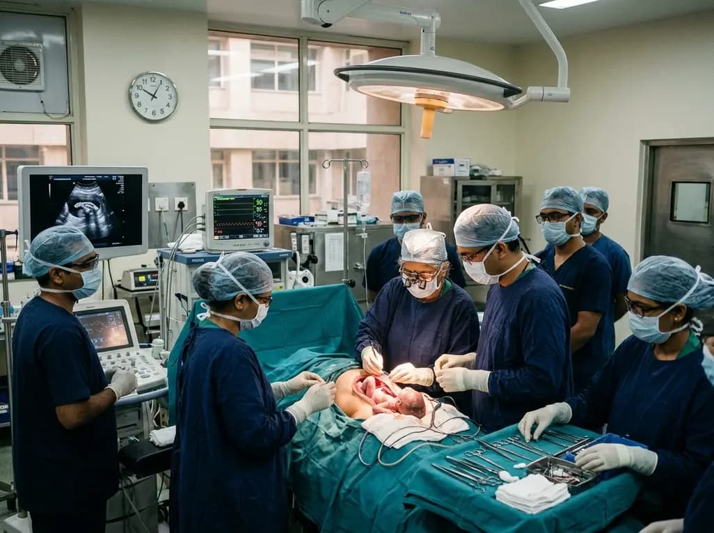
Doctors at AIIMS New Delhi have achieved a historic milestone by performing India's first open prenatal myelomeningocele repair on a 25-week unborn fetus. This landmark multidisciplinary procedure provides vital neuroprotective benefits, lowers hydrocephalus risks, and transforms maternal-fetal medicine nationwide.
Today
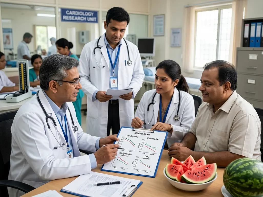
A randomized crossover trial reveals that daily fresh watermelon consumption significantly reduces baseline TMAO levels and favorably modulates lipid profiles, lowering triglycerides and LDL cholesterol in adults with overweight and obesity.
Today
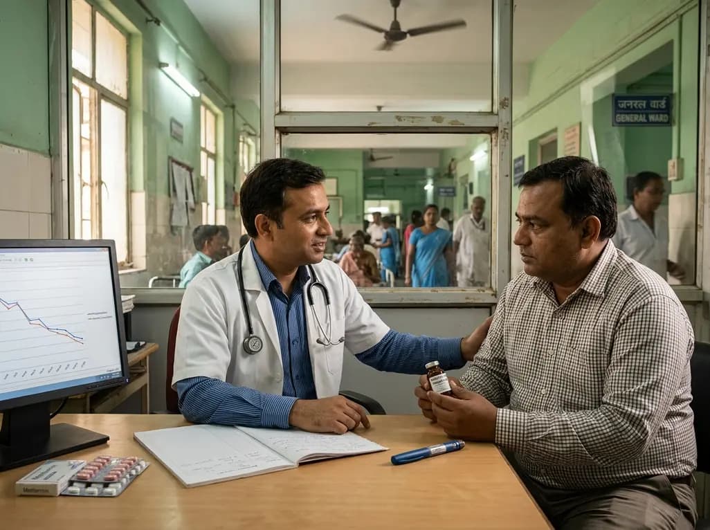
A large-scale retrospective cohort study demonstrates that GLP-1 receptor agonists significantly reduce all-cause hospitalizations, alcohol-related admissions, major cardiovascular events, and mortality in patients with overweight or obesity and alcohol use disorder, providing profound clinical insights.
Today
