

Loading, please wait...
Loading, please wait...

Malignant gliomas represent one of the most formidable operative challenges in modern neurosurgery. Achieving maximal cytoreductive resection directly correlates with prolonged progression-free survival and enhanced overall survival. However, surgeons frequently struggle to distinguish infiltrative tumor margins from functional brain parenchyma under standard illumination. Consequently, fluorescence-guided glioma surgery has emerged as an essential surgical modality designed to optimize tumor margin identification. By utilizing targeted optical contrast agents, surgical teams can visualize malignant tissue in real time. Nevertheless, clinicians historically harbored significant concerns regarding whether aggressive tumor removal might inadvertently trigger new postoperative neurological deficits.
The fundamental goal of neuro-oncological surgery remains maximal safe resection. High-grade gliomas, especially glioblastoma, exhibit an aggressive and infiltrative growth pattern that blurs anatomical borders. Under conventional white-light microscopy, tumor tissue often appears identical to adjacent edematous or normal brain cortex. Therefore, surgeons operating without optical adjuncts frequently leave behind microscopic or macroscopic residual disease. Residual malignant cells inevitably lead to rapid tumor progression and local recurrence.
Optical contrast agents overcome these critical limitations by inducing tumor-specific fluorescence. Agents such as 5-aminolevulinic acid selectively accumulate within neoplastic cells, synthesizing fluorescent protoporphyrin IX. Similarly, sodium fluorescein extravasates through areas with disrupted blood-brain barriers, highlighting abnormal vascular zones. As a result, fluorescence-guided glioma surgery dramatically enhances visualization of subtle tumor margins. Surgeons can identify malignant infiltrates that conventional white-light inspection consistently misses.
However, enhanced optical contrast also presents a double-edged sword in neurosurgical oncology. When margins fluoresce brightly near eloquent cortex or critical white matter tracts, surgeons might push resection boundaries too far. Therefore, neurosurgeons have urgently sought definitive meta-analytic evidence to determine whether this enhanced visualization increases postoperative neurological morbidity.
To resolve long-standing safety questions, investigators conducted a rigorous systematic review and meta-analysis following PRISMA guidelines. The researchers performed comprehensive searches across PubMed, Embase, and Scopus from database inception through March 2026. The systematic analysis evaluated comparative trials that examined fluorescence-guided techniques against conventional white-light surgery in adult patients undergoing glioma resection.
Ultimately, the systematic review included 27 distinct studies representing a pooled cohort of 3,241 patients. Among these individuals, 1,974 patients underwent fluorescence-guided resection, whereas 1,267 patients received conventional white-light resection. Furthermore, the authors identified 13 high-quality comparative studies suitable for quantitative meta-analysis of new postoperative neurological deficits.
The researchers assessed methodological quality across all included investigations. Although observational studies carried moderate risks of confounding bias, randomized controlled trials provided rigorous comparative data. Additionally, investigators performed predefined sensitivity analyses and subgroup evaluations to test the stability of their statistical findings. Specifically, they stratified outcomes based on study design, comparing randomized clinical trials directly against observational cohorts. Consequently, this comprehensive design provided the most robust statistical appraisal of surgical safety published to date.
The primary clinical objective of this meta-analysis was evaluating the incidence of new or worsening postoperative neurological deficits. Many neurosurgeons feared that increased extent of resection would inevitably damage adjacent eloquent cortex. However, the quantitative meta-analysis demonstrated no significant difference in neurological deficit risk between fluorescence-guided surgery and conventional white-light surgery. The pooled risk ratio was 1.02 with a 95% confidence interval of 0.75 to 1.40. Furthermore, the statistical heterogeneity across studies remained low to moderate, with an I-squared value of 39.3%.
To ensure robustness, the authors conducted a sensitivity analysis that excluded studies reporting zero adverse neurological events. Importantly, this sensitivity analysis yielded concordant results, showing a risk ratio of 1.03 (95% CI 0.71 to 1.51; I-squared = 54.4%). Moreover, subgroup analysis stratified by study methodology revealed no statistically significant divergence between randomized controlled trials and observational studies.
These consistent quantitative findings deliver immense reassurance to operating neurosurgeons. The data conclusively demonstrate that enhanced tumor visualization facilitates cytoreduction without causing collateral neurological impairment. Thus, optical guidance empowers surgeons to achieve aggressive tumor clearance while safely maintaining functional integrity.
Surgical teams globally utilize two primary optical agents for intraoperative tumor delineation: 5-aminolevulinic acid and fluorescein sodium. Both fluorophores offer distinct pharmacological profiles and operational benefits during tumor resection. 5-aminolevulinic acid functions as an oral biochemical precursor that tumor cells metabolize into photoactive protoporphyrin IX. Under blue-violet excitation light, high-grade glioma tissue emits a distinct violet-red fluorescence. This intracellular metabolic accumulation provides remarkable specificity for identifying malignant glial cells.
In contrast, fluorescein sodium serves as a vascular marker that surgeons administer intravenously after induction. Fluorescein leaks into the extracellular tumor matrix through regions with a disrupted blood-brain barrier. Under specialized yellow-light filters, the dye illuminates neoplastic tissue with intense yellow-green fluorescence. Because fluorescein requires no pre-ingestion lead time and costs significantly less, many resource-conscious surgical centers favor its adoption.
Both optical modalities effectively enhance intraoperative orientation and facilitate maximal safe resection. Clinical studies confirm that neither agent increases permanent neurological deficits when surgeons respect eloquent boundaries. Therefore, neurosurgical units can select the fluorophore that best matches their technical infrastructure and budgetary resources.
The adoption of optical guidance techniques is expanding rapidly across neurosurgical centers in India. High-grade gliomas represent a major operative workload in tertiary hospitals and academic institutions throughout the country. However, implementing advanced optical technologies requires careful planning regarding equipment acquisition, cost management, and staff training. Specialized surgical microscopes equipped with dedicated fluorescence filters represent a substantial capital investment for healthcare facilities.
In resource-limited settings, fluorescein sodium presents a practical and economical alternative to 5-ALA. Fluorescein is widely available across Indian operating theaters and integrates seamlessly with affordable filter systems. Furthermore, surgical teams must combine optical visualization with advanced intraoperative neurophysiological monitoring. Intraoperative monitoring, including motor evoked potentials and direct cortical stimulation, provides indispensable functional feedback during resection near eloquent speech and motor pathways.
Additionally, multidisciplinary collaboration enhances surgical safety. Neurosurgeons, neuro-anesthesiologists, and neuro-oncologists must establish standardized perioperative protocols. For example, patients receiving 5-ALA require strict light-protection precautions for 24 hours to prevent cutaneous phototoxicity. By establishing comprehensive institutional guidelines, neurosurgical centers in India can safely maximize tumor resection while protecting essential patient neurological function.
While current systematic evidence confirms the objective neurological safety of optical guidance, important research gaps remain. The included studies primarily quantified motor deficits, language impairment, and basic neurological scores such as the Karnofsky Performance Scale. However, conventional clinical deficit scales often fail to capture subtle cognitive decline, executive dysfunction, or nuanced changes in health-related quality of life.
Consequently, future prospective trials must incorporate standardized patient-reported outcome measures. Evaluating subjective cognitive performance, emotional wellbeing, and functional independence will provide a comprehensive understanding of patient recovery. Furthermore, rapid innovations in optical technologies continue to transform neuro-oncological surgery. Emerging techniques such as near-infrared fluorophores, targeted molecular probes, and quantitative fluorescence lifetime imaging offer superior tissue penetration and deeper margin contrast.
In addition, integrating optical guidance with artificial intelligence algorithms and advanced robotic visualization will further refine intraoperative margin definition. As these sophisticated technologies mature, surgical teams will achieve unprecedented precision in glioma resection. Ultimately, combining real-time optical guidance with functional brain mapping will solidify the standard of maximal safe resection worldwide.
No, fluorescence-guided glioma surgery does not increase postoperative neurological deficits compared to conventional white-light surgery. A meta-analysis of 27 studies and 3,241 patients demonstrated an equivalent neurological deficit risk (RR=1.02; 95% CI 0.75-1.40). Therefore, surgeons can achieve greater extent of resection without subjecting patients to heightened risks of functional impairment.
The two main fluorophores are 5-aminolevulinic acid and fluorescein sodium. 5-aminolevulinic acid is an oral agent metabolized intracellularly into fluorescent protoporphyrin IX, offering high tumor specificity. Conversely, fluorescein sodium is administered intravenously, accumulating in areas with disrupted blood-brain barriers to highlight abnormal tissues with bright yellow-green fluorescence.
Fluorescence guidance visually identifies neoplastic tissue, while intraoperative neurophysiological monitoring assesses real-time neuronal function. By combining optical imaging with direct cortical stimulation and evoked potential tracking, neurosurgeons determine exactly when to halt resection near eloquent cortex. This multimodal strategy prevents permanent neurological deficits during aggressive cytoreductive surgery.
Disclaimer: This content is for informational and educational purposes only... Refer to the latest local and national guidelines for clinical practice.
References

Read summarized clinical updates, watch expert medical content, and earn CME certifications right from your smartphone.
Open your smartphone camera to scan and install.





A comprehensive meta-analysis of 27 studies and 3,241 patients confirms that fluorescence-guided glioma surgery significantly improves tumor visualization and resection extent without increasing the risk of postoperative neurological deficits compared to conventional white-light surgery.
Today
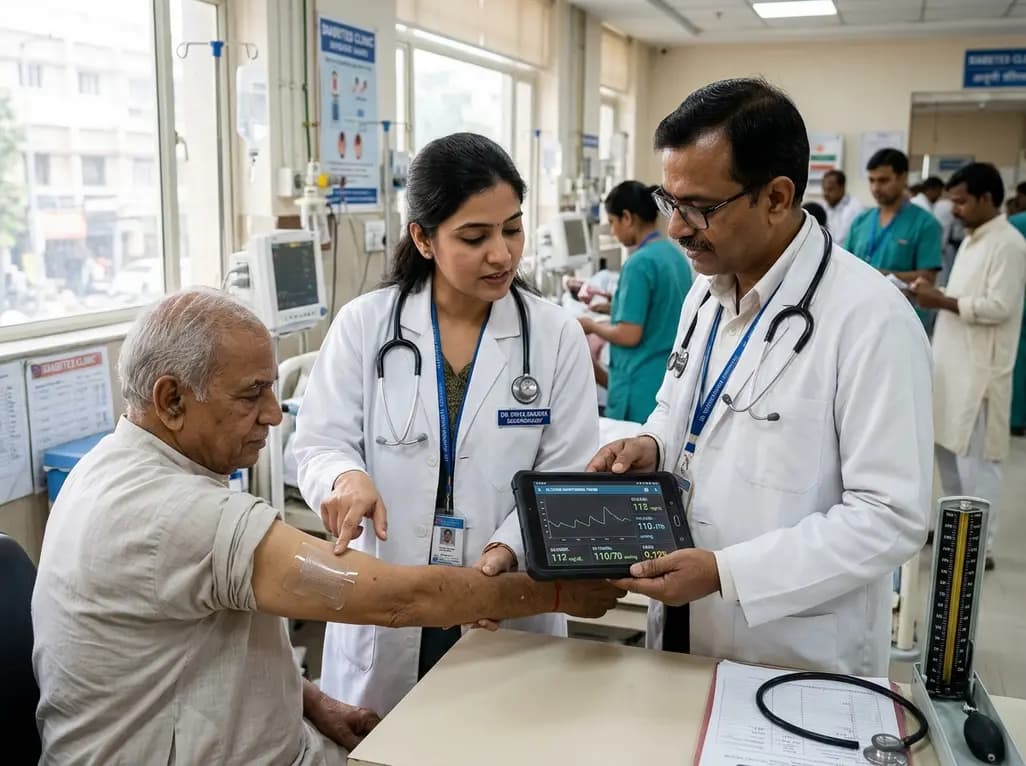
A breakthrough ultrathin multimodal sensor decouples physical hemodynamic monitoring from disposable electrochemical calibration, achieving long-term wearable glucose prediction with 81.36% accuracy and a 9.12% MARD. This offers a robust, drift-free solution for outpatient metabolic surveillance.
Today

A clinical review of menopause and neurology highlights the neurobiological impacts of midlife hormonal shifts on cognition, epilepsy, migraine, multiple sclerosis, and stroke, emphasizing safe hormone therapy prescribing.
Today

Co-administering antiseizure medications and DOACs frequently causes pharmacokinetic interactions, subtherapeutic drug levels, and thromboembolic failures. This review highlights enzyme induction risks, unpredictable outcomes with levetiracetam, and the vital role of therapeutic drug monitoring.
Today

A comparative review of tension band wiring and intramedullary screw fixation in thumb metacarpophalangeal arthrodesis, evaluating union rates, hardware complications, and clinical outcomes.
Today

Following regulatory scrutiny from FSSAI regarding promotional representations and lean muscle preservation claims, Dr. Reddy's Laboratories and Nestlé Health Science are currently reviewing the branding and marketing strategy for their joint nutritional supplement Celevida GLP+.
Today
