.webp&w=384&q=75)

Loading, please wait...
Loading, please wait...
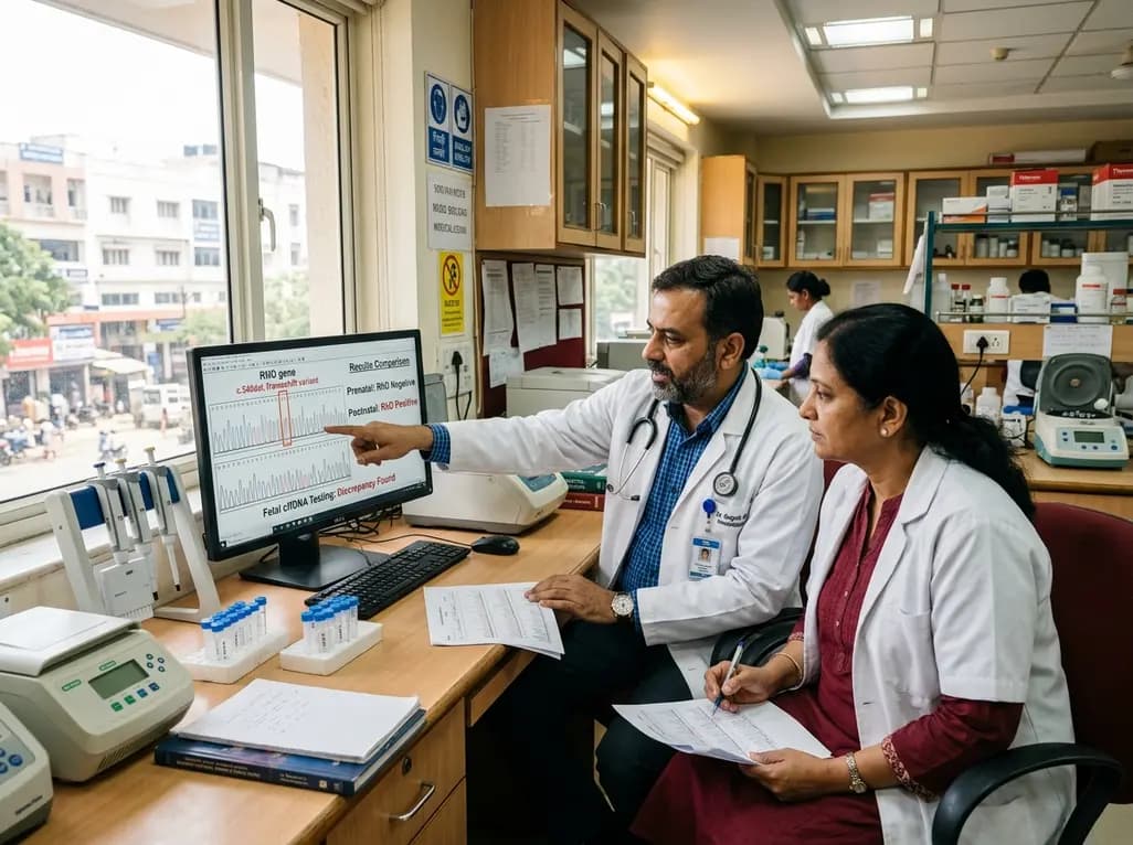
Non-invasive prenatal screening has transformed modern obstetric care by evaluating cell-free fetal DNA circulating in maternal plasma. Among its most vital applications, fetal RHD genotyping allows targeted antenatal immunoprophylaxis for RhD-negative pregnant individuals. This targeted protocol prevents unnecessary administration of human plasma-derived anti-D immunoglobulin in women who carry an RhD-negative fetus. Consequently, clinical laboratories rely heavily on high-throughput molecular assays to predict fetal blood groups accurately. However, discrepancies between prenatal molecular predictions and postnatal serological evaluations occasionally occur. A newly published multi-platform genetic investigation has characterized a novel non-functional allele (c.540del; p.Leu181CysfsTer48) that challenges conventional testing algorithms. Understanding the genetic origins of these discordances is essential for obstetricians, hematologists, and transfusion specialists navigating modern reproductive care.
Hemolytic disease of the fetus and newborn remains a major cause of perinatal morbidity and mortality worldwide. Routine antenatal anti-D immunoprophylaxis has significantly decreased alloimmunization rates in D-negative mothers. Nevertheless, universal prophylaxis exposes nearly forty percent of RhD-negative women to unnecessary plasma-derived products because their fetuses are also RhD-negative. Therefore, introducing non-invasive fetal RHD genotyping has optimized resource utilization and minimized biological exposure risks. By targeting specific exons within cell-free DNA, diagnostic platforms predict fetal antigen presence with high sensitivity. In addition, health systems benefit from reduced overall expenditure on donor-derived immunoglobulins. Despite these substantial advantages, clinicians must remember that molecular presence does not inherently guarantee structural protein expression on red blood cell membranes.
Recently, researchers investigated a discordant case where prenatal cell-free DNA screening predicted an RhD-positive fetus, but postnatal umbilical cord serology confirmed a D-negative phenotype. To resolve this diagnostic conundrum, investigators performed comprehensive molecular evaluations using commercial sequence-specific PCR kits, Sanger sequencing, and targeted next-generation sequencing. The analytical workflow successfully identified a single-nucleotide deletion within exon 4 of the RHD gene, designated as c.540del. This deletion creates a translational frameshift at codon 181, generating a premature stop codon forty-eight residues downstream (p.Leu181CysfsTer48). Genetic testing of the family confirmed paternal transmission, as the father was heterozygous for this frameshift mutation. Furthermore, genomic screening detected the identical allele in an unrelated RhD-negative individual of Swedish ancestry, indicating that this variant may circulate within specific regional populations.
The human RH locus is situated on chromosome 1p36 and contains two homologous, tandemly arranged genes: RHD and RHCE. Because these genes share substantial sequence homology, genetic recombination, gene conversions, and point mutations occur frequently. In standard assays, primer sets amplify specific exons, such as exon 4, exon 5, or exon 10, to verify the presence of the RHD gene. However, the c.540del frameshift truncation severely disrupts the normal polypeptide structure. The premature stop codon prevents the synthesis of the twelve-transmembrane-domain RhD protein, leading to rapid mRNA decay or non-functional polypeptide degradation. Consequently, no D antigen embeds within the erythroid membrane. Standard cell-free DNA assays amplify the intact exon fragments, thereby yielding a false-positive prediction of RhD positivity because the molecular screening pipeline cannot evaluate downstream translational competence.
Cell-free DNA testing relies primarily on quantitative real-time PCR or digital droplet PCR to amplify designated target segments. Most commercial assays interrogate multiple distinct exons to distinguish maternal DNA from fetal DNA and to mitigate interference from partial-D or pseudo-alleles. Nevertheless, these routine platforms possess inherent limitations when encountering rare structural mutations. For instance, single-nucleotide deletions residing outside primer-binding domains do not disrupt amplification, causing the platform to register positive gene signals. In contrast, comprehensive next-generation sequencing panels and long-read sequencing technologies can identify internal sequence truncations and complex hybrid genes. Therefore, when routine serological results contradict prenatal screening, laboratories must deploy advanced sequencing to differentiate true antigen suppression from technical collection errors.
Discordant blood typing results generate significant clinical dilemmas during the perinatal period. When non-invasive screening falsely predicts an RhD-positive fetus, the mother receives routine antenatal anti-D immunoprophylaxis. Fortunately, unnecessary prophylaxis carries minimal direct maternal morbidity beyond mild injection-site discomfort and low plasma-related exposure risks. However, the reverse scenario—a false-negative prenatal result where an RhD-positive fetus is missed—poses grave risks because omitted immunoprophylaxis can precipitate maternal alloimmunization and severe fetal anemia in subsequent pregnancies. Furthermore, accurate donor and recipient genotyping remains pivotal in blood banking. An individual carrying the c.540del variant has an RhD-negative serological phenotype and must receive D-negative red cell transfusions to prevent immune sensitization against foreign RhD epitopes.
Establishing robust laboratory algorithms is vital for detecting and resolving prenatal-postnatal typing discrepancies. First, transfusion services should always verify maternal and neonatal serology at delivery using potent monoclonal anti-D reagents. If discordance arises between prenatal cell-free DNA predictions and newborn cord blood serology, laboratories must immediately exclude clerical errors, maternal cell contamination, and weak-D phenotypes through adsorption-elution techniques. Subsequently, reference immunohematology laboratories should conduct Sanger sequencing or targeted NGS of both parents and the neonate. In addition, establishing centralized national registries of rare RHD alleles will refine regional primer designs and improve the interpretive accuracy of future non-invasive prenatal screening programs.
Discordant results typically arise from non-functional or variant RHD genes. Standard prenatal tests detect target genetic sequences within cell-free DNA but cannot assess whether the gene produces a functional, membrane-bound protein. Consequently, null alleles, premature stop codons, or frame-shift mutations like c.540del yield positive genetic signals despite a completely negative serological blood group at birth.
Administering anti-D immunoglobulin to an RhD-negative woman carrying an RhD-negative fetus does not cause significant clinical harm. The injected antibodies gradually clear from the maternal circulation without attacking maternal red blood cells. However, this unnecessary administration increases healthcare expenses and consumes donor-derived plasma supplies without providing any therapeutic benefit to the patient or pregnancy.
Transfusion services must classify individuals carrying the non-functional c.540del variant as RhD-negative for blood transfusions. Because their red blood cells do not express the RhD protein on the cell surface, transfusing RhD-positive blood could trigger anti-D alloantibody formation. Therefore, these patients should consistently receive RhD-negative packed red blood cells during elective transfusions and emergency resuscitations.
Disclaimer: This content is for informational and educational purposes only and does not constitute medical advice, diagnosis, or treatment. Healthcare professionals should exercise independent clinical judgment. Refer to the latest local and national guidelines for clinical practice.
References
1. Badri A et al. Discordant prenatal and postnatal RhD typing caused by a novel RHD frameshift variant (c.540del; p.Leu181CysfsTer48) in Swedish individuals. Vox Sang. 2026 Aug 20. doi: 10.1111/vox.70351. PMID: 42624819.
2. Runkel B, Bein G, Sieben W, Sow D, Polus S, Fleer D. Targeted antenatal anti-D prophylaxis for RhD-negative pregnant women: a systematic review. BMC Pregnancy Childbirth. 2020;20(1):83. doi:10.1186/s12884-020-2742-4.
3. Yang H, Llewellyn A, Walker R, Harden M, Saramago P, Griffin S, Simmonds M. High-throughput, non-invasive prenatal testing for fetal rhesus D status in RhD-negative women: a systematic review and meta-analysis. BMC Med. 2019;17(1):37. doi:10.1186/s12916-019-1254-4.
4. Sandler SG, Flegel WA, Westhoff CM, Denomme GA, Delaney M, Keller MA, et al. It's time to phase in RHD genotyping for patients with a serologic weak D phenotype. Transfusion. 2015;55(3):680-689. doi:10.1111/trf.12941.

Read summarized clinical updates, watch expert medical content, and earn CME certifications right from your smartphone.
Open your smartphone camera to scan and install.





A novel RHD frameshift variant (c.540del) highlights crucial limitations in cell-free fetal DNA testing, causing discordant prenatal and postnatal RhD typing and reinforcing the need for rigorous molecular follow-up in transfusion medicine.
last month

Pancreatic cancer cachexia affects over 80% of patients, driving severe muscle wasting, systemic inflammation, and treatment intolerance. This review highlights key pathophysiological drivers, exocrine insufficiency, objective CT imaging, and emerging therapies like GDF-15 inhibitors to improve clinical outcomes.
Today

A scoping review reveals how pregnancy affects phonation through laryngeal edema and reduced phonation time rather than major acoustic pitch alterations. Learn how to identify, evaluate, and manage vocal symptoms in expectant mothers.
Today

According to WHO data, average lifespan reached 73.3 years in 2023, recovering from pandemic lows. However, healthy life expectancy lags at 62.8 years due to surging noncommunicable conditions like cardiovascular disease and dementia, demanding proactive primary screening and sustained chronic care.
Today

Researchers have engineered an artificial sensory-pain integrated receptor using a multi-threshold organic synaptic transistor. The system distinguishes benign touch from noxious stimuli, enabling safer, more responsive prosthetic limbs with built-in memory and simplified hardware architecture.
Today

Medical robotics is transitioning beyond operative suites into diagnostics, cardiology, and endocrinology. Explore how innovations in tele-robotics, AI integration, and nanomedicine are overcoming geographical barriers and transforming precision healthcare delivery across India's evolving disease landscape.
Today
