.webp&w=384&q=75)

Loading, please wait...
Loading, please wait...
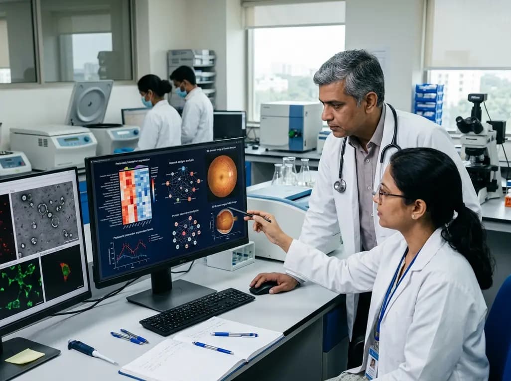
Age-related macular degeneration represents a leading cause of irreversible vision loss worldwide, placing a heavy burden on healthcare systems and elderly patients. Retinal pathology traditionally stems from advancing age, environmental stressors, and strong genetic predispositions. However, researchers increasingly recognize systemic blood circulation as a fundamental driver of retinal disease progression. Liquid biopsies provide non-invasive opportunities to detect pathological changes before severe structural damage occurs. In this context, circulating extracellular vesicles in AMD have emerged as potent carriers of molecular information. These nanoscale membrane-bound vesicles actively transport proteins, lipids, and nucleic acids across biological barriers, directly reflecting cellular homeostasis and systemic dysfunction. A groundbreaking multi-omics investigation analyzed plasma-derived extracellular vesicle-enriched samples from individuals with age-related macular degeneration and matched control subjects. The investigators sought to determine whether circulating vesicular cargo mirrors the complex biochemical disturbances seen within degenerating retinal tissue. Consequently, the findings demonstrated that peripheral blood vesicles carry distinctive signatures of systemic inflammation, oxidative damage, and metabolic disruption. This discovery offers promising avenues for earlier diagnosis, accurate disease staging, and personalized therapeutic interventions in clinical ophthalmology.
Comprehensive proteomic analysis of plasma-derived vesicles identified numerous significantly altered proteins between patient cohorts. Interestingly, the vast majority of these differentially expressed vesicular proteins demonstrated marked downregulation in affected individuals compared to healthy controls. Bioinformatic analysis uncovered a dense molecular interaction network dominated by complement cascade components and crucial endopeptidase inhibitors. The complement system plays a central role in retinal homeostasis, and its chronic overactivation drives drusen accumulation and retinal pigment epithelial atrophy. Moreover, endopeptidase inhibitors modulate local extracellular matrix turnover and tissue remodeling within the choroid and subretinal space. When vesicular transport of these protective regulators decreases, proteolytic balance deteriorates rapidly across vascular beds. Furthermore, many altered vesicular proteins directly participate in immune cell modulation, coagulation pathways, and systemic redox regulation. These molecular perturbations closely align with established candidate biomarkers previously linked to macular disease onset and choroidal neovascularization. Thus, the altered vesicular proteome does not merely represent passive bystander debris. Instead, it reflects widespread systemic proteolysis dysregulation and inflammatory imbalance that actively contribute to ongoing retinal microvascular damage.
In addition to distinctive proteomic remodeling, quantitative lipidomic evaluation demonstrated striking metabolic alterations within circulating vesicles. Researchers observed a significant elevation in the total sphingomyelin to ceramide ratio among individuals diagnosed with macular degeneration. Sphingolipids maintain plasma membrane integrity, regulate signal transduction cascades, and direct cellular stress responses. Ceramide generation typically promotes controlled apoptosis, whereas sphingomyelin abundance influences membrane fluidity and vesicular budding kinetics. Therefore, an abnormal sphingomyelin to ceramide ratio indicates profound disruption in systemic sphingolipid metabolism and enzymatic turnover. Furthermore, altered lipid ratios modify the biophysical properties of vesicle membranes, impairing normal intercellular communication between vascular endothelial cells and neural targets. These systemic lipid defects closely mirror the metabolic stress observed within aging retinal pigment epithelial cells during disease initiation. Consequently, defective lipid processing promotes subretinal lipid accumulation, contributing directly to basal laminar deposits and drusen development. By capturing these systemic lipid shifts, plasma vesicle analysis provides critical insights into metabolic vulnerability, enabling clinicians to monitor systemic lipid homeostasis alongside localized retinal imaging findings.
To complement mass spectrometry investigations, scientists utilized high-resolution Raman spectroscopy to evaluate the structural integrity of vesicular cargo. This advanced vibrational technique provided label-free biochemical fingerprints of plasma-derived samples, confirming widespread macromolecular changes. Specifically, Raman spectral signatures demonstrated pronounced oxidative modifications in both vesicular proteins and structural lipids among patients. Reactive oxygen species induce extensive cross-linking, carbonyl formation, and lipid peroxidation, severely degrading normal biomolecular function. Furthermore, the spectral data revealed significant compositional abnormalities, highlighting a persistent failure of systemic redox defenses in macular degeneration. Photoreceptor outer segments and retinal pigment epithelial cells endure continuous light-induced oxidative stress under physiological conditions. However, when systemic antioxidant buffering fails, oxidative damage spreads beyond the ocular environment into the peripheral vascular circulation. Thus, Raman spectroscopy confirms that circulating vesicles directly carry the physical footprints of oxidative stress. Detecting these molecular modifications non-invasively allows clinicians to evaluate systemic oxidative burden, providing an objective metric to track therapeutic responses to antioxidant interventions and lifestyle modifications.
The convergence of proteomic, lipidomic, and spectroscopic findings establishes a coherent pathophysiological profile in macular degeneration. Systemic complement activation, impaired redox balance, and disrupted lipid catabolism coalesce within circulating vesicles to reflect active retinal disease mechanisms. Clinicians have long sought robust blood-based biomarkers to supplement conventional optical coherence tomography and fundus photography. Although structural imaging reliably detects established anatomical defects, molecular biomarkers can identify subclinical metabolic distress far earlier. Consequently, profiling plasma vesicles opens unprecedented opportunities for precision medicine in geriatric eye care. Risk stratification models can incorporate vesicular complement proteins and lipid ratios to predict rapid disease progression or conversion to neovascular stages. Furthermore, identifying specific molecular phenotypes enables targeted therapeutic selection, matching individual patients with complement inhibitors, antioxidant therapies, or lipid-modulating agents. As precision ophthalmology advances, circulating vesicles will serve as dynamic liquid biopsies, bridging systemic biochemical health with localized ocular pathology and enhancing long-term visual outcomes.
Integrating extracellular vesicle diagnostics into routine clinical practice requires standardized biobanking, automated isolation platforms, and rigorous analytical validation. Currently, size-exclusion chromatography and microfluidic technologies allow efficient isolation of plasma vesicles from standard venipuncture samples. Clinical laboratories must establish reproducible reference ranges for vesicular complement components, sphingolipids, and oxidation markers across diverse populations. Moreover, longitudinal clinical trials should evaluate whether serial vesicle measurements correlate with treatment response following anti-VEGF therapy or geographic atrophy interventions. Multidisciplinary collaboration among ophthalmologists, geriatricians, and laboratory specialists will accelerate the adoption of these non-invasive diagnostic tools. Ultimately, blood-derived vesicular profiling will transform preventive eye care, empowering practitioners to intervene before irreversible photoreceptor loss occurs and preserving patient quality of life.
Circulating extracellular vesicles in AMD carry molecular cargo originating from diverse tissues, including vascular and neural cells. These vesicles transport dysregulated complement proteins, oxidized lipids, and altered enzymes that mirror the systemic inflammation and oxidative stress occurring within the degenerating retina. Consequently, analyzing plasma vesicles provides a non-invasive window into localized retinal pathophysiological processes without requiring intraocular tissue sampling.
Lipidomic profiling reveals a significant elevation in the total sphingomyelin to ceramide ratio within plasma vesicles from affected patients. This shift indicates impaired sphingolipid metabolism, altering vesicle membrane fluidity and cell signaling mechanisms. Furthermore, these lipid abnormalities reflect defective metabolic processing in retinal pigment epithelial cells, which actively contributes to subretinal lipid accumulation and drusen formation.
Yes, plasma vesicle profiling holds strong potential for monitoring disease progression and identifying high-risk patients. Distinct vesicular proteomic signatures and oxidation markers correlate with complement overactivation and tissue remodeling stages. Therefore, tracking these biochemical changes over time enables clinicians to stratify patient risk, predict conversion to neovascular forms, and personalize antioxidant or complement-targeted interventions effectively.
Disclaimer: This content is for informational and educational purposes only and does not constitute medical advice. Consult qualified healthcare professionals for diagnosis and management. Refer to the latest local and national guidelines for clinical practice.
References

Read summarized clinical updates, watch expert medical content, and earn CME certifications right from your smartphone.
Open your smartphone camera to scan and install.





A comprehensive multi-omics analysis reveals that plasma-derived extracellular vesicles in age-related macular degeneration carry molecular signatures of oxidative stress, altered sphingolipid metabolism, and complement dysregulation, presenting new avenues for non-invasive liquid biopsy diagnostics.
4 weeks back

Pancreatic cancer cachexia affects over 80% of patients, driving severe muscle wasting, systemic inflammation, and treatment intolerance. This review highlights key pathophysiological drivers, exocrine insufficiency, objective CT imaging, and emerging therapies like GDF-15 inhibitors to improve clinical outcomes.
Today

A scoping review reveals how pregnancy affects phonation through laryngeal edema and reduced phonation time rather than major acoustic pitch alterations. Learn how to identify, evaluate, and manage vocal symptoms in expectant mothers.
Today

According to WHO data, average lifespan reached 73.3 years in 2023, recovering from pandemic lows. However, healthy life expectancy lags at 62.8 years due to surging noncommunicable conditions like cardiovascular disease and dementia, demanding proactive primary screening and sustained chronic care.
Today

Researchers have engineered an artificial sensory-pain integrated receptor using a multi-threshold organic synaptic transistor. The system distinguishes benign touch from noxious stimuli, enabling safer, more responsive prosthetic limbs with built-in memory and simplified hardware architecture.
Today

Medical robotics is transitioning beyond operative suites into diagnostics, cardiology, and endocrinology. Explore how innovations in tele-robotics, AI integration, and nanomedicine are overcoming geographical barriers and transforming precision healthcare delivery across India's evolving disease landscape.
Today
