

Loading, please wait...
Loading, please wait...

Modern clinical oncology relies heavily on precise histological characterization and comprehensive molecular profiling. Consequently, endoscopists consider EUS-guided tissue acquisition indispensable when evaluating solid pancreatic lesions. Historically, diagnostic algorithms depended primarily on fine-needle aspiration for cytological examination. However, cytology frequently lacks the architectural integrity necessary for definitive classification. Today, individualized treatment paradigms demand cohesive core tissue for immunohistochemistry and genomic sequencing. Therefore, novel needle geometries and refined procedural strategies have emerged, transforming diagnostic endoscopy into an essential driver of personalized cancer therapy.
Historically, endoscopists utilized standard aspiration needles for sampling pancreatic masses. Although these conventional devices achieved acceptable diagnostic sensitivity, they yielded predominantly aspirates with disrupted stroma. In response, bioengineers designed cutting fine-needle biopsy devices to preserve architectural context. Early reverse-bevel needles offered moderate tissue yield improvements. However, crown-cut Franseen needles and opposing-bevel fork-tip needles revolutionized procedural performance. These innovative end-cutting instruments shear tissue cleanly along multiple planes, acquiring intact histological cores.
Furthermore, extensive clinical trials confirm that end-cutting biopsy devices outperform traditional aspiration needles in diagnostic accuracy. In addition, these specialized needles procure adequate core architecture in significantly fewer needle passes. As a result, operators achieve definitive diagnoses faster while reducing procedure duration and sedation requirements. Therefore, current international practice guidelines recommend end-cutting biopsy needles over conventional aspiration needles as the preferred primary modality for pancreatic tumors. Moreover, modern biopsy needles facilitate ancillary testing that cytology cannot support. Pathologists can reliably perform immunohistochemical assays to differentiate ductal adenocarcinomas from neuroendocrine neoplasms, lymphoma, or autoimmune pancreatitis. Thus, modern needle geometry directly addresses the diagnostic challenges of heterogeneous pancreatic lesions.
Procuring diagnostic specimens requires both optimal needle geometry and deliberate technical maneuvers. Solid pancreatic neoplasms typically feature prominent central necrosis, cellular heterogeneity, and dense desmoplasia. Consequently, sampling along a single static puncture track frequently collects acellular debris rather than viable malignant cells. To overcome this challenge, endoscopists routinely execute the fanning technique during acquisition. Specifically, the operator adjusts the ultrasound elevator and scope dial to steer the needle tip across diverse areas during each pass.
Moreover, clinicians combine the fanning technique with rotational torque maneuvers to maximize cellular procurement. By applying subtle rotational torque to the echoendoscope shaft during advancement, the cutting tip shaves fresh tissue cylinders along uncompromised tissue planes. Accordingly, clinical trials confirm that fanning and torque strategies significantly elevate diagnostic adequacy compared to conventional single-trajectory passes. Furthermore, these maneuvers decrease the cumulative number of needle passes needed to secure definitive histology. Thus, employing multiplanar fanning alongside gentle torque ensures comprehensive tumor sampling without causing excessive local mechanical trauma.
The choice of suction applied during acquisition profoundly affects histological specimen quality. Historically, clinicians routinely applied high negative pressure using a locked 10- or 20-milliliter vacuum syringe. However, intense negative suction often draws excessive peripheral blood into the channel, diluting diagnostic cellular elements and obscuring tissue architecture. Consequently, endoscopists have transitioned toward lower-pressure and fluid-assisted aspiration techniques to preserve core tissue integrity.
Notably, the slow-pull technique involves gradually withdrawing the internal stylet while oscillating the needle within the tumor. This controlled maneuver creates continuous low negative pressure, procuring clean histological cylinders with minimal hemorrhagic contamination. Alternatively, endoscopists employ the wet suction technique, preloading the needle with sterile saline before applying suction. Saline eliminates air compressibility, transmitting negative pressure evenly throughout the needle bore. Furthermore, clinicians increasingly utilize heparinized wet suction to prevent clot formation inside the needle channel. As a result, heparinized aspiration yields cohesive, intact tissue cores free of distracting fibrin or red blood cells. Therefore, selecting an optimal aspiration technique substantially elevates histological yield.
Rapid on-site cytopathological evaluation has long represented the gold standard for confirming specimen adequacy during biopsies. Nevertheless, many clinical centers cannot provide dedicated on-site pathologists during routine endoscopy schedules due to logistical and financial constraints. To bridge this gap, endoscopists increasingly adopt macroscopic on-site evaluation immediately following tissue procurement. Specifically, the proceduralist inspects the expelled specimen visually under good lighting without microscopic equipment.
Extensive clinical studies demonstrate that identifying a distinct whitish-yellow core cylinder measuring at least four millimeters correlates strongly with microscopic adequacy. Furthermore, obtaining a cumulative core length exceeding ten millimeters provides sufficient cellular material for next-generation sequencing and comprehensive molecular analysis. In the era of precision oncology, molecular profiling determines targeted therapeutic options for advanced pancreatic adenocarcinoma. For instance, genomic sequencing uncovers actionable mutations, microsatellite instability, or BRCA alterations. Thus, macroscopic inspection allows endoscopists to confirm sample adequacy reliably, avoid redundant passes, and safeguard sufficient tissue for advanced genomic profiling.
Endoscopic tissue acquisition demonstrates an exceptional safety record, with serious adverse events occurring in fewer than two percent of cases. However, clinicians must actively prevent recognized complications, including post-procedure pancreatitis, localized hemorrhage, peritoneal infections, and needle-tract seeding. Endoscopists minimize acute pancreatitis risks by avoiding normal pancreatic parenchyma and utilizing direct, short needle paths. Moreover, operators routinely apply color Doppler sonography prior to needle puncture to avoid traversing major vascular structures.
Importantly, major clinical guidelines do not endorse routine prophylactic antibiotics prior to sampling solid pancreatic tumors. Because solid lesions lack fluid collections, post-procedural bacteremia and infection risks remain negligible. Therefore, physicians should withhold prophylactic antibiotics unless evaluating cystic lesions or managing concomitant biliary obstruction. Meanwhile, needle-tract seeding remains a rare but critical concern, particularly during transgastric puncture of pancreatic body and tail lesions. Consequently, multidisciplinary teams must document the biopsy route meticulously. Surgical teams can then incorporate the gastric puncture site into the definitive en bloc surgical resection field.
Franseen and fork-tip needles feature precision-engineered cutting edges designed to slice intact tissue cylinders rather than merely aspirating isolated cells. Consequently, they preserve histologic architecture, cellular cohesion, and surrounding tumor stroma. This structural preservation enables accurate immunohistochemical analysis and extensive genomic sequencing. Furthermore, these end-cutting needles consistently demonstrate higher diagnostic accuracy, require fewer passes, and decrease procedural duration compared to conventional fine-needle aspiration devices in daily clinical practice.
Current international guidelines do not recommend routine prophylactic antibiotic administration before sampling solid pancreatic lesions. Because solid tumors do not contain stagnant fluid, the absolute risk of post-procedural bacteremia or retroperitoneal infection remains negligible. Indiscriminate antibiotic prophylaxis increases healthcare costs and drives antimicrobial resistance unnecessarily. However, endoscopists should administer targeted prophylactic coverage if the patient has an obstructed biliary tract, undergoes concurrent cystic lesion aspiration, or presents with significant systemic immunosuppression.
Macroscopic on-site evaluation allows the endoscopist to assess specimen adequacy immediately at the bedside without requiring a dedicated cytopathologist. By visually inspecting the extruded material for visible whitish-yellow core tissue cylinders, the proceduralist accurately estimates diagnostic quality. This immediate visual feedback helps endoscopists terminate the procedure once they obtain adequate core tissue. Consequently, this approach reduces redundant needle passes, decreases procedure times, lowers complication rates, and guarantees sufficient tissue volume for molecular profiling.
Disclaimer: This content is for informational and educational purposes only and should not be considered medical advice or relied upon for clinical decisions. Refer to the latest local and national guidelines for clinical practice.
References

Read summarized clinical updates, watch expert medical content, and earn CME certifications right from your smartphone.
Open your smartphone camera to scan and install.





A comprehensive clinical review on endoscopic ultrasound-guided tissue acquisition for pancreatic solid tumors, highlighting Franseen and fork-tip needles, technical sampling maneuvers, suction strategies, macroscopic on-site evaluation, and complication management.
Today
A rare case of homozygous deletional hereditary persistence of fetal hemoglobin (HPFH) in Ghanaian twins reveals 100% HbF and erythrocytosis with normal red cell indices, providing key insights into globin gene regulation, differential diagnosis from delta-beta thalassemia, and clinical implications.
Today
The 2026 KASID guidelines provide comprehensive clinical recommendations on sexuality, fertility, pregnancy, and lactation for patients with inflammatory bowel disease, prioritizing sustained remission and multidisciplinary care to optimize maternal and neonatal outcomes.
Today

A comparative non-contact study reveals that high-power radiofrequency delivery with the QDOT MICRO catheter induces an artifactual contact force rise. Electrophysiologists must understand this power-dependent thermal and electrical phenomenon to avoid inadvertent catheter withdrawal and ineffective ablation.
Today
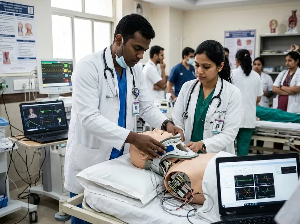
A novel proof-of-concept study presents a six-degree-of-freedom articulated manikin neck equipped with linear potentiometers. The system provides real-time kinematic motion feedback during cervical immobilisation, delivering sub-degree tracking accuracy to improve pre-hospital trauma training and patient safety.
Today

Novel glucose-responsive mPEG-PSA nanocarriers deliver insulin aspart on demand, achieving dual postprandial and basal glycemic control without hypoglycemia in diabetic models. This biomaterial breakthrough offers promising clinical implications for physiologic diabetes management.
Today
