

Loading, please wait...
Loading, please wait...

Historically, clinicians regarded bronchiectasis as an exclusively neutrophilic disease driven by recurrent microbial infection and impaired mucociliary clearance. However, modern translational evidence clearly establishes that eosinophilic bronchiectasis represents a distinct and clinically meaningful inflammatory phenotype. Up to thirty percent of individuals with non-cystic fibrosis bronchiectasis demonstrate systemic or airway eosinophilia. Consequently, researchers now recognize that heterogeneous biological pathways govern airway damage in these patients. In the multicentric Turkish cohort evaluated by Tümüklü and colleagues, investigators validated peripheral eosinophil distribution patterns previously observed across Western European cohorts. By identifying patients through routine complete blood counts, clinicians can rapidly stratify individuals without requiring invasive bronchoscopic sampling. Therefore, distinguishing between eosinophilic and non-eosinophilic subsets provides essential insights into disease biology, exacerbation frequency, and long-term functional decline.
Furthermore, recognizing this phenotype transforms conventional treatment models from empirical antibiotic cycling to targeted biological interventions. Because airway inflammation damages bronchial architecture progressively, early phenotyping prevents irreversible pulmonary remodelling. Indian clinicians frequently encounter post-infectious bronchiectasis, where chronic inflammation persists even after pathogen eradication. Thus, tracking blood eosinophils helps clinicians identify secondary type 2 inflammation promptly. When physicians adopt this systematic phenotyping approach, they can personalize patient care and optimize clinical outcomes effectively.
The biological mechanisms underlying eosinophil recruitment in bronchiectasis involve complex interactions between airway epithelial cells and adaptive immune pathways. Specifically, damaged bronchial epithelial cells release innate alarmins such as interleukin-33, interleukin-25, and thymic stromal lymphopoietin in response to environmental triggers or chronic insults. Subsequently, these alarmins stimulate type 2 innate lymphoid cells and helper T cells, which secrete large volumes of interleukin-5, interleukin-4, and interleukin-13. Interleukin-5 plays a pivotal role because it controls eosinophil maturation, terminal differentiation, and survival in peripheral circulation. In addition, interleukin-13 drives goblet cell metaplasia, which worsens mucus hypersecretion and accelerates airway plugging. As activated eosinophils migrate into the bronchial wall, they release cytotoxic granule proteins, including eosinophil peroxidase and major basic protein.
Moreover, these cationic proteins cause extensive tissue damage and disrupt the epithelial barrier. Interestingly, eosinophilic inflammation frequently coexists with low-grade bacterial colonization rather than mutual exclusion. Therefore, dual-pathway activation often triggers concurrent neutrophilic proteolysis and eosinophil-mediated oxidative tissue destruction. Consequently, patients develop accelerated bronchomalacia, persistent bronchorrhea, and severe airflow limitation over time.
Differentiating eosinophilic bronchiectasis from non-eosinophilic presentations requires careful clinical, physiological, and high-resolution computed tomography evaluations. Non-eosinophilic cohorts generally exhibit classical neutrophilic pathology characterized by persistent mucopurulent sputum, recurrent bacterial infections, and frequent colonization by organisms like Pseudomonas aeruginosa. In contrast, individuals with eosinophilic traits often present with dry cough, recurrent wheezing, and pronounced bronchial hyperreactivity. Clinicians frequently note that these patients report prominent diurnal symptom variation resembling severe asthma. Furthermore, spirometry reveals accelerated declines in forced expiratory volume in one second among eosinophilic individuals, reflecting active peripheral airway inflammation.
Radiologically, high-resolution computed tomography demonstrates distinct morphological features between these inflammatory subsets. Although non-eosinophilic disease shows severe cystic changes across lower lobes, eosinophilic cohorts show prominent bronchial wall thickening and mucus impaction. Additionally, eosinophilic patients often exhibit multi-lobar or upper lobe predilection, which mirrors allergic fungal airway disorders. Radiologists also identify centrilobular nodules and mosaic attenuation secondary to small airway air trapping. Thus, correlating chest imaging findings with inflammatory markers sharpens diagnostic accuracy and directs therapy.
Clinicians assess systemic inflammation using reliable, widely accessible hematological biomarkers during stable outpatient visits. The primary cut-off for defining the eosinophilic phenotype rests at an absolute blood eosinophil count of 300 cells per microliter. Multiple longitudinal investigations confirm that this threshold reliably correlates with airway eosinophilia and elevated sputum eosinophil cationic protein. However, physicians must check baseline counts during stability because systemic steroids or bacterial infections suppress peripheral eosinophils temporarily. Therefore, experts recommend obtaining at least two separate complete blood count measurements before establishing a permanent phenotypic diagnosis.
In addition to absolute eosinophil numbers, complementary biomarkers significantly enhance clinical stratification. For instance, fractional exhaled nitric oxide measurements above 25 parts per billion reflect proximal epithelial cytokine activation driven by interleukin-13. Similarly, serum total immunoglobulin E and specific fungal testing exclude conditions like allergic bronchopulmonary aspergillosis. Meanwhile, composite hematological ratios such as the neutrophil-to-lymphocyte ratio provide valuable insights into background neutrophilic activity. By integrating these simple biological tests, physicians construct an accurate inflammatory profile for each individual without incurring excessive laboratory expenditures.
In India, bronchiectasis represents a substantial yet under-recognized clinical challenge across tertiary and community respiratory clinics. Historically, post-tubercular sequelae constitute the single largest etiology of bronchiectasis in Indian patients. However, persistent symptoms like chronic cough and wheeze often stem from unrecognized eosinophilic airway disease rather than recurring mycobacterial infection. Furthermore, high environmental exposure to biomass smoke, particulate air pollution, and airborne fungal spores commonly induces secondary allergic inflammation. Because tropical climates foster fungal growth, clinicians must systematically screen every patient for Aspergillus sensitization alongside standard eosinophil profiling.
Moreover, empirical antibiotic prescribing remains rampant in outpatient settings when treating persistent bronchiectatic exacerbations. When Indian physicians mistakenly attribute eosinophilic flares to acute bacterial surges, patients receive repeated, unnecessary courses of broad-spectrum antibiotics. Consequently, antimicrobial resistance rises while untreated airway eosinophilia causes ongoing pulmonary destruction. Therefore, routine complete blood count assessment offers an affordable, immediate tool for Indian doctors to separate bacterial exacerbations from eosinophilic flare-ups. Implementing this structured diagnostic approach will substantially reduce antimicrobial misuse and improve long-term respiratory care nationwide.
Recognizing inflammatory phenotypes directly influences clinical management and guides personalized respiratory therapy. In non-eosinophilic bronchiectasis, treatment prioritizes aggressive airway clearance, hypertonic saline nebulization, and targeted antimicrobial eradication for persistent pathogens. Conversely, patients with eosinophilic bronchiectasis benefit substantially from anti-inflammatory therapies that target type 2 cytokine cascades. Inhaled corticosteroids, combined with long-acting beta-agonists, effectively suppress bronchial mucosal swelling and decrease exacerbation rates in this specific subgroup. However, clinicians must avoid indiscriminate corticosteroid use in pure neutrophilic disease due to elevated mycobacterial infection risks.
In severe, refractory eosinophilic cases, monoclonal antibodies targeting interleukin-5, interleukin-5 receptor alpha, or interleukin-4 and interleukin-13 pathways offer promising therapeutic alternatives. Although randomized trials continue in non-cystic fibrosis bronchiectasis, real-world data show marked reductions in exacerbations and improved quality of life. In addition, novel oral inhibitors targeting dipeptidyl peptidase 1 show great promise for dampening mixed neutrophilic-eosinophilic inflammation. As translational research progresses, integrating biomarker-driven care will redefine pulmonary practice and transform outcomes for patients worldwide.
Most clinical studies define eosinophilic bronchiectasis using an absolute blood eosinophil count of 300 cells per microliter or higher during stable clinical states. Alternatively, some guidelines consider a threshold of 3 percent on differential leukocyte counts. However, clinicians must confirm this elevation across multiple routine testing intervals. Active bacterial exacerbations or concurrent oral corticosteroid therapies frequently suppress circulating eosinophil counts, which can mask underlying type 2 airway inflammation during acute clinical presentations.
Classic bronchiectasis management focuses primarily on daily airway clearance physiotherapy, mucolytics, and targeted antimicrobial treatments against chronic pathogen colonization. In contrast, eosinophilic bronchiectasis features prominent type 2 inflammatory cascades that respond favorably to anti-inflammatory therapies. Patients frequently experience reduced exacerbation rates and improved airflow when clinicians add inhaled corticosteroids to their standard regimen. Furthermore, refractory individuals with persistent eosinophilia may benefit from targeted biological therapies directed against interleukin-5 pathways.
Post-tubercular lung damage remains the most common predisposing factor for bronchiectasis across India. Many recovered tuberculosis patients suffer from persistent respiratory symptoms that clinicians misinterpret as persistent bacterial infections or disease reactivation. Evaluating blood eosinophil counts uncovers secondary type 2 airway inflammation, allergic bronchopulmonary aspergillosis, or asthma overlap. Identifying these phenotypes prevents unnecessary antibiotic cycles, prevents progressive pulmonary architectural damage, and enables physicians to initiate timely anti-inflammatory therapies that preserve lung function.
Disclaimer: This content is for informational and educational purposes only... Refer to the latest local and national guidelines for clinical practice.
References

Read summarized clinical updates, watch expert medical content, and earn CME certifications right from your smartphone.
Open your smartphone camera to scan and install.





Eosinophilic bronchiectasis represents a critical inflammatory phenotype affecting up to 30% of patients. Learn how identifying blood eosinophils guides targeted therapy and improves respiratory outcomes in clinical practice.
Today
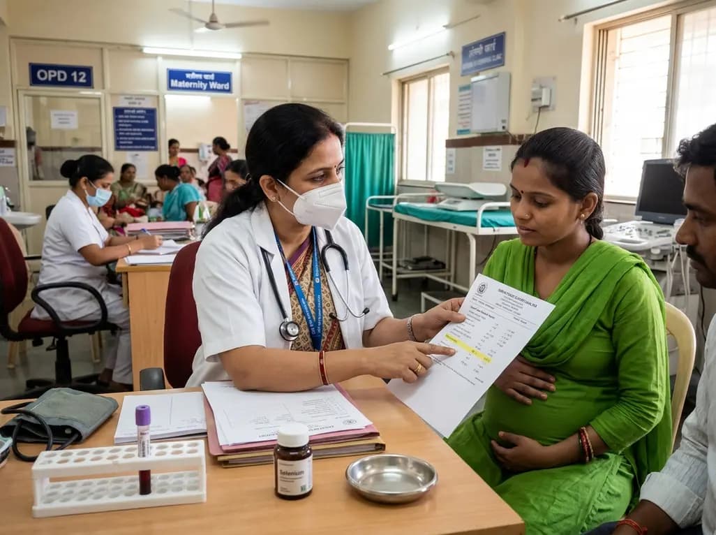
A prospective study reveals significant first-trimester trace-element alterations in pregnant women with epilepsy. Lower serum selenium independently predicted subsequent seizure recurrence despite prior remission, highlighting the vital clinical role of micronutrient profiling during antenatal care.
Today

With hypertension affecting over 300 million adults in India and household monitor penetration remaining under 5%, local manufacturing expansion of validated oscillometric devices marks a pivotal shift. This overview explores clinical protocols, diagnostic thresholds, and strategies to improve hypertension control.
Today

Atraumatic subcapital femoral neck fracture can emerge as the initial manifestation of osteonecrosis of the femoral head. A comprehensive systematic review highlights key diagnostic features, the indispensable role of MRI, and critical considerations when choosing between arthroplasty and internal fixation.
Today
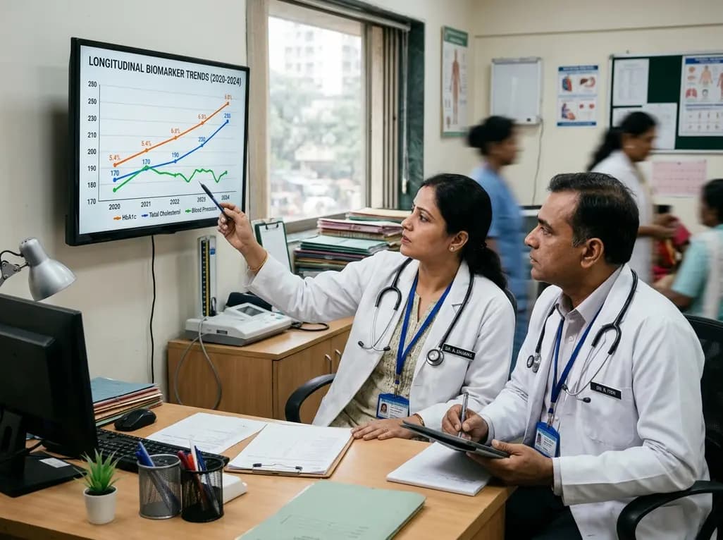
Annual health checks only offer static snapshots. Real-world evidence from 2.1 million insured Indians reveals that tracking dynamic biomarker trends over time enables clinicians to spot evolving cardiometabolic risks early, reverse borderline pathology, and deliver tailored preventive interventions.
Yesterday
