.webp&w=384&q=75)

Loading, please wait...
Loading, please wait...

Extracellular vesicles serve as vital mediators of intercellular communication and hold immense promise as non-invasive biomarkers for numerous pathological conditions. However, characterizing nanoscale membrane heterogeneity remains a formidable technical challenge in modern laboratory medicine. Liquid biopsy platforms frequently struggle with biological variability and sample batch effects during molecular quantification. To resolve these measurement inconsistencies, researchers developed a standardized analytical platform utilizing engineered nanovesicles alongside machine learning-optimized impedance spectroscopy. This breakthrough framework establishes calibrated biophysical standards for surface protein detection. Consequently, this innovation bridges the gap between basic membrane biophysics and translational diagnostic applications, particularly for complex neurodegenerative disorders.
Clinical diagnostics increasingly rely on extracellular vesicles to detect early pathologic alterations in cellular states. These lipid-bilayer capsules shuttle critical proteins, nucleic acids, and signaling lipids between distant tissues. However, analyzing vesicle surface proteins with conventional methods presents substantial technical hurdles. Standard techniques like enzyme-linked immunosorbent assays and conventional flow cytometry often require extensive chemical labeling. Furthermore, these optical assays demand substantial sample volumes and struggle to detect low-abundance surface epitopes accurately. Natural vesicle populations also exhibit significant size diversity and membrane heterogeneity, which distorts quantitative readouts across different diagnostic laboratories. Therefore, clinicians encounter difficulty when comparing biomarker data from disparate testing platforms. The absence of reliable, uniform reference standards has historically prevented regulatory standardization in vesicle-based liquid biopsies. In addition, conventional methods cannot easily capture dynamic changes in protein conformation or early oligomeric clustering on intact membranes. Consequently, investigators needed a robust methodology capable of characterizing native membrane interactions without altering molecular integrity. Developing standardized synthetic or semi-synthetic reference materials represents a decisive step toward eliminating analytical ambiguity. By addressing these physical constraints, clinicians can unlock reliable liquid biopsy diagnostics for neurodegenerative diseases and systemic illnesses.
To establish reliable analytical reference points, researchers designed biomimetic calibrators using genetically modified mammalian cellular systems. Specifically, investigators engineered HeLa cells to express precise copy numbers of amyloid-β 42 peptides on their outer membranes. By expressing one, three, or nine copies of the target protein, the team established distinct molecular densities. Subsequently, researchers extruded these modified cells through micro-porous membrane filters to generate standardized engineered nanovesicles. This mechanical extrusion process reliably preserves the native lipid orientation while replicating natural vesicular dimensions. Moreover, this approach generates high yields of stable vesicles with precisely controlled oligomeric surface configurations. These cell-derived vesicles provide an ideal reference standard for calibrating diagnostic instruments across multiple operational settings. Consequently, laboratory teams can now eliminate batch-to-batch variation by testing clinical samples against well-defined, reproducible biophysical controls. Furthermore, the synthetic calibrators mimic native membrane environments far better than artificial polystyrene beads or isolated recombinant proteins. This physiological fidelity ensures that surface-bound proteins maintain authentic folding kinetics and spatial interactions during downstream analysis. Ultimately, this scalable bioengineering approach delivers the consistent physical benchmarks necessary for rigorous clinical assay validation.
Electrical impedance spectroscopy measures how biological particles resist and store alternating electrical current across varying frequencies. However, raw spectral data contains complex multidimensional variables that obscure subtle membrane-protein interactions. To overcome this limitation, researchers implemented advanced machine learning algorithms to analyze impedance signatures systematically across frequencies ranging from 10 Hertz to 1 Megahertz. The computational model evaluated thousands of data points to identify the exact electrical parameters correlated with distinct protein oligomeric states. Notably, the machine learning workflow identified reactance changes at precisely 1 kilohertz as the optimal diagnostic feature. This specific frequency window effectively distinguished single-copy peptides from higher-order oligomers on the vesicular surface. Therefore, the algorithm streamlined data collection by eliminating non-informative electrical noise across other frequencies. In addition, this focused measurement significantly accelerated assay processing times, making rapid point-of-care evaluation practical. The integration of artificial intelligence transformed raw multi-frequency spectra into a straightforward quantitative readout of surface protein clustering. Consequently, this computational approach eliminates manual interpretation errors and standardizes impedance diagnostics. By coupling algorithmic feature selection with electrical sensing, clinicians gain a reliable tool for high-throughput biomolecular characterization.
Understanding the physical mechanisms driving electrical changes requires detailed biophysical and mathematical modeling. In this study, investigators constructed an equivalent-circuit model to elucidate how surface proteins alter vesicular electrical properties. The circuit analysis demonstrated that membrane capacitance directly correlates with protein oligomerization density on the lipid bilayer. Furthermore, structural predictions confirmed that larger protein assemblies displace surface hydration layers and rearrange local membrane dipoles. These nanoscale molecular rearrangements alter the dielectric characteristics of the vesicle surface in a predictable manner. Importantly, the platform operates entirely without fluorescent dyes, radioactive tracers, or antibody conjugations. This label-free capability allows researchers to monitor the time-resolved aggregation of amyloid-β peptides directly on intact membrane surfaces. As a result, scientists can track intermediate oligomer formation in real time under physiological buffer conditions. Because early oligomers represent the most neurotoxic species in amyloid pathologies, tracking their assembly dynamics provides crucial mechanistic knowledge. Thus, biophysical modeling bridges macroscopic impedance measurements with microscopic molecular phenomena. This versatile analytical framework enables researchers to study diverse membrane-associated protein misfolding pathways with unprecedented precision.
The development of this standardizable impedance platform introduces profound clinical implications for early neurodegenerative disease detection. Currently, diagnosing conditions like Alzheimer's disease relies on invasive cerebrospinal fluid draws or expensive positron emission tomography scans. However, neural cells continuously shed extracellular vesicles into peripheral circulation, providing a minimally invasive window into central nervous system pathology. By utilizing standardizable engineered nanovesicles as calibrators, clinical laboratories can accurately quantify circulating neurotoxic oligomers from routine blood samples. Furthermore, the high sensitivity of 1 kilohertz reactance measurements enables detection of pathological protein aggregates well before extensive neuronal death occurs. Early detection creates an invaluable therapeutic window for disease-modifying interventions and clinical trial recruitment. Additionally, pharmaceutical developers can employ this label-free platform to screen drug candidates designed to inhibit amyloid aggregation or dissociate existing oligomers. The standardized framework also supports therapeutic monitoring by tracking vesicle-associated protein dynamics over time during clinical treatments. Ultimately, this scalable, cost-effective bioelectronic technology could democratize neurodegenerative biomarker screening in hospital and community settings globally.
Engineered nanovesicles act as uniform biomimetic reference standards with defined surface protein configurations. Unlike synthetic plastic beads or recombinant proteins, these extruded vesicles accurately replicate native lipid bilayer environments and membrane curvature. Consequently, they establish reproducible biophysical baselines, allowing clinical diagnostic laboratories to eliminate analytical batch variability and standardize surface biomarker measurements across diverse liquid biopsy testing platforms reliably.
Electrical impedance spectroscopy provides rapid, label-free detection of biomolecular changes directly on vesicular membranes. Traditional optical assays require complex chemical labeling, antibody tags, and large sample volumes, which can distort delicate protein conformations. In contrast, impedance spectroscopy measures intrinsic dielectric alterations, enabling real-time, time-resolved monitoring of protein aggregation dynamics without disrupting native membrane structure or introducing chemical artifacts.
This innovative bioelectronic platform enables highly sensitive quantification of pathogenic amyloid-beta oligomers within peripheral blood-derived extracellular vesicles. Because it detects subtle membrane capacitance changes at 1 kilohertz, the system identifies toxic protein clustering at ultra-low concentrations. Therefore, clinicians can identify early neurodegenerative processes years before clinical dementia symptoms manifest, facilitating timely interventions and streamlined therapeutic clinical trials.
Disclaimer: This content is for informational and educational purposes only and should not be considered medical advice or a substitute for direct consultation with a qualified healthcare professional. Refer to the latest local and national guidelines for clinical practice.
References

Read summarized clinical updates, watch expert medical content, and earn CME certifications right from your smartphone.
Open your smartphone camera to scan and install.





A novel platform combining engineered cell-derived nanovesicles and machine learning-optimized impedance spectroscopy enables standardizable, label-free profiling of extracellular vesicle surface proteins and amyloid-beta oligomerization for diagnostic applications.
Today

A new study reveals critical factors driving ALS disease progression, survival disparities, and escalating caregiver burden, highlighting the power of multidisciplinary care.
Today

A multicenter Italian registry study evaluated 153 pregnancies in women with multiple sclerosis exposed to anti-CD20 monoclonal antibodies, demonstrating excellent maternal disease control and reassuring fetal safety without heightened risk of major congenital anomalies.
Today
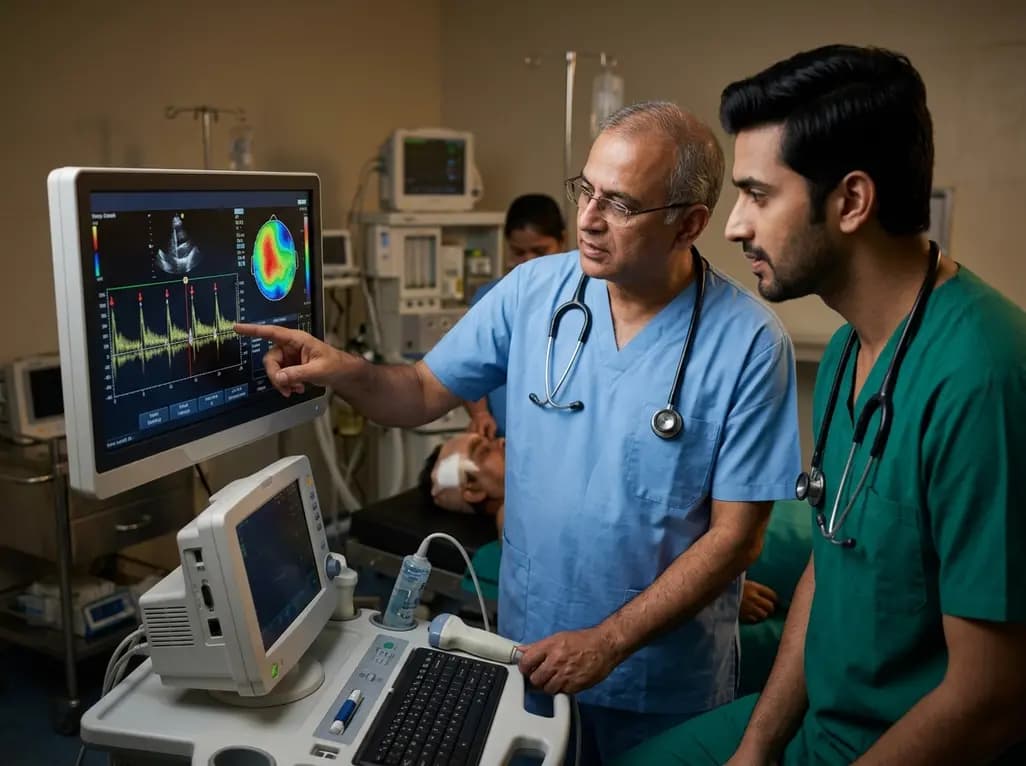
Recent evidence shows that cerebral microemboli can trigger cortical spreading depolarization in humans, presenting as post-surgical migraine aura. Real-time transcranial Doppler detection and prompt antiplatelet therapy offer vital diagnostic and therapeutic pathways for clinicians.
Today
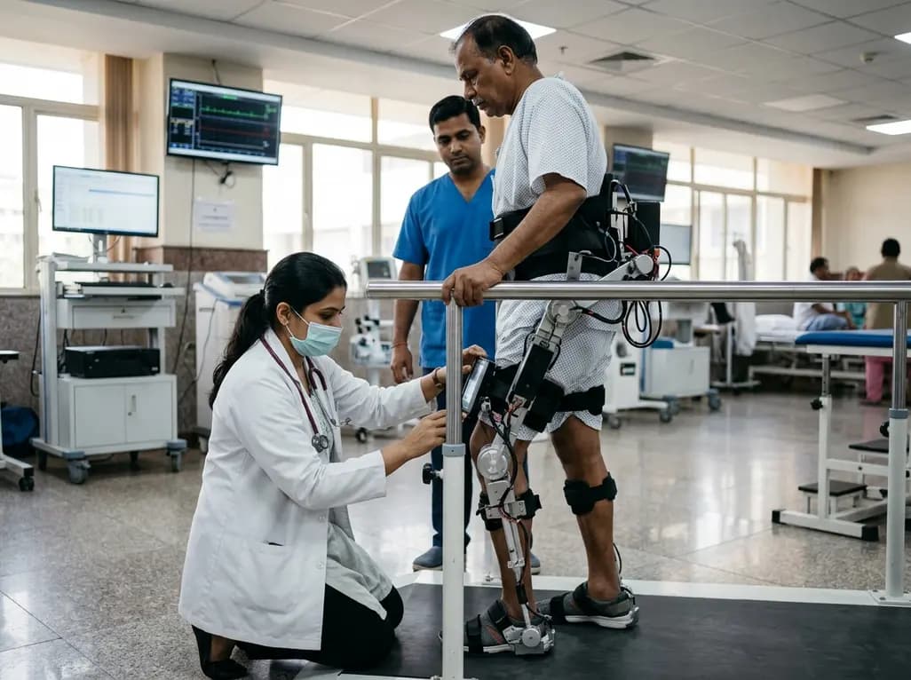
A breakthrough study introduces an AI-optimized Second-Order Sliding Mode Controller for lower extremity exoskeletons, improving tracking accuracy and disturbance rejection for safer, more effective neuro-rehabilitation.
Today

A 49-year-old man with uncontrolled type 2 diabetes developed a severe MSSA thigh abscess after inserting a continuous glucose monitor on his upper thigh. This case highlights the risks of off-label device placement and the critical role of interdisciplinary care in preventing cutaneous complications.
Today
