.webp&w=384&q=75)

Loading, please wait...
Loading, please wait...
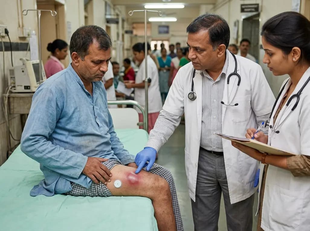
Continuous glucose monitoring (CGM) technology has revolutionized glycemic control for millions of patients living with diabetes mellitus worldwide. By providing real-time interstitial glucose readings, trend arrows, and customizable alerts, these devices improve time-in-range and reduce dangerous glycemic fluctuations. However, device-related cutaneous adverse events remain an underappreciated clinical challenge. While the majority of dermatological reactions consist of mild contact dermatitis or erythema, invasive bacterial complications can occur. A severe CGM sensor skin infection presenting as a deep subcutaneous abscess underscores the need for strict adherence to approved anatomical insertion sites, particularly in patients with severe baseline hyperglycemia.
A 49-year-old male with poorly controlled type 2 diabetes mellitus (glycated hemoglobin of 11.3%) was referred to an ambulatory care clinical pharmacist for intensive glycemic management. Because he had experienced recurrent sensor dislodgement from his posterior upper arm during daily physical activities, the patient elected to insert a new CGM sensor into his anterior upper thigh. Three days following placement, he presented to an outpatient clinic with intense localized pain, swelling, and difficulty bearing weight.
Clinical examination revealed an exquisitely tender, erythematous, fluctuant mass measuring 4.5 by 5.0 cm directly beneath the sensor insertion site. The clinical team diagnosed an acute subcutaneous abscess and promptly removed the sensor. The primary provider performed an initial bedside incision and drainage (I&D), yielding copious purulent material. The patient received a prescription for oral sulfamethoxazole/trimethoprim to provide empirical coverage for common skin pathogens. However, four days later, the patient returned with deteriorating pain, expanding induration, and significantly impaired ambulation. A repeat surgical incision and drainage was required to evacuate re-accumulated fluid. Wound cultures confirmed heavy growth of methicillin-susceptible Staphylococcus aureus (MSSA). The team initiated wound packing, applied topical mupirocin, and continued systemic antibiotics, leading to clinical resolution.
Manufacturers and regulatory authorities approve CGM sensors for specific anatomical zones, most notably the posterior upper arm and the abdomen. These validated locations offer predictable subcutaneous tissue depth, adequate interstitial fluid flow, and minimal mechanical disturbance. Conversely, when patients choose non-approved alternate sites such as the thigh, they inadvertently expose the insertion canal to heightened biomechanical stressors.
First, the thigh undergoes repetitive muscular contraction, continuous flexion during ambulation, and constant friction against clothing. This repeated shear stress can cause micro-pistoning of the transcutaneous sensor filament. Consequently, microscopic epidermal tears develop around the insertion cannula, disrupting the skin barrier. Second, the groin and upper thigh maintain higher surface temperatures, greater sweat production, and denser moisture accumulation. These environmental factors weaken adhesive dressings, trap moisture against the stratum corneum, and promote rapid proliferation of cutaneous microbiota. When an indwelling foreign body breaches the damaged barrier under these conditions, a localized CGM sensor skin infection can rapidly evolve into an invasive, deep-space abscess.
Chronic, uncontrolled hyperglycemia represents a major independent risk factor for pyogenic soft tissue infections. Patients with markedly elevated glycated hemoglobin levels exhibit profound polymorphonuclear neutrophil dysfunction, including impaired chemotaxis, defective phagocytosis, and diminished intracellular bactericidal activity. Furthermore, persistent microvascular disease reduces localized tissue perfusion and impairs early inflammatory recruitment.
In this clinical scenario, the patient’s severely elevated HbA1c of 11.3% created an ideal metabolic environment for pyogenic bacteria. Staphylococcus aureus is the predominant commensal organism colonizing the human skin and anterior nares. Once the continuous glucose sensor breached the stratum corneum, the organism accessed deep subcutaneous adipose layers. Bacteria readily establish dense biofilms on synthetic polyurethane and metallic device components. These biofilms shield pathogens from host phagocytic cells and systemic antibiotics alike. Therefore, even a seemingly minor transcutaneous insertion can escalate into extensive tissue necrosis, suppurative collections, and debilitating cellulitis if the host defense mechanism is metabolically compromised.
The management of device-associated soft tissue infections requires an aggressive, structured approach. When a patient presents with localized fluctuance, erythema, and severe focal tenderness at an insertion site, clinicians must immediately remove the indwelling foreign material. Source control remains the single most critical intervention. Antimicrobial therapy alone cannot effectively sterilize an undrained subcutaneous fluid collection or overcome established biofilm architecture.
Bedside incision and drainage must be thorough, ensuring complete disruption of internal loculations and comprehensive evacuation of purulence. In complex or recurrent cases, wound packing facilitates continuous drainage and prevents premature superficial closure. Clinicians should consistently obtain purulent material for Gram stain and microbiological culture prior to administering antimicrobial therapy. Although empirical oral regimens often target community-acquired methicillin-resistant Staphylococcus aureus (MRSA), definitive therapy must align with isolated susceptibility profiles. For confirmed MSSA, targeted oral agents such as cephalexin, dicloxacillin, or continuing susceptible agents like co-trimoxazole ensure optimal clearance. Concurrently, clinicians must closely assess glycemic stability, as systemic inflammation frequently triggers marked insulin resistance.
Preventing invasive skin complications requires rigorous patient education and standardized device handling protocols. Clinicians and diabetes educators must emphasize that patients should never alter manufacturer-recommended insertion locations without explicit medical guidance. Approved sites provide validated safety margins that mitigate mechanical trauma and microbial translocation.
Healthcare professionals should instruct patients to clean the intended insertion area thoroughly using isopropyl alcohol or chlorhexidine swabs, allowing the skin to dry completely before device application. Clinicians must advise against placing sensors over broken skin, active rashes, severe scar tissue, or areas subjected to high friction from waistbands. Additionally, patients who struggle with adhesive failure or premature dislodgement should utilize medical-grade over-bandages, skin-prep barrier wipes, or specialized adhesive patches rather than migrating the sensor to off-label anatomical sites. Patients must also inspect the insertion site daily through clear adhesive windows or during sensor changes, seeking prompt evaluation if localized redness expands beyond the device border.
The growing adoption of continuous glucose monitoring in primary care and outpatient endocrinology emphasizes the indispensable value of an interdisciplinary diabetes management team. Ambulatory care pharmacists, nurse educators, and clinical specialists frequently serve as the primary point of contact for routine device troubleshooting, dose titrations, and ongoing patient education.
During routine clinical encounters, clinicians should actively inspect device attachment sites and review practical application techniques. When patients report recurrent sensor detachment, educators must investigate the root cause, such as excessive perspiration, rigorous exercise, or incorrect skin preparation. By proactively identifying improper off-label device usage, clinicians can redirect patients toward safe retention strategies before complications occur. Furthermore, pharmacists and triage clinicians possess the clinical acumen to distinguish benign contact dermatitis from early bacterial soft tissue infection. Prompt referral for surgical drainage and tailored antibiotic therapy prevents extensive tissue destruction, reduces emergency department visits, and preserves patient confidence in essential diabetes technologies.
While some patients place sensors on their thighs to prevent accidental displacement, regulatory approvals primarily specify the posterior upper arm or abdomen. The thigh experiences frequent mechanical shear, friction from clothing, and moisture accumulation from sweat. Consequently, placing sensors in alternate non-validated anatomic zones substantially increases the risk of mechanical dislodgement, skin barrier disruption, and bacterial colonization, which can precipitate serious cutaneous infections requiring invasive drainage.
Clinicians and patients should actively monitor for progressive erythema extending beyond one centimeter, localized warmth, severe tenderness, fluctuance, and purulent discharge. Unlike common allergic contact dermatitis, which manifests primarily with diffuse pruritus and superficial scaling, a pyogenic infection produces worsening localized pain, throbbing discomfort, and systemic manifestations such as fever or sudden, unexplained elevations in interstitial glucose readings due to acute systemic inflammation.
Treatment begins with immediate removal of the indwelling sensor and careful clinical assessment for fluctuance. If an abscess is present, prompt incision and drainage serves as the cornerstone of effective management. Clinicians should obtain wound cultures to guide pathogen-directed antimicrobial therapy, such as oral sulfamethoxazole/trimethoprim or clindamycin for staphylococcal species, supplemented with wound packing, topical mupirocin, and rigorous glycemic optimization to accelerate recovery.
Disclaimer: This content is for informational and educational purposes only. It is not intended to provide medical advice or to be a substitute for professional medical advice, diagnosis, or treatment. Patients should always consult their physician or other qualified healthcare provider with any questions they may have regarding a medical condition. Healthcare professionals should exercise their own independent clinical judgment when interpreting and applying this information. Refer to the latest local and national guidelines for clinical practice.
References
Shaeer KM et al. Thigh abscess following alternate-site continuous glucose monitoring sensor placement. Am J Health Syst Pharm. 2026 Aug 28. doi: undefined. PMID: 42664480.
Attalla J, Maletkovic J. MSSA Cellulitis and Abscess as a Complication of the Use of Continuous Glucose Monitoring. J Endocr Soc. 2021;5(Suppl 1):A395-A396.
Heinemann L, Freckmann G. CGM Versus FGM; or, Continuous Glucose Monitoring Is Not Continuous Glucose Monitoring. J Diabetes Sci Technol. 2015;9(5):947-950.

Read summarized clinical updates, watch expert medical content, and earn CME certifications right from your smartphone.
Open your smartphone camera to scan and install.





A 49-year-old man with uncontrolled type 2 diabetes developed a severe MSSA thigh abscess after inserting a continuous glucose monitor on his upper thigh. This case highlights the risks of off-label device placement and the critical role of interdisciplinary care in preventing cutaneous complications.
4 weeks back

Red blood cell transfusion is critical in sickle cell disease management but brings significant immunological complexity, including alloimmunisation and iron overload. Extended RBC phenotyping and tailored protocols optimize clinical safety.
Today
Discover the crucial distinctions between mesonephric and mesonephric-like proliferations of the female genital tract. Learn the key histomorphologic, immunohistochemical, and molecular differences essential for accurate diagnosis and clinical management.
Today

A large UK Biobank study reveals that plasma proteomic signatures capture preclinical organ damage and improve multiorgan risk prediction across early CKM syndrome stages 0 to 2 beyond traditional PREVENT clinical models.
Today

A retrospective CBCT study reveals that superior TMJ space asymmetry correlates moderately with occipital condyle-atlas-axis asymmetry in unilateral TMJ osteoarthritis, highlighting an imaging correlate between jaw biomechanics and upper cervical alignment rather than a standalone diagnostic marker.
Today

A Bayesian multilevel meta-analysis reveals that aerobic training combined with moderate carbohydrate restriction modestly lowers HbA1c in type 2 diabetes. However, sparse data and very low certainty leave incremental benefits over exercise or diet alone unproven, highlighting the need for individualized care.
Today
