.webp&w=384&q=75)

Loading, please wait...
Loading, please wait...
Cortical spreading depolarization represents a profound electrophysiological wave that propagates slowly across the cerebral cortex. Historically recognized as the primary substrate underlying migraine aura, this phenomenon involves massive neuronal and glial depolarization accompanied by marked ionic shifts. However, clinicians increasingly recognize that cortical spreading depolarization is not confined solely to primary headache conditions. Emerging clinical evidence demonstrates that physical vascular perturbations, such as microemboli, can directly initiate these self-propagating cortical events. Consequently, understanding this mechanism provides vital insight into transient neurological deficits occurring after cardiothoracic and vascular procedures.
Electrophysiologically, cortical spreading depolarization is characterized by a nearly complete breakdown of transmembrane ion gradients. Neurons and astrocytes release substantial amounts of intracellular potassium and glutamate into the extracellular space. This biochemical surge triggers widespread cellular depolarization. Subsequently, this wave travels slowly across cortical gray matter at a rate of approximately two to five millimeters per minute. As the depolarization wave progresses, it temporarily shuts down normal synaptic transmission, resulting in transient focal neurological symptoms.
Furthermore, extensive translational studies demonstrate that diverse physical and metabolic triggers can elicit these propagating waves. Localized hypoxia, cortical mechanical stimulation, and microvascular occlusions reliably generate depolarization events. In well-perfused cortical tissue, these waves typically resolve without producing permanent structural damage. However, when metabolic reserves become compromised, recurring depolarization waves can accelerate ischemic injury. Therefore, recognizing the broad clinical spectrum of spreading depolarization helps clinicians identify unusual transient neurological deficits in postoperative settings.
A recent milestone clinical report evaluated a 68-year-old woman who developed daily episodes of typical visual migraine aura following mitral valve surgery. Clinicians performed real-time transcranial Doppler monitoring during active aura episodes to investigate potential vascular triggers. Remarkably, transcranial Doppler ultrasonography demonstrated high-intensity transient signals, confirming the active passage of microemboli through the posterior cerebral circulation.
Notably, the patient experienced sequential, retinotopically spreading positive visual phenomena during the exact periods of microembolic transit. Following the initiation of antiplatelet therapy, the recurrent daily aura attacks completely ceased. Furthermore, repeated transcranial Doppler examinations confirmed the total disappearance of microembolic signals in the posterior circulation. This case provides direct evidence that particulate or gaseous microemboli can immediately precipitate cortical spreading depolarization in humans without causing permanent infarction on brain imaging.
The biological bridge connecting particulate vascular occlusion and neuroelectrical excitation involves acute microvascular mismatch. When microemboli travel through distal cerebral arterioles, they cause momentary focal hypoperfusion. Although these tiny occlusions may not persist long enough to produce permanent ischemic necrosis, they acutely impair local oxidative phosphorylation. Consequently, sodium-potassium pumps fail in the immediate microenvironment, leading to localized extracellular potassium accumulation.
Moreover, this focal ion imbalance reaches the critical threshold necessary to ignite a full cortical spreading depolarization event. Once ignited, the depolarization wave propagates beyond the original territory of the microvascular occlusive event. Additionally, the propagating depolarization prompts reactive hyperemia followed by prolonged oligemia across the affected cortex. Importantly, particulate thrombi and air microemboli both successfully lower the threshold for depolarization. As a result, patients without a history of migraine can abruptly develop typical aura symptoms in the presence of an active embolic source.
Clinicians frequently struggle to distinguish between transient ischemic attacks and atypical migraine aura symptoms. Traditional transient ischemic attacks typically present with sudden, simultaneous negative neurological deficits, such as hemiparesis, aphasia, or monocular blindness. In contrast, cortical spreading depolarization produces characteristic gradual, sequential spreading symptoms that involve both positive and negative features.
For instance, patients experiencing depolarization-mediated visual episodes describe scintillating scotomas, zigzag flashing lines, or expanding visual field defects that spread retinotopically over twenty to thirty minutes. Similarly, somatosensory manifestations spread sequentially over minutes from fingers to hand, forearm, and perioral areas. Therefore, evaluating the tempo and progression of symptoms remains the most powerful clinical differentiator.
However, the realization that microemboli directly induce spreading depolarization blurs the traditional boundary between structural ischemic events and functional migraine auras. Misdiagnosing these episodes as benign primary migraines can lead to dangerous delays in identifying cardiac or vascular sources of emboli. Clinicians should therefore treat new-onset aura in older adults as a potential thromboembolic warning sign.
Managing microemboli-induced cortical spreading depolarization requires immediate identification and elimination of the underlying embolic source. Because these transient spells stem from physical vascular microemboli rather than primary cortical hyperexcitability, standard migraine prophylactics often provide inadequate relief. Instead, antithrombotic therapies represent the definitive treatment strategy.
Specifically, clinicians should perform comprehensive cardiovascular and neurovascular evaluations when patients develop unexplained late-onset aura episodes. Diagnostic assessments must include echocardiography to evaluate valve function and rule out intracardiac shunts, alongside transcranial Doppler monitoring. When microembolic signals appear on transcranial Doppler, optimizing antiplatelet or anticoagulant regimens quickly eliminates microembolic shower activity.
Furthermore, antiplatelet agents such as aspirin and clopidogrel suppress platelet aggregation on mechanical prostheses and atheromatous plaques. Achieving complete microembolic clearance results in total cessation of aura symptoms. In addition, secondary stroke risk diminishes substantially once therapeutic antithrombotic coverage stabilizes the vulnerable endothelium. Therefore, prompt initiation of antiplatelet therapy protects both cortical neurophysiology and long-term vascular health.
Cardiothoracic surgeries, structural heart interventions, and endovascular procedures carry inherent risks of particulate and gaseous microembolization. During cardiopulmonary bypass and valve manipulation, microscopic particles frequently enter the arterial circulation. Although modern arterial filters capture larger emboli, subclinical microemboli often reach the cerebral microvasculature.
Consequently, many postoperative patients report transient visual obscurations, sensory tingling, or cognitive clouding that standard computed tomography scans fail to explain. Understanding that cortical spreading depolarization mediates these post-surgical symptoms transforms how multidisciplinary teams approach postoperative neurovascular care. Rather than dismissing transient visual anomalies as anesthesia aftereffects or migraine mimics, surgical teams should initiate prompt neurovascular workups.
Moreover, transcranial Doppler ultrasonography provides a practical, non-invasive bedside tool to monitor cerebral microembolism continuously. Integrating microemboli detection protocols in high-risk cardiac recovery units enables clinicians to titrate anticoagulant and antiplatelet medications dynamically. Ultimately, translating benchside neurophysiology into clinical bedside management improves diagnostic accuracy, prevents unnecessary diagnostic delays, and safeguards patients against progressive cerebrovascular ischemia.
Cerebral microemboli lodge temporarily within distal microvessels, producing localized microvascular hypoperfusion and energy depletion. This transient metabolic stress impairs ATP-dependent sodium-potassium pumps in cortical neurons and glia. Consequently, rapid intracellular potassium efflux and glutamate release occur within the microenvironment. When extracellular potassium surpasses a critical threshold, it triggers a self-propagating wave of cortical spreading depolarization across adjacent healthy brain tissue, producing characteristic spreading aura symptoms.
Classic transient ischemic attacks typically produce sudden, simultaneous negative neurological deficits, such as sudden weakness or numbness. In contrast, microembolism-induced cortical spreading depolarization generates sequential, gradually spreading symptoms over five to thirty minutes. Patients typically report positive features, including scintillating scotomas, zigzag patterns, or marching paresthesias, reflecting the physical propagation of depolarization across cortical somatotopic or retinotopic maps rather than fixed focal territorial ischemia.
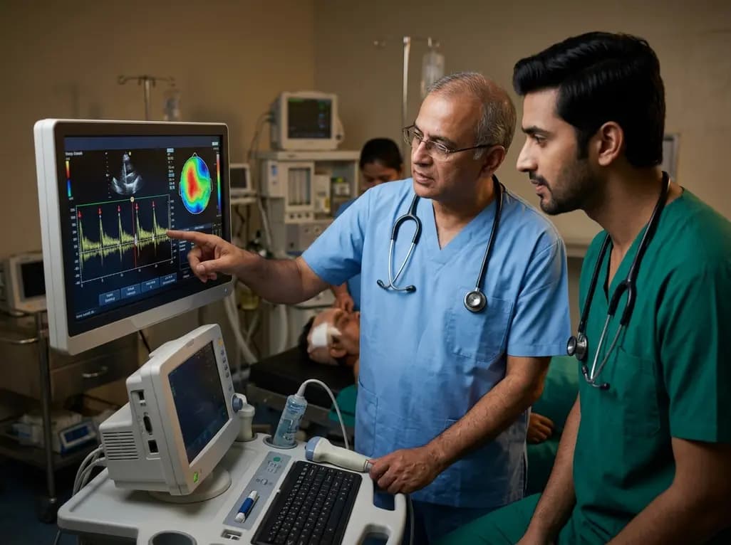
Transcranial Doppler ultrasonography serves as a vital non-invasive bedside diagnostic tool for detecting microembolic signals in real time. Following cardiac or vascular procedures, transcranial Doppler can identify high-intensity transient signals within the middle or posterior cerebral arteries during active neurological episodes. Demonstrating microemboli helps clinicians confirm a thromboembolic trigger, distinguish secondary aura from primary headache disorders, and assess therapeutic response to antiplatelet or anticoagulant interventions.
Disclaimer: This content is for informational and educational purposes only and is intended for healthcare professionals. It does not constitute medical advice, diagnosis, or treatment recommendations. Always consult clinical judgment and refer to the latest local and national guidelines for clinical practice.
References

Read summarized clinical updates, watch expert medical content, and earn CME certifications right from your smartphone.
Open your smartphone camera to scan and install.





Recent evidence shows that cerebral microemboli can trigger cortical spreading depolarization in humans, presenting as post-surgical migraine aura. Real-time transcranial Doppler detection and prompt antiplatelet therapy offer vital diagnostic and therapeutic pathways for clinicians.
4 weeks back

A systematic review of 41 preclinical studies reveals that baicalin dampens systemic inflammation, inhibits NF-κB and NLRP3 pathways, and improves organ preservation in sepsis models. However, substantial study heterogeneity and a complete lack of human clinical trials highlight the need for further translational research.
Today
Discover the crucial distinctions between mesonephric and mesonephric-like proliferations of the female genital tract. Learn the key histomorphologic, immunohistochemical, and molecular differences essential for accurate diagnosis and clinical management.
Today

A large UK Biobank study reveals that plasma proteomic signatures capture preclinical organ damage and improve multiorgan risk prediction across early CKM syndrome stages 0 to 2 beyond traditional PREVENT clinical models.
Today
A narrative review investigates whether the articularis genus muscle functions as an independent anatomical entity or blends with the vastus intermedius. We evaluate its morphology, role in retracting the suprapatellar bursa, and direct clinical significance in anterior knee pain and arthroplasty.
Today

A Bayesian multilevel meta-analysis reveals that aerobic training combined with moderate carbohydrate restriction modestly lowers HbA1c in type 2 diabetes. However, sparse data and very low certainty leave incremental benefits over exercise or diet alone unproven, highlighting the need for individualized care.
Today
