

Loading, please wait...
Loading, please wait...

Neurosurgical procedures in the posterior fossa require exceptional precision to protect eloquent neural structures. The endoscopic trans-Magendie approach has emerged as an innovative technique for accessing lesions of the fourth ventricle. Historically, surgical teams rely on telovelar or transvermian corridors to explore this cavity. However, traditional microscopic visualization imposes physical constraints and rigid lines of sight. A recent anatomical study by Leone and colleagues introduces a retractorless endoscopic corridor through a suboccipital burr hole. This strategy provides effective visualization while preserving craniocervical stability.
Accessing the fourth ventricle has historically presented major technical dilemmas in neurosurgery. The classic transvermian route requires incising the cerebellar vermis. Consequently, patients frequently face postoperative cerebellar mutism syndrome and severe equilibrium disturbances. To prevent such complications, neurosurgeons developed the telovelar approach. This corridor enters the ventricle through the cerebellomedullary fissure by opening the tela choroidea and inferior medullary velum.
Although the telovelar corridor protects vermian tissue, it still presents distinct line-of-sight limitations under microsurgical illumination. The microscopic view restricts the cranial angle of attack, which hampers inspection of the upper ventricular chamber. Moreover, evaluating the patency of the cerebral aqueduct remains challenging with a straight microscopic trajectory. To achieve an adequate upward angle, operators frequently resect the posterior arch of the first cervical vertebra (C1). While this laminectomy expands the working corridor, it introduces additional operative trauma. Furthermore, extensive bone removal increases the risk of craniocervical instability and persistent postoperative neck pain. Therefore, minimally invasive corridors that avoid C1 bone resection offer clear clinical advantages.
The endoscopic trans-Magendie approach addresses operative limitations by utilizing the natural central aperture of the fourth ventricle. In this anatomical feasibility study, investigators evaluated four formalin-fixed, vascularly injected cadaveric specimens. Specifically, the authors developed a retractorless trajectory using high-definition rigid endoscopes through a limited suboccipital burr hole craniotomy.
Instead of requiring an extensive suboccipital craniectomy, this operative strategy utilizes a minimal keyhole opening. The surgical trajectory navigates directly through the cisterna magna toward the foramen of Magendie. Because the neuroendoscope brings light and wide-angle optics directly to the target, surgeons do not require mechanical brain retractors. Retractorless surgery substantially decreases local tissue ischemia and avoids contusion of the cerebellar tonsils. In addition, the endoscope provides angled visualization across complex anatomical corners. Consequently, the surgical team can clearly inspect the inferior medullary velum, the tela choroidea, and the lateral recesses. Most importantly, entering through the natural foramen preserves critical surrounding architecture. As a result, this refined corridor creates an unobstructed avenue to deep ventricular pathology while respecting delicate regional neuroanatomy.
A primary finding of this feasibility analysis centers on eliminating the traditional C1 laminectomy. When performing conventional microsurgery, the surgeon requires a wide cranial trajectory to view the ventricular roof and aqueduct. In contrast, the endoscopic trans-Magendie approach circumvents this geometric constraint through close-up, angled optic visualization.
During anatomical dissections, the researchers achieved adequate cranial and lateral visualization of the fourth ventricle floor without disturbing the posterior C1 arch. The endoscopic shaft enters through the suboccipital burr hole and passes smoothly above the C1 ring. Thus, the optical instrument directly reaches the inferior two-thirds of the fourth ventricle. Furthermore, angled endoscopes offer panoramic views of the caudalmost portion of the cerebral aqueduct. Surgeons can confirm aqueductal patency without applying traction to the brainstem. In addition, the visualization extends laterally toward the foramina of Luschka, exposing vital colliculi safely. Because the surgical team eliminates bone removal at the craniocervical junction, patients preserve the suboccipital muscular attachments. Therefore, this targeted strategy reduces operative morbidity and safeguards structural stability.
Transitioning from large suboccipital craniotomies to keyhole access marks an essential milestone in cranial surgery. In this technique, the operator creates a single, precisely positioned suboccipital burr hole rather than a wide craniotomy. This limited opening minimizes blood loss and shortens operative duration considerably.
Additionally, smaller bone openings dramatically diminish soft tissue disruption and postoperative wound complications. Conventional wide suboccipital craniotomies frequently cause troublesome cerebrospinal fluid leaks and pseudomeningoceles. By keeping the craniotomy minimal and dural opening small, the trans-Magendie corridor reduces the incidence of fluid collections. Furthermore, the retractorless nature of neuroendoscopy protects the posterior inferior cerebellar arteries and their delicate perforating branches. Surgeons navigate working instruments alongside the endoscope, using dynamic bimanual manipulation. In addition, neurophysiological monitoring remains fully compatible with this minimally invasive corridor. Operating through a small burr hole preserves muscular attachments, accelerating patient postoperative mobilization. By uniting miniature bone removal with advanced visualization, this approach delivers an elegant pathway for neurosurgical intervention.
The findings of this anatomical study carry direct clinical relevance for managing diverse intraventricular lesions. Pathologies situated in the fourth ventricle include ependymomas, medulloblastomas, choroid plexus papillomas, cavernous malformations, and epidermoid cysts. Traditionally, accessing these deep-seated lesions required significant cerebellar retraction.
Fortunately, the full-endoscopic trans-Magendie corridor permits targeted tumor biopsy, lesion debulking, or complete microdissection through a natural anatomical conduit. For non-infiltrative or soft tumors, such as epidermoid cysts or cavernomas, endoscopic visualization allows excellent dissection off the delicate ventricular floor. Moreover, clear visualization of the obex and the vagal and hypoglossal triangles prevents disastrous brainstem injury. In patients presenting with obstructive hydrocephalus, the surgeon can inspect the aqueduct of Sylvius and verify adequate cerebrospinal fluid outflow. However, operating in deep, narrow spaces under endoscopic visualization alters standard depth perception. Surgeons must overcome the loss of binocular stereoscopic vision through dedicated training. Moving forward, clinical trials and prospective registries must corroborate these cadaveric observations. Ultimately, merging endoscopic optics with advanced neuronavigation will ensure the safest patient outcomes.
The endoscopic trans-Magendie approach is a minimally invasive neurosurgical corridor directed into the fourth ventricle. It utilizes the natural foramen of Magendie via a suboccipital burr hole craniotomy. This retractorless technique provides clear illumination and wide-angle visualization without requiring cerebellar vermian division or extensive bone resection.
Avoiding resection of the posterior arch of C1 prevents disruptions to craniocervical structural stability and reduces operative trauma. Preserving the C1 bone maintains vital ligamentous attachments, lowers the incidence of persistent postoperative neck pain, and diminishes the long-term risk of spinal deformity or suboccipital muscle atrophy.
This approach accommodates lesions located within the inferior two-thirds of the fourth ventricle. Suitable pathologies include low-grade ependymomas, cavernous malformations, epidermoid cysts, and subependymomas. Additionally, surgeons utilize this corridor to evaluate aqueductal patency, resolve cerebrospinal fluid pathway obstructions, and conduct targeted biopsies with minimal neurological morbidity.
Disclaimer: This content is for informational and educational purposes only. It is not intended to be a substitute for professional medical advice, diagnosis, or treatment. Always seek the advice of your physician or other qualified health provider with any questions you may have regarding a medical condition. Refer to the latest local and national guidelines for clinical practice.
References

Read summarized clinical updates, watch expert medical content, and earn CME certifications right from your smartphone.
Open your smartphone camera to scan and install.





An anatomical feasibility study demonstrates that the full-endoscopic trans-Magendie approach provides clear visualization of the fourth ventricle and cerebral aqueduct through a minimally invasive burr hole, eliminating the need for C1 laminectomy and extensive suboccipital craniectomy.
Today

The 2025 Australian meningococcal surveillance report shows a 13% decline in invasive meningococcal disease notifications to 118 cases. Genogroup B accounted for 82.2% of typed infections, predominantly impacting infants and young adults, highlighting critical vaccination and rapid diagnostic imperatives.
Today
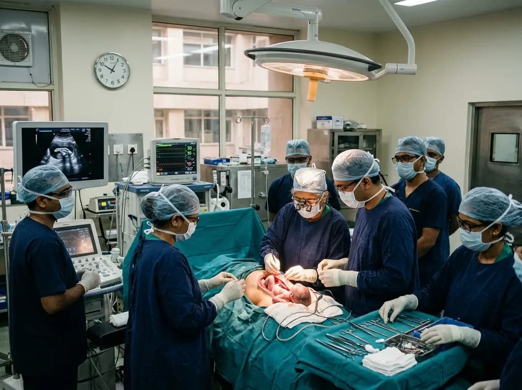
Doctors at AIIMS New Delhi have achieved a historic milestone by performing India's first open prenatal myelomeningocele repair on a 25-week unborn fetus. This landmark multidisciplinary procedure provides vital neuroprotective benefits, lowers hydrocephalus risks, and transforms maternal-fetal medicine nationwide.
Today

The ACE-PAS registry evaluated 21,612 patients fitted with the ASSURE wearable cardioverter defibrillator. The study demonstrated a 100% ventricular arrhythmia conversion rate, an inappropriate shock incidence of 0.0065 per patient-month, and high daily compliance of 23.1 hours, confirming real-world safety.
Today

A clinical overview of resveratrol supplementation in mitigating exercise-induced muscle damage. This narrative review explores physiological mechanisms, reductions in soreness markers like creatine kinase, dosage strategies, and bioavailability enhancements for sports medicine and nutrition practitioners.
Today

Childhood insulinoma is a rare cause of recurrent hyperinsulinemic hypoglycemia that often eludes early diagnosis. Advanced GLP-1 receptor-targeted PET/CT imaging using 68Ga-exendin-4 complements anatomic MRI to localize occult pancreatic lesions, facilitating focused, curative surgical enucleation.
Today
