.webp&w=384&q=75)

Loading, please wait...
Loading, please wait...
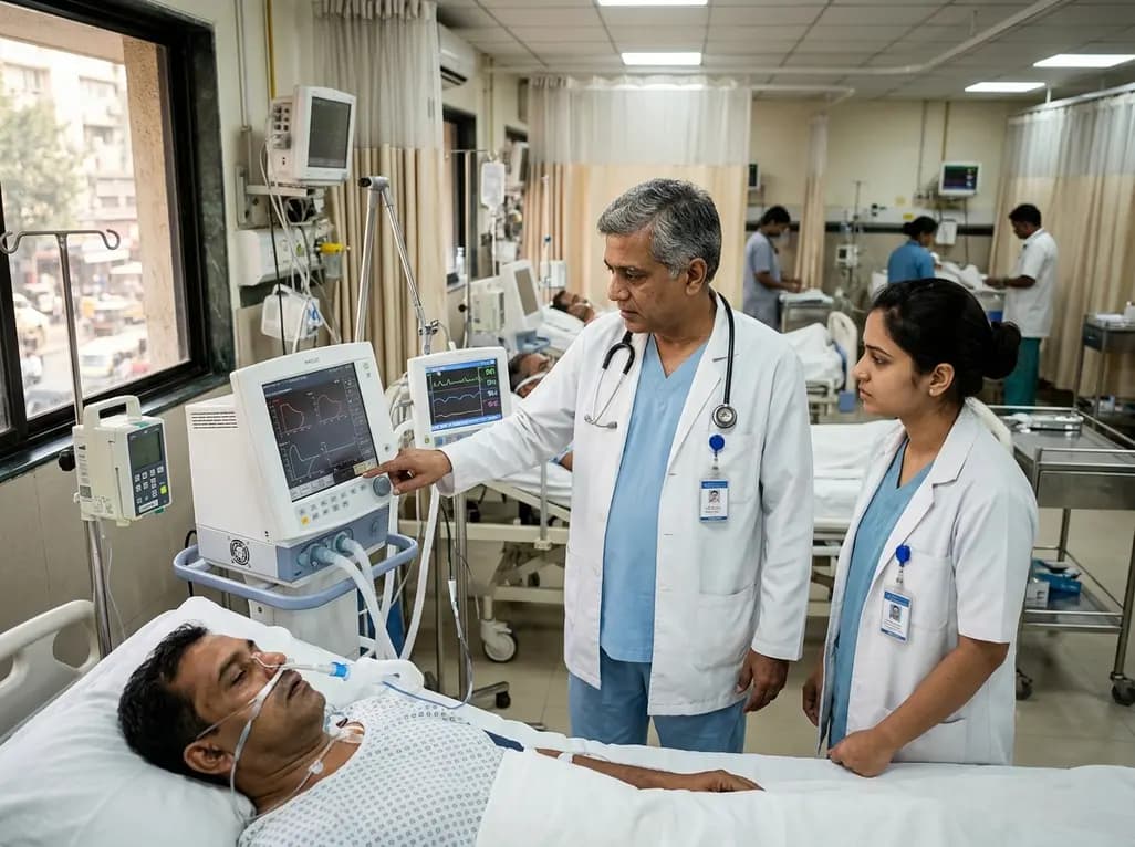
Mechanical ventilation remains a cornerstone of life support in intensive care units, yet optimizing ventilatory parameters for patients suffering from acute respiratory failure presents persistent clinical challenges. A major physiological obstacle during positive pressure ventilation is the dynamic collapse of conducting airways, which can occur during both inspiration and expiration. Recent observational research highlights how elevated pleural pressures serve as a primary driver of airway instability in critically ill patients. When pleural pressure rises due to chest wall stiffness, abdominal distension, or pulmonary edema, it exerts compressive forces on fragile distal airways. Understanding these mechanics is essential for clinicians striving to personalize mechanical ventilation and prevent ventilator-induced lung injury.
Airway collapse occurs when extraluminal pressures exceed intraluminal pressure, leading to anatomical closure of small airways. During inspiration, airway closure creates a threshold phenomenon known as airway opening pressure. Consequently, the mechanical ventilator must generate sufficient initial airway pressure to overcome this threshold before gas flow into alveoli can begin. Conversely, during expiration, cyclic airway narrowing or premature closure produces expiratory flow limitation. This phenomenon traps gas inside distal alveoli, generating intrinsic positive end-expiratory pressure, commonly termed auto-PEEP.
Both inspiratory airway opening pressure and expiratory flow limitation significantly impair gas exchange efficiency and increase the work of breathing. Furthermore, repetitive opening and closing of collapsed airways generates cyclic shear stress. This repetitive mechanical strain damages delicate alveolar-capillary membranes and accelerates inflammatory injury. Clinicians often miss these subtle mechanical abnormalities during routine bedside assessments because traditional ventilator graphics do not always display explicit closure dynamics. Therefore, advanced physiological monitoring techniques are required to detect hidden airway instability promptly. Recognizing these underlying physical mechanisms allows intensive care physicians to move beyond standardized ventilation protocols toward customized, patient-specific respiratory support strategies that minimize pulmonary trauma and optimize alveolar recruitment.
Pleural pressure represents the extraluminal force acting directly upon the outer surface of the pulmonary parenchyma and small airways. Under physiological conditions, pleural pressure remains negative throughout the normal respiratory cycle. However, critically ill patients frequently experience severe pathophysiological alterations that cause marked increases in intrathoracic chest wall stiffness. Conditions such as severe chest wall edema, pleural effusion, massive fluid resuscitation, obesity, and intra-abdominal hypertension directly contribute to elevated pleural pressures.
When pleural pressure increases, transpulmonary pressure—the net pressure distending the lung tissue—decreases dramatically at any given airway opening pressure. Consequently, the surrounding chest wall compresses the lung parenchyma and vulnerable conducting airways. This compressive force promotes total airway closure at higher lung volumes than expected. Studies using esophageal pressure monitoring confirm that higher esophageal pressures, which accurately mirror pleural pressures, directly correlate with greater inspiratory airway opening pressures and severe expiratory flow limitation. Furthermore, elevated pleural pressures elevate auto-PEEP levels after zero-PEEP washouts, confirming that elevated extrinsic forces physically prevent complete alveolar emptying. Managing these mechanical burdens requires targeted interventions aimed at counteracting extrinsic chest wall compressions while maintaining adequate transpulmonary pressure gradients.
Accurate assessment of respiratory mechanics requires precise measurement of pleural pressure in critically ill, mechanically ventilated patients. Because direct pleural pressure measurement is invasive and technically difficult, clinicians routinely utilize esophageal pressure as a reliable surrogate. An esophageal balloon catheter positioned in the lower third of the esophagus measures local pressure changes, allowing bedside estimation of pleural pressure and calculation of transpulmonary pressure.
To evaluate airway collapse dynamics, clinicians can perform slow-flow pressure-volume curve maneuvers after a washout period at zero positive end-expiratory pressure. During low-flow inflation, an abrupt inflection point in the pressure-volume curve identifies the airway opening pressure, marking the precise threshold required to recruit collapsed airways. Similarly, expiratory flow limitation can be identified through visual inspection of pressure-volume loops, monitoring PEEP absorber behavior, or applying advanced mathematical techniques such as the Rex method. Clinical trial data demonstrate that patients exhibiting airway opening pressure display significantly higher esophageal pressures compared to non-affected individuals. Additionally, patients with expiratory flow limitation show substantially higher esophageal pressures, confirming that esophageal manometry provides crucial diagnostic insight into extrinsic airway compression. Integrating these diagnostic tools allows clinicians to identify patients requiring tailored PEEP settings.
Clinical research in mechanically ventilated patients with acute respiratory failure provides strong empirical evidence linking pleural pressure to airway collapse. In prospective cohort evaluations of critically ill adults, inspiratory airway opening pressure was observed in a majority of ventilated patients, with a high proportion simultaneously exhibiting expiratory flow limitation. Statistically significant associations confirm that patients with inspiratory airway closure are far more likely to present with expiratory flow limitation than those without initial closure.
Quantitative analysis reveals strong positive correlations between baseline esophageal pressure following zero-PEEP washouts and measured airway opening pressure. Similarly, auto-PEEP values strongly correlate with both esophageal pressure and airway opening pressure thresholds. Patients presenting with airway closure demonstrate mean esophageal pressures exceeding 11 cmH2O, whereas non-affected patients show significantly lower baseline esophageal pressures around 7 cmH2O. Likewise, patients exhibiting expiratory flow limitation demonstrate significantly higher esophageal pressures than those maintaining continuous expiratory flow. These quantitative findings demonstrate that elevated pleural pressures directly cause airway collapse during both phases of respiration. Consequently, critical care practitioners must recognize that elevated airway opening pressure and expiratory flow limitation reflect abnormal chest wall mechanics rather than isolated intrinsic lung disease.
Identifying airway opening pressure and expiratory flow limitation carries profound therapeutic implications for mechanically ventilated patients. Applying positive end-expiratory pressure (PEEP) above the measured airway opening pressure prevents end-expiratory airway closure and cyclic recruitment-derecruitment. Consequently, setting PEEP appropriately counterbalances elevated pleural pressures, stabilizes small airways, and reduces shear stress on lung tissues.
Additionally, identifying expiratory flow limitation prevents inappropriate PEEP reductions that could exacerbate airway closure and gas trapping. When managing patients with elevated pleural pressures, clinicians must carefully balance ventilator parameters to avoid excessive alveolar overdistension while maintaining adequate open-lung conditions. Transpulmonary pressure-guided PEEP titration offers a personalized approach, ensuring that positive airway pressure effectively overcomes extrinsic chest wall compression without inducing volutrauma. Moreover, managing extrapulmonary causes of elevated pleural pressure—such as reducing intra-abdominal pressure, draining abdominal ascites, or optimizing fluid balance—can significantly reduce compressive forces on the lungs. Ultimately, combining physiological diagnostic techniques with customized mechanical ventilation strategies improves lung compliance, optimizes oxygenation, and protects critically ill patients from ventilator-induced pulmonary damage. Continuous bedside reassessment ensures that mechanical settings adapt dynamically as pleural pressure dynamics evolve during acute illness recovery.
Esophageal pressure serves as a clinical surrogate for pleural pressure because the esophagus lies within the thoracic cavity between the lungs and chest wall. A thin balloon catheter placed in the lower esophagus senses surrounding thoracic pressure changes. By subtracting esophageal pressure from airway pressure, clinicians calculate transpulmonary pressure. This calculation accurately reflects the net force distending alveolar tissue, helping clinicians adjust ventilator settings for patients with stiff chest walls.
Airway opening pressure is the minimal pressure required to reopen collapsed conducting airways at the start of inspiration. Clinically, identifying this pressure threshold is critical because ventilating below this level fails to recruit alveoli and causes cyclic airway injury. By setting positive end-expiratory pressure above the airway opening pressure, intensive care clinicians keep conducting airways patent throughout respiration, improving gas exchange and protecting delicate lung parenchyma from severe shear stress.
Clinicians detect expiratory flow limitation at the bedside using several specialized diagnostic methods. These include visual evaluation of continuous flow-volume loops during mechanical ventilation, observing PEEP absorber behavior, and performing zero-PEEP washout maneuvers with low-flow pressure-volume curves. Advanced mathematical algorithms, such as the Rex method, also analyze continuous flow signals. Detecting expiratory flow limitation enables clinicians to identify gas trapping and optimize ventilator parameters to prevent dynamic airway collapse.
Disclaimer: This content is for informational and educational purposes only and does not constitute medical advice, diagnosis, or treatment. Always seek the advice of a qualified healthcare provider with any questions you may have regarding a medical condition or ventilator management. Refer to the latest local and national guidelines for clinical practice.
References
1. Messias Hirano Padrao E et al. The association of elevated pleural pressures with airway collapse during inspiration and expiration on mechanically ventilated patients. J Appl Physiol (1985). 2026 Jul 22. doi: 10.1152/japplphysiol.00136.2026. PMID: 42485068.
2. Grieco DL, Chen L, Brochard L. Transpulmonary pressure: importance and measurement. Curr Opin Crit Care. 2017 Feb;23(1):17-23.
3. Yoshida T, Amato MBP, Grieco DL, et al. Esophageal pressure monitoring in acute respiratory distress syndrome. Intensive Care Med. 2018 Sep;44(9):1538-1541.

Read summarized clinical updates, watch expert medical content, and earn CME certifications right from your smartphone.
Open your smartphone camera to scan and install.





A study in mechanically ventilated patients reveals that elevated pleural pressures strongly correlate with airway collapse during inspiration (airway opening pressure) and expiration (expiratory flow limitation). Monitoring esophageal pressure helps optimize positive end-expiratory pressure titration.
3 months ago

Following regulatory scrutiny from FSSAI regarding promotional representations and lean muscle preservation claims, Dr. Reddy's Laboratories and Nestlé Health Science are currently reviewing the branding and marketing strategy for their joint nutritional supplement Celevida GLP+.
Today

Demanding exhaustive hormonal profiling can inadvertently discourage clinical studies involving females. Methodological standards must align with the specific scientific question to expand, not restrict, equitable evidence for women.
Today

The Directorate General of Health Services directs Indian states to halt the hazardous reuse and reprocessing of single-use medical devices. Discover regulatory implications under the Drugs and Cosmetics Act, critical infection risks, legal liabilities, and operational changes required across clinical specialties.
Today
A novel calcium polyphosphate coacervate enables injectable bone implants with in situ hierarchical porosity via phase separation and gas foaming, significantly enhancing cell proliferation, energy metabolism, and osteogenic differentiation for minimally invasive bone defect repair.
Today

India is advancing preventive healthcare by integrating genomics, artificial intelligence, and quantum technologies. With the SANGAM programme sequencing one million Indian genomes, clinicians can unlock personalized therapies, detect cardiometabolic risks early, and curb the burden of diabetes and heart disease.
Today
