

Loading, please wait...
Loading, please wait...

Accurate diagnosis of early-stage pulmonary sarcoidosis represents one of the most demanding clinical hurdles in modern respiratory medicine. When patients present with atypical intrathoracic lymphadenopathy or subtle parenchymal infiltrates, clinicians frequently face ambiguous clinical pictures. This diagnostic dilemma intensifies markedly when initial laboratory evaluations demonstrate normal serum angiotensin-converting enzyme activity. Consequently, physicians must explore innovative non-invasive tools to establish timely, reliable differential diagnoses.
Clinicians regularly encounter patients with bilateral hilar lymphadenopathy and vague respiratory symptoms who present significant diagnostic uncertainties. Although radiographic imaging provides essential anatomical clues, chest computed tomography cannot definitively distinguish noncaseating granulomas from malignant or infectious pathologies. For instance, lymphoma, bronchogenic carcinoma, and occupational pneumoconioses frequently mimic benign granulomatous syndromes. In addition, mediastinal lymph node tuberculosis exhibits radiographic patterns that appear nearly identical to sarcoidosis.
Consequently, clinicians frequently order serum biomarkers to clarify ambiguous radiological findings. However, classical biomarkers often lack sufficient sensitivity during the nascent stages of granulomatous development. When initial imaging results appear atypical, physicians face heightened pressure to perform invasive tissue samplings. Such invasive procedures carry recognized procedural risks, including pneumothorax, pulmonary hemorrhage, and anesthetic complications. Furthermore, specialized diagnostic facilities such as endobronchial ultrasound are not universally available across peripheral healthcare centers. Therefore, patients frequently experience prolonged diagnostic delays or receive inappropriate empirical therapies. Developing reliable non-invasive diagnostic strategies has thus become an urgent clinical priority for chest physicians worldwide.
For decades, clinicians have utilized serum angiotensin-converting enzyme as a frontline diagnostic biomarker for systemic and pulmonary sarcoidosis. Granulomatous epithelioid cells actively produce this peptidyl dipeptidase, theoretically elevating circulating enzyme concentrations above standard baseline parameters. Nevertheless, extensive clinical studies consistently reveal that normal serum enzyme activity occurs in up to forty percent of confirmed sarcoidosis cases. In early or localized presentations, the total anatomical granuloma burden remains relatively small. Consequently, enzyme release into systemic circulation often fails to exceed diagnostic laboratory thresholds.
Moreover, individual genetic factors significantly alter baseline enzyme levels. Polymorphisms within the ACE gene, particularly insertion and deletion alleles, account for substantial variations in physiological enzyme activity across diverse ethnic populations. Concomitant medications, such as angiotensin-converting enzyme inhibitors, also suppress circulating enzyme levels and mask underlying granulomatous disease. Conversely, elevated enzyme levels occur in several non-sarcoid conditions, including fungal infections, active tuberculosis, hyperthyroidism, and hepatic disorders. Therefore, relying exclusively on serum angiotensin-converting enzyme produces unacceptable rates of diagnostic misclassification, necessitating more comprehensive multi-analyte diagnostic frameworks.
To overcome these longstanding clinical limitations, recent research has explored artificial intelligence algorithms trained on routine peripheral blood indicators. Rather than relying on a solitary biomarker, modern machine learning paradigms evaluate multidimensional immunological interactions across standard hematological and biochemical panels. Researchers analyzed hundreds of patients presenting with atypical thoracic imaging and normal serum enzyme concentrations. By applying rigorous feature selection methods like LASSO regression, investigators isolated highly predictive peripheral blood indicators.
These algorithms examine complex patterns across complete blood counts, coagulation profiles, liver biochemistry, and systemic inflammatory indices. Specifically, variations in absolute lymphocyte counts, neutrophil-to-lymphocyte ratios, platelet indices, and serum globulin levels reflect distinct pathological processes. Machine learning models, including random forests and gradient boosting classifiers, synthesize these subtle physiological alterations into unified predictive probabilities. In clinical validation cohorts, these computational architectures achieved outstanding discriminatory accuracy, significantly outperforming individual traditional biomarkers. Because routine blood counts and metabolic panels are inexpensive and universally accessible, algorithmic analysis offers an equitable diagnostic solution for decentralized clinical environments.
In high-burden infectious environments, differentiating pulmonary sarcoidosis from mediastinal lymph node tuberculosis represents a paramount diagnostic challenge. Both clinical conditions present with subacute constitutional symptoms, persistent cough, and intrathoracic lymphadenopathy. Furthermore, standard histopathological examination can show granulomatous inflammation, yet acid-fast bacilli smears and mycobacterial cultures from needle aspirates frequently yield negative results. Because treating sarcoidosis requires systemic corticosteroid immunosuppression, misdiagnosing tuberculosis as sarcoidosis can trigger catastrophic mycobacterial dissemination.
Conversely, mistaking sarcoidosis for tuberculosis exposes patients to prolonged, toxic courses of empirical anti-tubercular therapy. Machine learning models address this critical dilemma by capturing systemic immune divergence between infectious and non-infectious granulomatous syndromes. Active tuberculosis characteristically stimulates pronounced monocytosis, accelerated catabolism, and acute-phase reactant elevations due to persistent bacterial antigen stimulation. In contrast, sarcoidosis typically demonstrates compartmentalized pulmonary inflammation with distinct peripheral immune modulation. By integrating these nuanced hematological divergence patterns, computational models provide high diagnostic specificity. Consequently, clinicians can avoid unnecessary empirical anti-tubercular regimens and expedite appropriate anti-inflammatory therapies.
Implementing artificial intelligence models into frontline clinical practice can streamline existing diagnostic pathways for complex pulmonary cases. When patients present with atypical lymphadenopathy and normal serum enzymes, clinicians input standard laboratory values into a decision-support tool. The algorithm analyzes routine complete blood counts, metabolic markers, and inflammatory indices, instantly computing a validated risk probability for pulmonary sarcoidosis. Consequently, this non-invasive probability assessment assists clinicians in categorizing patients into distinct risk cohorts.
Individuals with high model-predicted probability for sarcoidosis can proceed directly to confirmatory endobronchial ultrasound or targeted biopsy, avoiding extensive, costly exclusion workups. Conversely, patients identified with high risk for tuberculosis or malignancy receive rapid microbiologic testing or oncologic referrals without delay. Furthermore, in remote healthcare centers lacking advanced interventional bronchoscopy suites, algorithmic scoring provides valuable objective guidance for timely patient transfer. While algorithms cannot replace histopathology, they provide an invaluable cognitive safety net. They optimize procedural decisions, reduce medical expenses, and accelerate definitive therapy.
Serum angiotensin-converting enzyme is produced by epithelioid cells within granulomas. Consequently, its circulating concentration directly reflects total granuloma burden across the body. In early or mild disease stages, the overall granuloma mass remains relatively limited. Therefore, enzyme secretion often fails to exceed standard laboratory reference ranges. Furthermore, ACE gene polymorphisms significantly influence baseline physiological enzyme concentrations. As a result, relying solely on serum ACE produces frequent false negatives during early disease evaluations.
Tuberculosis and sarcoidosis induce distinct systemic inflammatory responses that alter routine peripheral blood parameters. Specifically, active tuberculosis generally provokes marked systemic catabolism, profound neutrophilia, and significant monocytosis. In contrast, sarcoidosis displays distinctive shifts in lymphocyte subsets, platelet indices, and albumin levels. Therefore, multivariable algorithmic models evaluate these intricate multi-parameter ratios simultaneously. Consequently, the algorithms detect subtle immunological footprints that clearly separate infectious mycobacterial disease from noncaseating granulomatous inflammation without requiring tissue confirmation.
Currently, artificial intelligence tools cannot completely replace histopathological confirmation, which remains the definitive gold standard. However, these validated models serve as exceptional clinical triage instruments. Clinicians can utilize these non-invasive scores to identify patients with very high pre-test probability. Furthermore, this approach reduces unnecessary diagnostic delays and prevents empirical anti-tubercular therapy in resource-limited settings. Consequently, computational models guide safer, targeted procedural referrals, minimizing invasive endobronchial ultrasound biopsies for indeterminate presentations.
Disclaimer: This content is for informational and educational purposes only. It is not intended to substitute for professional medical advice, diagnosis, or treatment. Always seek the advice of your physician or other qualified health provider with any questions you may have regarding a medical condition or before undertaking any diet, supplement, fitness, or other health program. Refer to the latest local and national guidelines for clinical practice.
References

Read summarized clinical updates, watch expert medical content, and earn CME certifications right from your smartphone.
Open your smartphone camera to scan and install.





Machine learning models using routine peripheral blood markers provide high diagnostic accuracy for early-stage pulmonary sarcoidosis in patients presenting with normal serum ACE activity, aiding differentiation from tuberculosis and malignancy.
Today

A KGMU study reveals over half of women with breast abnormalities fail to undergo diagnostic confirmation, leading to advanced-stage detection. Discover the socio-cultural barriers, clinical risks of incomplete resection, and community-level solutions needed to bridge the critical screening-to-diagnosis gap in India.
Today
A meta-analysis of 18 studies reveals that bariatric surgery does not significantly alter circulating homocysteine levels, regardless of follow-up duration. Clinicians must maintain vigilance over post-surgical micronutrient supplementation to prevent hyperhomocysteinemia.
Today
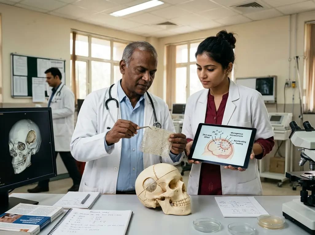
Researchers have engineered a self-powered Zn/C micro-galvanic electrospun membrane that couples sustained zinc release with endogenous microcurrents. This bioelectric scaffold stimulates M2 macrophage polarization and accelerates bone deposition to achieve robust cranial defect regeneration.
Today
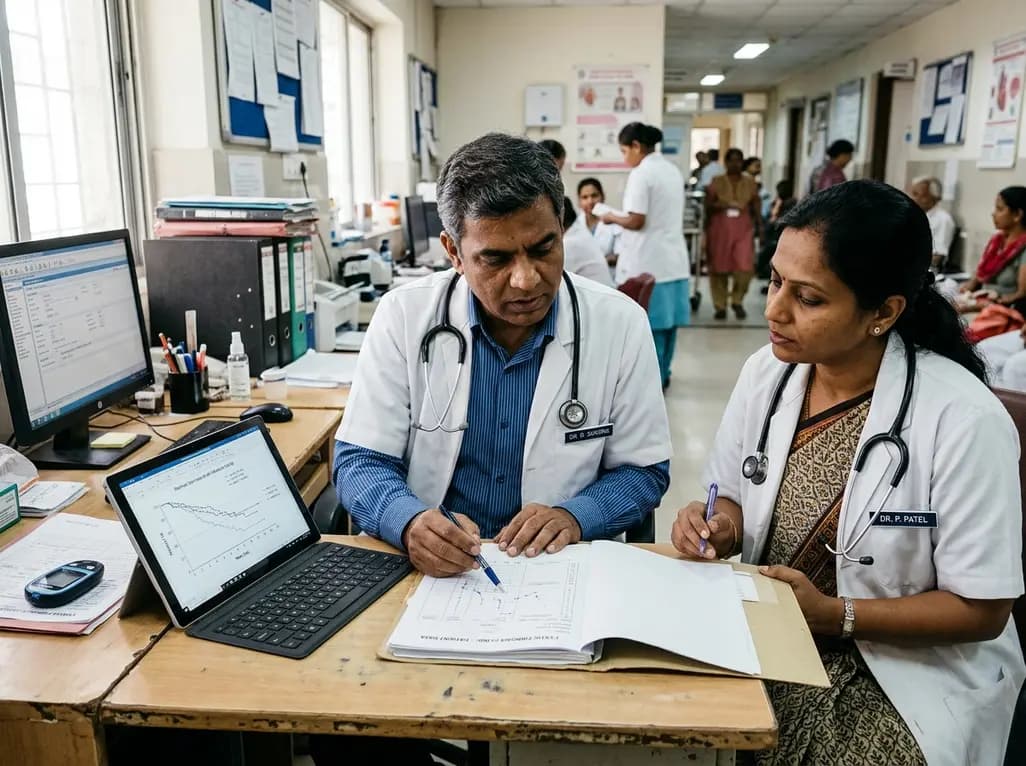
A nationwide UK Cystic Fibrosis Registry study demonstrates that higher updated mean HbA1c levels correlate with a graded, significant increase in all-cause mortality among individuals with cystic fibrosis-related diabetes, reinforcing the critical need for proactive glycemic monitoring and intervention.
Today
