

Loading, please wait...
Loading, please wait...

Stereotactic radiosurgery serves as an effective definitive and adjuvant treatment for intracranial meningiomas in contemporary neurosurgical oncology. Following radiation delivery, clinicians routinely schedule serial neuroimaging to monitor tumor regression and detect potential adverse radiation effects. However, neurosurgeons continue to debate the clinical necessity and optimal timing of routine early MRI follow-up within the first twelve months post-treatment. Intensive surveillance schedules frequently generate substantial patient distress and strain healthcare resources. A recent landmark study investigated whether early surveillance scans genuinely alter medical decision-making in clinical practice. By identifying specific patient cohorts that truly benefit from early imaging, medical teams can safely streamline surveillance protocols and optimize healthcare delivery.
Historically, post-radiosurgical surveillance schedules for solitary meningiomas have reflected institutional habit rather than robust empirical evidence. Many neurosurgical centers reflexively perform contrast-enhanced magnetic resonance imaging at three, six, and twelve months following stereotactic radiosurgery. Radiation oncologists often justify this rigorous surveillance schedule by citing concerns regarding post-radiation edema and pseudoprogression. Consequently, patients undergo multiple detailed brain scans throughout their initial treatment year. However, benign solitary meningiomas typically exhibit slow cellular proliferation and respond gradually to targeted stereotactic radiation. Therefore, marked volumetric changes or acute tumor progression rarely manifest within the first twelve months after treatment. In fact, early scans often detect asymptomatic, self-limiting peritumoral changes that resolve spontaneously without therapeutic intervention. These ambiguous radiographic findings frequently prompt diagnostic uncertainty and unnecessary clinical anxiety rather than definitive intervention. Moreover, repeated clinic visits and serial imaging impose significant cumulative costs upon patients and healthcare institutions. Thus, neurosurgical teams are critically questioning whether unselected early scanning provides genuine clinical utility.
To rigorously evaluate imaging yield, researchers conducted a comprehensive cohort study analyzing solitary meningiomas treated with radiosurgery. Specifically, the team examined one-hundred and twenty-five radiosurgical treatments across one-hundred and fifteen adult patients who completed imaging within twelve months. The investigators tracked whether early neuroimaging altered therapeutic decisions or prompted concrete clinical actions. They classified clinical actions into major events, such as surgical resection or repeat radiation, and minor adjustments, like steroid administration. Overall, early scans prompted clinical actions in only twelve percent of the entire treated cohort. Major therapeutic interventions occurred in five percent of cases, while minor changes occurred in seven percent. Remarkably, patients presenting with primary, treatment-naive benign solitary meningiomas required no major clinical interventions during the initial follow-up window. This finding proves that unselected early neuroimaging rarely alters clinical management for typical benign lesions. Consequently, routine scanning before one year yields minimal therapeutic benefit for the vast majority of patients. Clinicians can safely reconsider rigid early surveillance timelines.
Distinguishing between major and minor clinical interventions clarifies the true utility of post-treatment imaging. In the cohort, major therapeutic actions occurred exclusively in patients experiencing treatment failure or progressive symptoms. These cases required urgent craniotomy for surgical decompression or salvage stereotactic radiotherapy to re-establish control. Conversely, minor actions comprised benign clinical decisions that rarely altered long-term oncological outcomes. Clinicians primarily prescribed brief, tapering courses of dexamethasone to alleviate mild peri-tumoral edema or transient headache. In other cases, minor action simply entailed scheduling the next routine magnetic resonance appointment slightly earlier than planned. Furthermore, patients who developed significant complications almost universally experienced progressive neurological symptoms that prompted medical contact before scheduled imaging. Truly asymptomatic individuals rarely displayed unexpected radiographic abnormalities requiring emergent medical or surgical management. Therefore, routine early scans in asymptomatic patients with benign tumors primarily verify anticipated, stable intracranial anatomy. Consequently, neurosurgeons should rely primarily on clinical symptom monitoring rather than unselected serial imaging to detect early post-treatment complications.
Multivariable analysis identified two decisive risk factors predicting early clinical action: prior tumor-directed treatment and aggressive histological grade. Specifically, patients who underwent radiosurgery for residual or recurrent meningiomas following previous surgery or irradiation demonstrated nearly eight-fold higher odds of clinical action. Furthermore, individuals with World Health Organization Grade 2 or Grade 3 meningiomas exhibited over thirteen-fold greater odds of requiring therapeutic adjustments. In stark contrast, treatment-naive tumors and benign Grade 1 lesions demonstrated negative predictive values of ninety-six percent and ninety-three percent, respectively. Therefore, baseline disease characteristics provide reliable guidance for individualizing post-radiosurgical surveillance schedules. High-grade meningiomas harbor elevated mitotic activity, aggressive cellular infiltration, and higher propensities for early recurrence. Accordingly, these high-risk cohorts warrant vigilant surveillance with contrast neuroimaging at three and six months post-treatment. Conversely, treatment-naive benign solitary meningiomas maintain extremely low risks of early therapeutic failure. By replacing blanket surveillance practices with precise risk stratification, multidisciplinary neuro-oncology teams can focus diagnostic resources on patients facing genuine recurrence hazards.
Rationalizing surveillance timelines yields major psychological, economic, and operational benefits. Frequent neuroimaging generates considerable emotional distress, widely described as scanxiety, for recovering brain tumor patients. Anticipatory worry surrounding surveillance scans diminishes quality of life and exacerbates somatic complaints. Furthermore, frequent magnetic resonance scans impose heavy financial burdens on patients through procedure charges, lost wages, and travel expenses. This consideration holds special relevance in developing healthcare environments like India, where patients predominantly pay out-of-pocket for neuro-oncology care. In addition, neuroimaging centers in high-volume public hospitals face severe backlogs and lengthy waiting lists for diagnostic appointments. Postponing baseline scans to twelve months for low-risk, treatment-naive benign meningiomas safely alleviates patient anxiety while conserving valuable diagnostic scanner capacity. Meanwhile, clinical teams can prioritize immediate scanning slots for acute neurological emergencies and complex high-grade malignancies. Clinicians must accompany this deferred imaging approach with structured patient counseling and accessible teleconsultation. Patients instructed on specific red-flag symptoms can promptly seek medical care if unexpected changes arise. Thus, personalized surveillance fosters cost-effective, compassionate, and patient-centered neuro-oncology practice.
Benign, treatment-naive solitary meningiomas exhibit indolent biology and respond slowly to stereotactic radiosurgery. Evidence demonstrates that early surveillance scans within twelve months rarely alter patient management or mandate surgical re-intervention. Therefore, clinicians can safely defer routine neuroimaging to one year post-treatment in asymptomatic individuals without compromising clinical safety or long-term tumor control.
Patients diagnosed with high-grade meningiomas, such as World Health Organization Grade 2 or 3 lesions, require intensive early monitoring. Furthermore, individuals treated for recurrent tumors following prior surgery or radiation face significantly higher odds of early therapeutic failure. These high-risk cohorts benefit from targeted three-month to six-month scans to promptly detect progression.
Clinicians should implement structured clinical follow-up visits and comprehensive patient education instead of relying entirely on early scans. Specifically, medical teams must counsel patients to report warning symptoms immediately, including new-onset seizures, worsening headaches, or focal motor deficits. Rapid symptomatic assessment promptly identifies the rare individuals who need unscheduled, urgent imaging evaluations.
Disclaimer: This content is for informational and educational purposes only. It is not intended to be a substitute for professional medical advice, diagnosis, or treatment. Always seek the advice of your physician or other qualified health provider with any questions you may have regarding a medical condition. Never disregard professional medical advice or delay in seeking it because of something you have read here. Refer to the latest local and national guidelines for clinical practice.
References

Read summarized clinical updates, watch expert medical content, and earn CME certifications right from your smartphone.
Open your smartphone camera to scan and install.





A retrospective study reveals that routine early MRI follow-up within 12 months after radiosurgery rarely changes management in benign, treatment-naive solitary meningiomas, supporting a safe deferral of surveillance imaging to one year post-treatment.
Today

Standardizing continuous physiological waveforms in EHRs has long challenged clinical teams. The novel TIDE framework leverages HL7 FHIR R4 to decouple queryable clinical metadata from massive raw signal repositories, unlocking scalable telemetry exchange for bedside care and predictive research.
Today

A secondary analysis of the MOMS clinical trial reveals that infant sex does not influence neonatal opioid withdrawal syndrome severity in buprenorphine-exposed newborns. Maternal co-exposures and clinical factors drive outcomes, highlighting the need for objective assessment and supportive breastfeeding.
Today
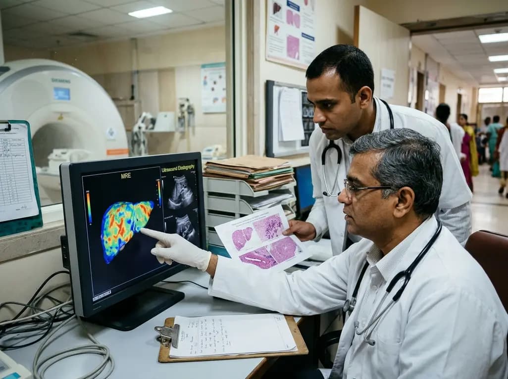
A comparative study reveals magnetic resonance elastography outperforms ultrasound elastography in assessing liver fibrosis in Fontan-associated liver disease, demonstrating stronger histological agreement and superior discrimination.
Today

A systematic review and meta-analysis highlights the substantial impact of plyometric jump training on jump performance, reactive strength, and landing kinematics in female team-sport athletes, providing evidence-based programming guidelines for sports medicine and orthopedics.
Today

A comprehensive review of the role of growth hormone in diabetic kidney disease and podocytopathy. Learn how elevated GH activates ZEB2, Notch, and TGF-beta1, driving podocyte loss and glomerulosclerosis, and explore novel targeted therapeutic approaches.
Today
